
For decades, two-dimensional (2D) cell culture has been a fundamental tool in biomedical research. Growing cells on flat plastic surfaces has enabled discoveries in cell biology, disease mechanisms, drug development, and toxicology, however, biology does not happen on a flat surface.
Inside the human body, cells exist within complex three-dimensional environments. They interact with neighboring cells, extracellular matrix (ECM), soluble factors, mechanical forces, and tissue-specific microenvironments. These interactions influence cell morphology, differentiation, signaling, gene expression, metabolism, and response to therapeutic compounds. This is where 3D cell culture is becoming increasingly valuable.
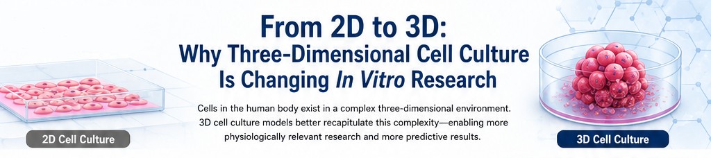
By allowing cells to organize into three-dimensional structures such as spheroids, 3D cultures can provide a more physiologically relevant model of human tissue while retaining many of the experimental advantages of in vitro research.
Why Does 3D Cell Culture Matter?
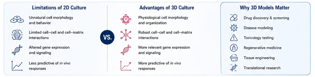
In conventional 2D culture, cells attach to a rigid, flat surface. Their interactions are primarily limited to neighboring cells within the same plane, and the artificial geometry can substantially alter cellular behavior. In contrast, 3D culture allows cells to interact in multiple dimensions and, in many models, to produce and organize their own extracellular matrix. This difference can have significant biological consequences.
Three-dimensional cultures can support:
- More physiologically relevant cell-cell interactions
- Cell-ECM interactions
- Tissue-like cellular organization
- More representative cellular morphology
- Development of tissue-specific phenotypes
- More complex signaling between different cell types
- Physiological gradients of oxygen, nutrients, and metabolites
- More biologically relevant responses to drugs and other treatments
These characteristics make 3D models particularly attractive for drug discovery, disease modeling, toxicity testing, regenerative medicine, tissue engineering, and translational research.
Spheroids: A Practical Bridge Between 2D Culture and Complex Tissue Models
Among the different approaches to 3D cell culture, spheroids have emerged as one of the most accessible and versatile platforms. Spheroids can be generated from a single cell type or from multiple cell types. In co-culture models, different populations can interact within the same three-dimensional microenvironment, enabling researchers to investigate biological processes that are difficult to reproduce using conventional monolayers.
For example, interactions between endothelial cells, pericytes, and astrocytes can be incorporated into a three-dimensional blood-brain barrier model. Similarly, co-culturing hepatocytes with supporting cells can provide a more representative system for investigating liver biology and disease.
The challenge, however, is that building a reliable 3D model from scratch can be technically demanding and time-consuming. Researchers may need to optimize cell ratios, aggregation conditions, culture media, plate formats, incubation conditions, spheroid size, uniformity, and downstream assay compatibility. This is where ready-to-use 3D models can provide a major advantage.
Ready-to-Use 3D Spheroids: Simplifying the Transition to 3D
Our Ready-to-Use 3D Spheroid portfolio is designed to remove much of the complexity associated with establishing 3D cultures. Instead of starting with individual cells and spending days or weeks developing and optimizing a spheroid-generation workflow, researchers can begin with cryopreserved, pre-formed 3D spheroids.
This can provide several practical benefits:
1. Save valuable research time
Developing a new 3D model can require substantial optimization. Ready-to-use spheroids allow researchers to move more rapidly from experimental planning to data generation.
2. Improve experimental consistency
Standardized, pre-formed spheroids can help reduce variability associated with manual spheroid formation and optimization.
3. Reduce the technical barrier to 3D culture
Researchers who are highly experienced in 2D cell culture but are new to 3D models can incorporate 3D systems without first developing an entirely new culture platform.
4. Support higher-throughput studies
Ready-to-use spheroids can be plated into 24-, 48-, or 96-well formats, making them suitable for screening and comparative experimental designs.
5. Start with biologically relevant multicellular systems
Many models incorporate primary human cells and, where appropriate, multiple cell types, providing an opportunity to investigate cellular interactions that cannot be adequately captured in simple monocultures.
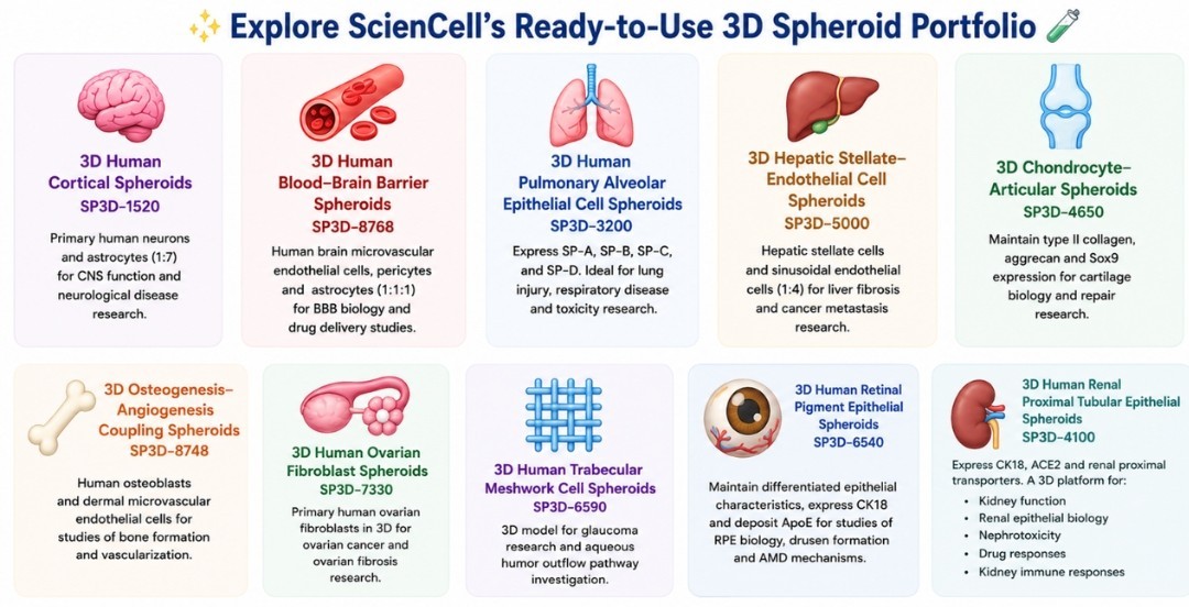
Explore Our Ready-to-Use 3D Models
Making 3D More Accessible
One of the most important developments in 3D cell culture is not simply the ability to create more sophisticated models, it is the ability to make those models practical and reproducible for everyday research.
A sophisticated model has limited value if researchers spend weeks troubleshooting how to generate it before they can perform their actual experiment.
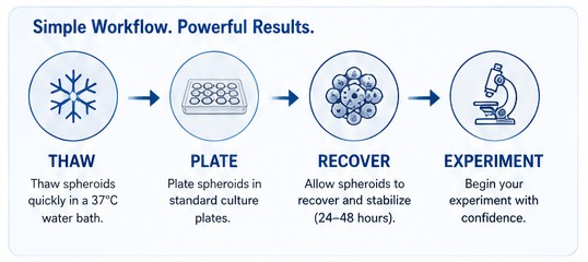
Ready-to-use spheroids address this bottleneck by providing researchers with a standardized starting point and making the workflow remarkably straightforward.
Bringing More Relevant Biology to the Bench
The fundamental advantage of 3D cell culture is simple:
Cells behave differently when they are allowed to interact in three dimensions.
By incorporating cell-cell interactions, extracellular matrix, tissue-like architecture, and multicellular communication, 3D models can provide biological information that conventional monolayer cultures may not capture. Our ready-to-use 3D spheroids take this concept one step further by making pre-formed, standardized models accessible without requiring researchers to build the entire 3D culture workflow themselves.
From cortical and blood-brain barrier models to liver, lung, bone, cartilage, ovarian, and ophthalmic models, these systems provide researchers with practical tools for exploring increasingly complex biological questions. The future of in vitro research isn't necessarily 2D versus 3D. It's choosing the right model for the biological question—and increasingly, that model may need three dimensions.