Sciencell Research Team
-
Posted: August 11, 2025
 Read more »
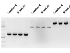
Human primary cardiomyocytes and cardiac tissue are invaluable for heart disease studies, drug discovery, and toxicity testing, often surpassing animal models or immortalized cell lines in relevance. However, obtaining adult human primary cardiomyocytes is challenging due to limited tissue availability,
-
Posted: July 30, 2025
.jpg) Read more »
Read more »Access Primary Cell Material Without the Hassle
If your research depends on DNA, RNA, or protein lysates from human primary cells, you know how challenging and expensive it can be to isolate these materials yourself. That’s where ScienCell’s Human Cell Pellets come in, offering an opportunity to
-
Posted: July 28, 2025
.jpg) Read more »
Read more »July'25 Edition-Seamless Protein Processing Starts Here: Explore Our Advanced Proteases!
Introducing Advanced Proteases for Your Research Needs:
At Sciencell, we are committed to providing high-quality tools to support groundbreaking research. This month, we are excited to showcase two powerful proteases:
-
Posted: July 06, 2025
.jpg) Read more »
Read more »Accelerate Your Research with ScienCell's High-Quality Recombinant Proteins!
Unlock precise control over cellular processes with ScienCell's extensive range of recombinant human proteins. Engineered for stability and biological activity, our products are essential tools for studying cell proliferation,
-
Posted: June 10, 2025Categories: Newsletter
.jpg) Read more »
Read more »Preserve the Future of Your Research with ScienCell's StemCryo®!
Unlocking the full potential of human pluripotent stem cells (hPSCs) often hinges on effective cryopreservation techniques.
ScienCell provides StemCryo®, a cutting-edge solution designed to ensure the viability and pluripotency of these
-
Posted: June 09, 2025Categories: Newsletter
.jpg) Read more »
Read more »Unraveling Cellular Dynamics: Explore Dermal Fibroblasts from ScienCell!
Fibroblasts, fundamental mesenchymal cells derived from the embryonic mesoderm, are widely utilized in cellular and molecular research due to their ease of culture and robust nature. These versatile cells are key secretors of a
-
Posted: June 09, 2025Categories: Newsletter
.jpg) Read more »
Read more »Accelerate your Stem Cell Research with Growth Factors and Recombinant Proteins from ScienCell!
Stem cells are multipotent cells with the ability to self-renew, self-replicate, and differentiate into multiple cell types, including mesenchymal stem cells (MSCs), hematopoietic stem cells (HSCs), neural
-
Posted: June 05, 2025
.jpg) Read more »
Read more »Save Time. Reduce Variability. Get Results.
Ready-to-Use Molecular Biology Products from Primary Cells
Working with primary cells is essential for biologically relevant data, but sourcing tissues, isolating cells, and extracting high-quality RNA, DNA, or protein can be time-consuming, expensive, and
-
Posted: May 28, 2025Categories: Newsletter
.jpg) Read more »
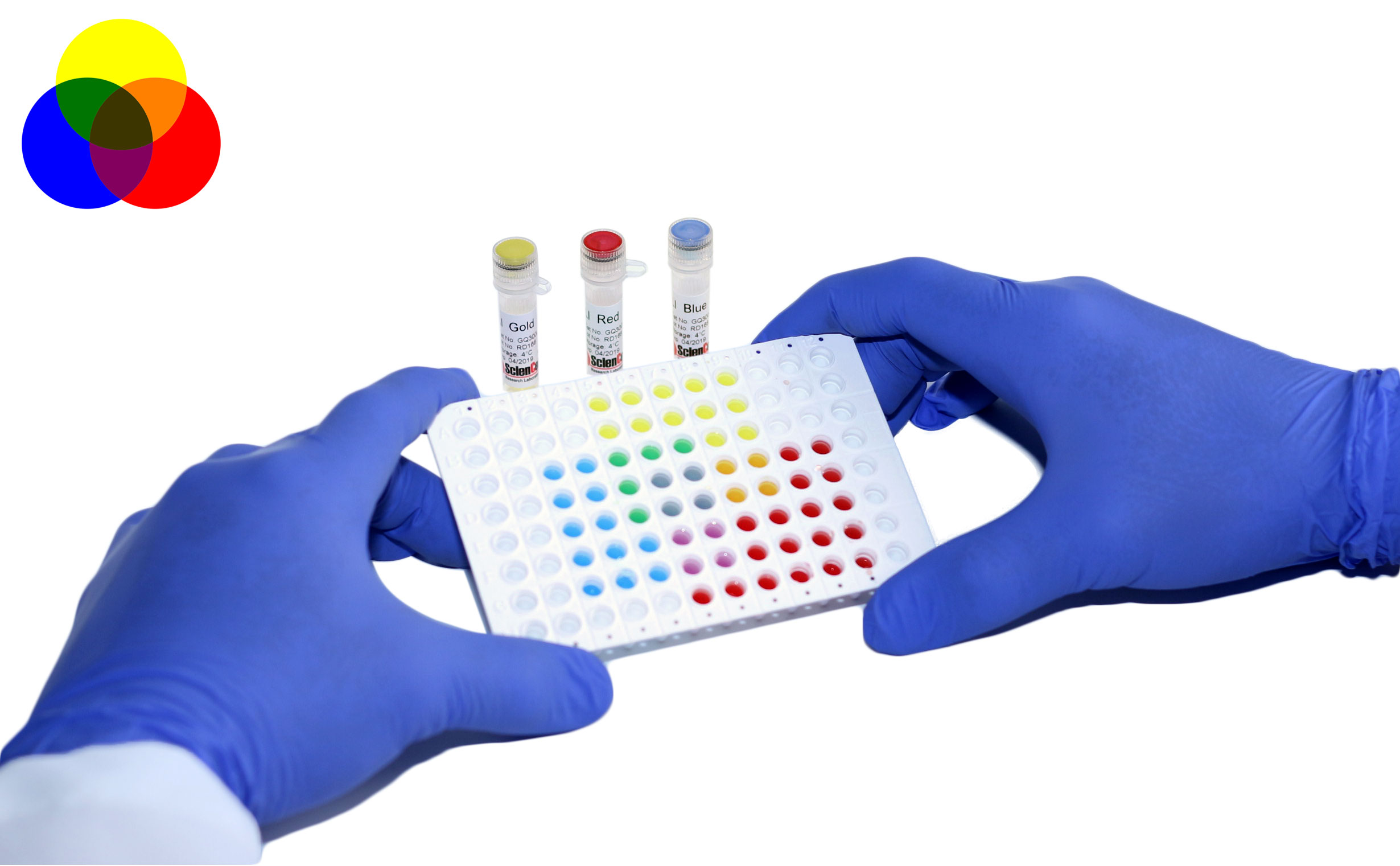
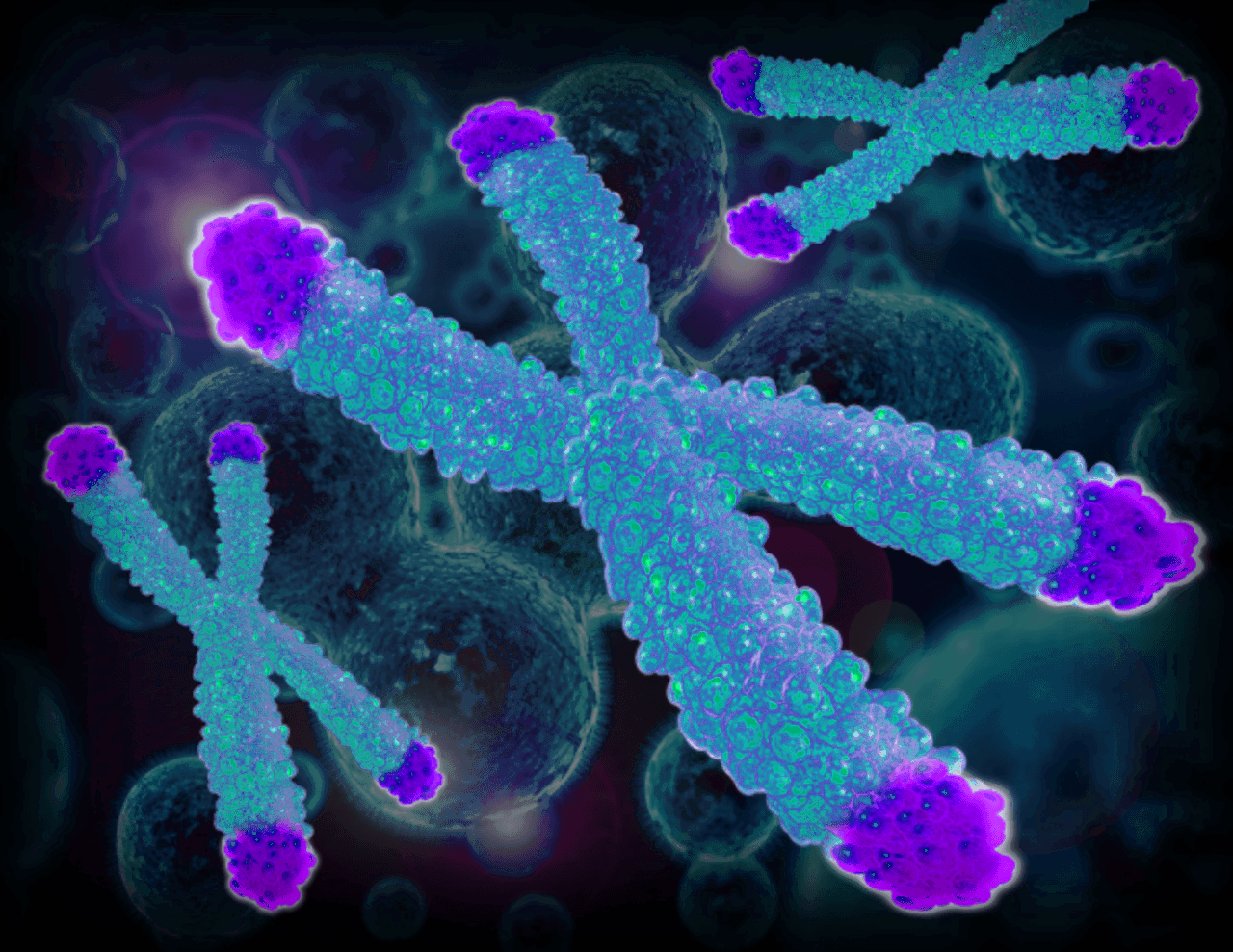
Read more »Unlock Hidden Insights: Pseudogene Analysis with GeneQuery™ qPCR Kits!
Pseudogenes are DNA segments similar to functional genes but with lost function. They were first described in 1977. Based on origin, they are classified as processed (e.g., human PTENP1), non-processed (e.g., human ARHGAP27P1), or
-
Posted: May 28, 2025Categories: Newsletter
.jpg) Read more »
Read more »Unlock Your Research Potential with ScienCell's High-Quality Macrophages!
Macrophages are crucial cells in the body, known for their role in removing cellular debris and destroying invading pathogens. Their main functions include phagocytizing invading microorganisms and scavenging dead, damaged cells,
-
Posted: May 27, 2025
.jpg) Read more »
Read more »Why Every Researcher Should Care About Metabolite Assays?
Several key metabolites play crucial roles in cellular bioenergetic homeostasis. ScienCell has developed sensitive assays to accurately quantify L-lactate, glucose, glutamate, and pyruvate.
L-Lactate is a key intermediate in glucose metabolism
-
Posted: May 26, 2025
.jpg) Read more »
Read more »Unlock the Power of Differentiation with Mesenchymal Stem Cells from Adipose, Liver, Bone, and Umbilical Cord!
Why Choose Our MSCs? Our Mesenchymal Stem Cells are:
- Multipotent—capable of differentiating into a variety of lineages, such as osteoblasts, chondrocytes, endothelial cells, hepatocyte-like
-
Posted: May 21, 2025Categories: Newsletter
.jpg) Read more »
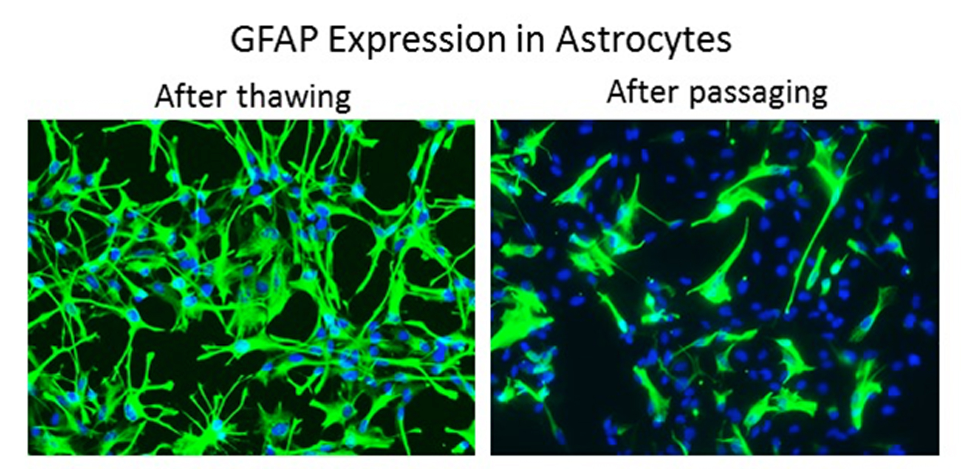
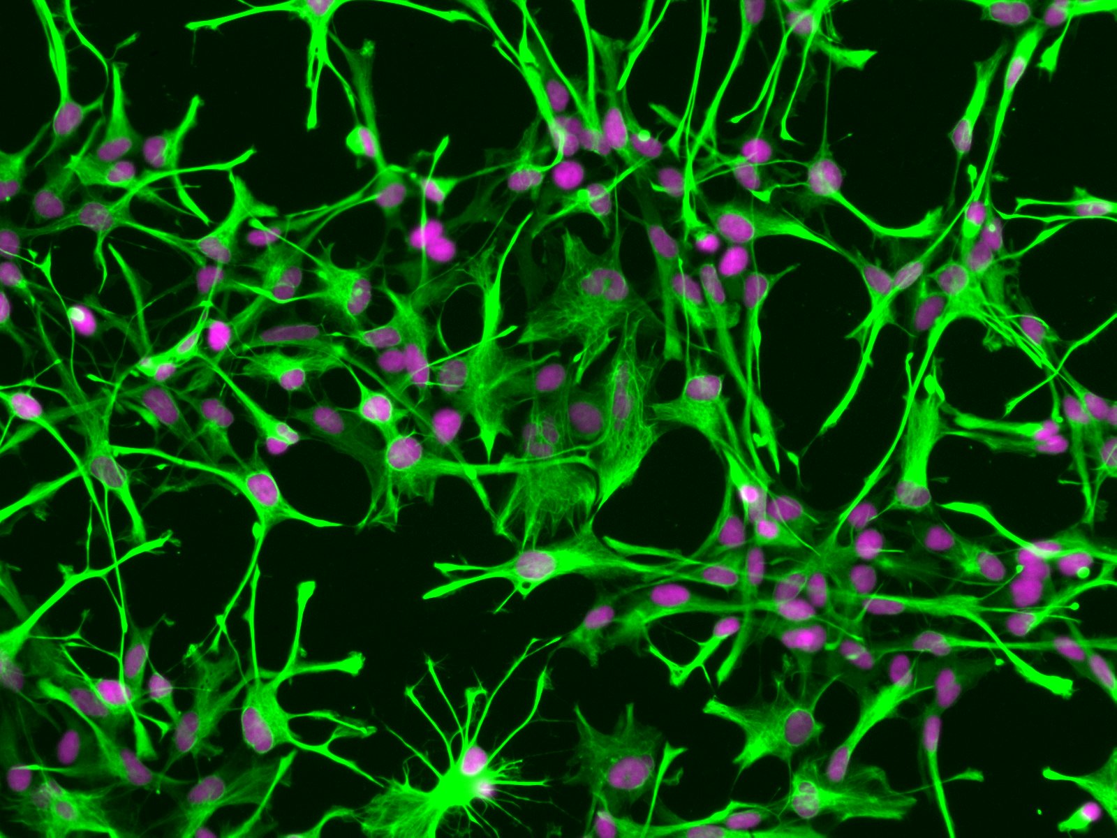
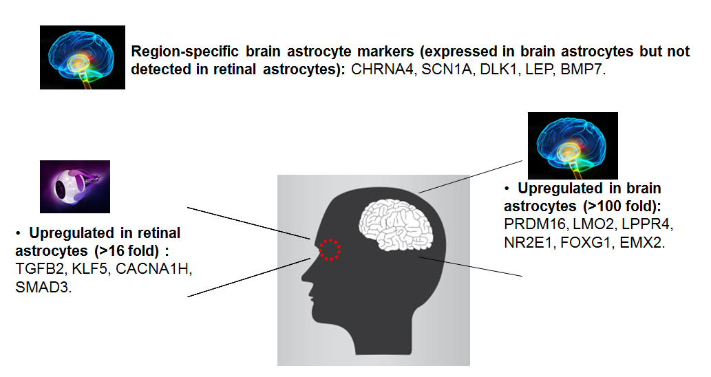
Read more »Astrocytes are the most abundant cell type in the central nervous system (CNS). They serve a variety of vital functions including supporting neuronal transmission and survival, regulating the blood-brain barrier, contributing to anti-inflammatory responses, and aiding in wound repair.
Astrocytes also
-
Posted: May 19, 2025Categories: Newsletter
.jpg) Read more »
Read more »GeneQuery™ Disease Models qPCR Array Kits are powerful tools for understanding the molecular mechanisms underlying disease onset and progression. By measuring the activity levels of specific genes, researchers can gain crucial insights into disease pathways, identify potential biomarkers, and evaluate
-
Posted: May 18, 2025
 Read more »
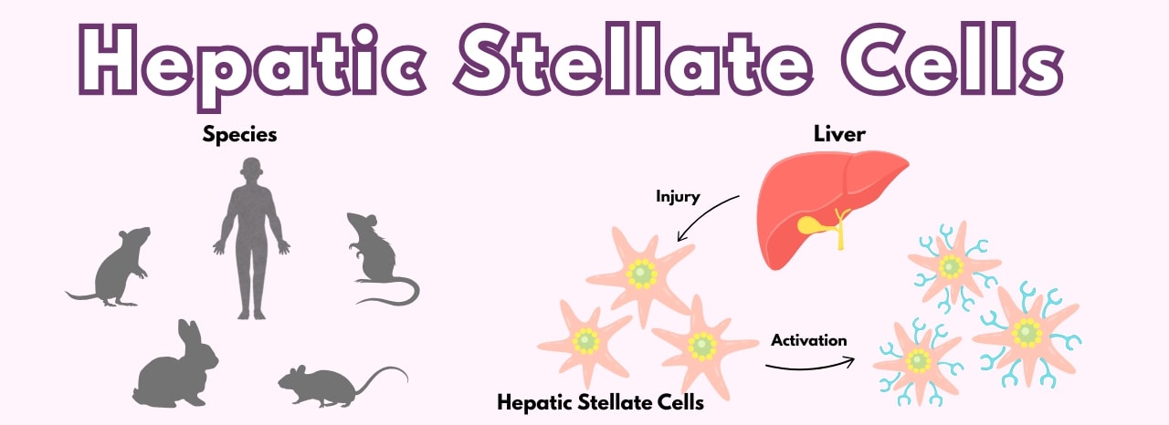
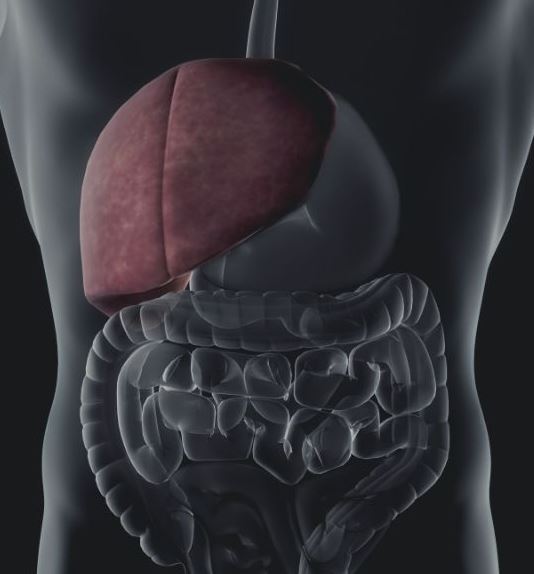
Drive Liver Research Forward with Hepatic Stellate Cells
Hepatic stellate cells (HSteC) are essential intralobular connective tissue cells that perform diverse functions in the liver, including extracellular matrix homeostasis, repair, regeneration, fibrosis, and control of retinol metabolism, storage,
-
Posted: May 15, 2025
 Read more »
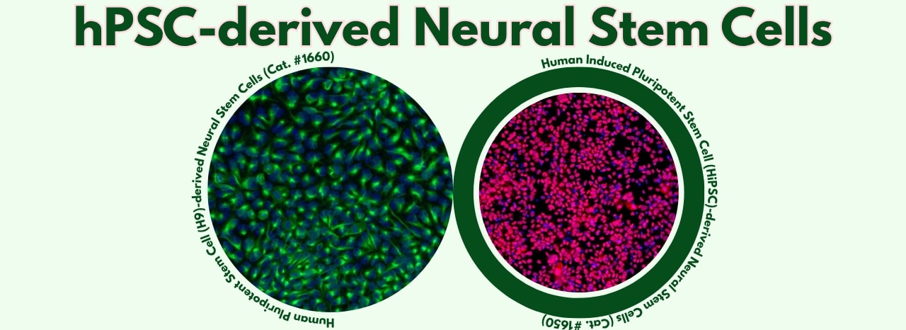
Leading the way in providing advanced tools for life science research, ScienCell Research Laboratories offers high-quality, well-characterized neural stem cell products derived from pluripotent stem cell sources, along with supporting reagents.
These products provide unique advantages for diverse applications,
-
Posted: May 15, 2025Categories: Newsletter
 Read more »
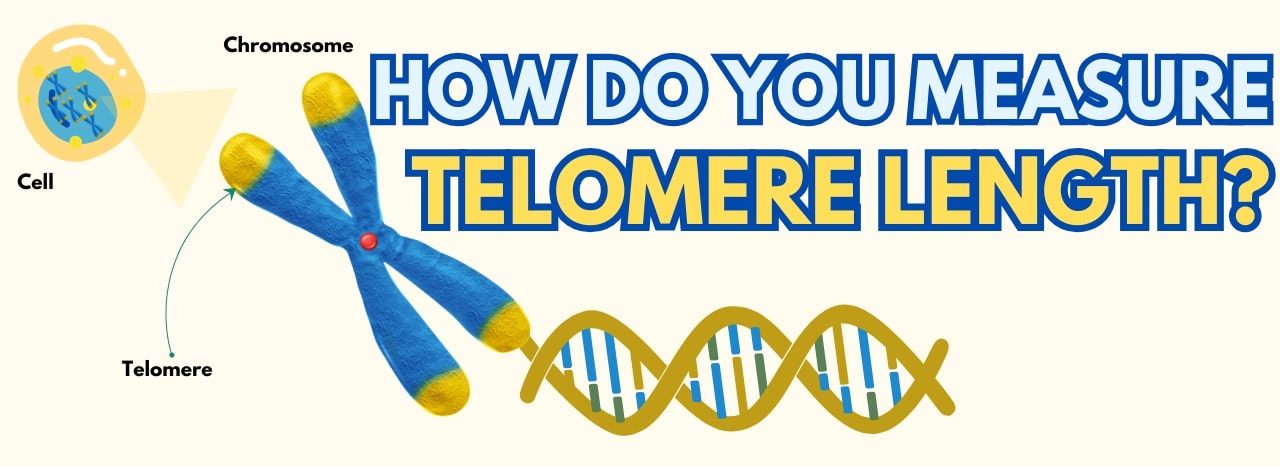
Advance Your Telomere Research Using ScienCell’s Absolute Telomere Length Quantification qPCR Kits!
Understanding Telomeres and Their Role in Genomic Stability
Telomeres are nucleoprotein structures that cap chromosome ends, protecting them from degradation and ensuring genomic stability. Each cell
-
Posted: May 14, 2025
 Read more »
In cellular and molecular research, the quality and reliability of your reagents are paramount to achieving reproducible and meaningful results. ScienCell offers a selection of high-quality
cell culture reagents designed to support critical steps in your experimental workflows!
Essential Solutions
-
Posted: May 14, 2025
 Read more »
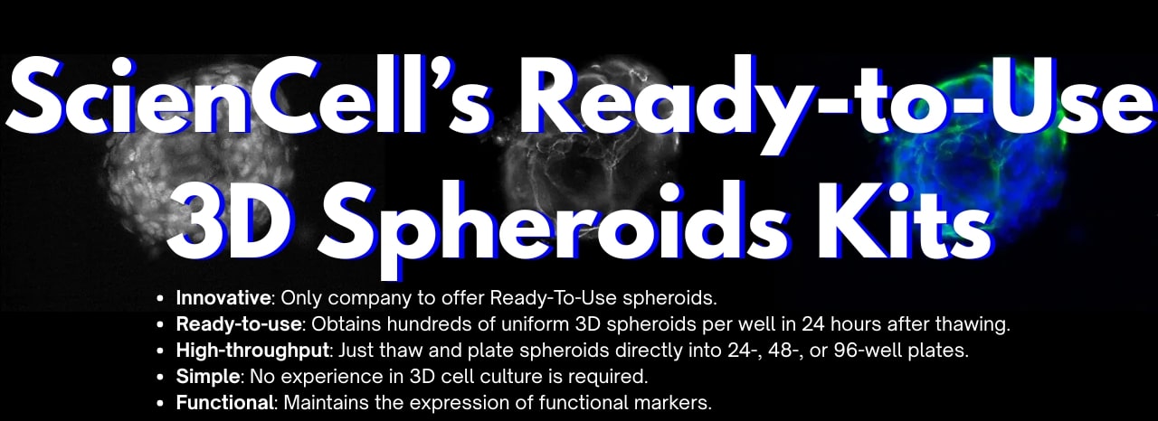
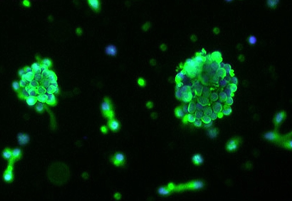
Building and maintaining consistent 3D spheroid models can traditionally involve long and complex workflows sometimes described as tedious and rate limiting.
Our featured ready-to-use 3D spheroid kits address this challenge. These innovative products allow researchers to obtain highly homogenous and
-
Posted: May 12, 2025
.jpg) Read more »
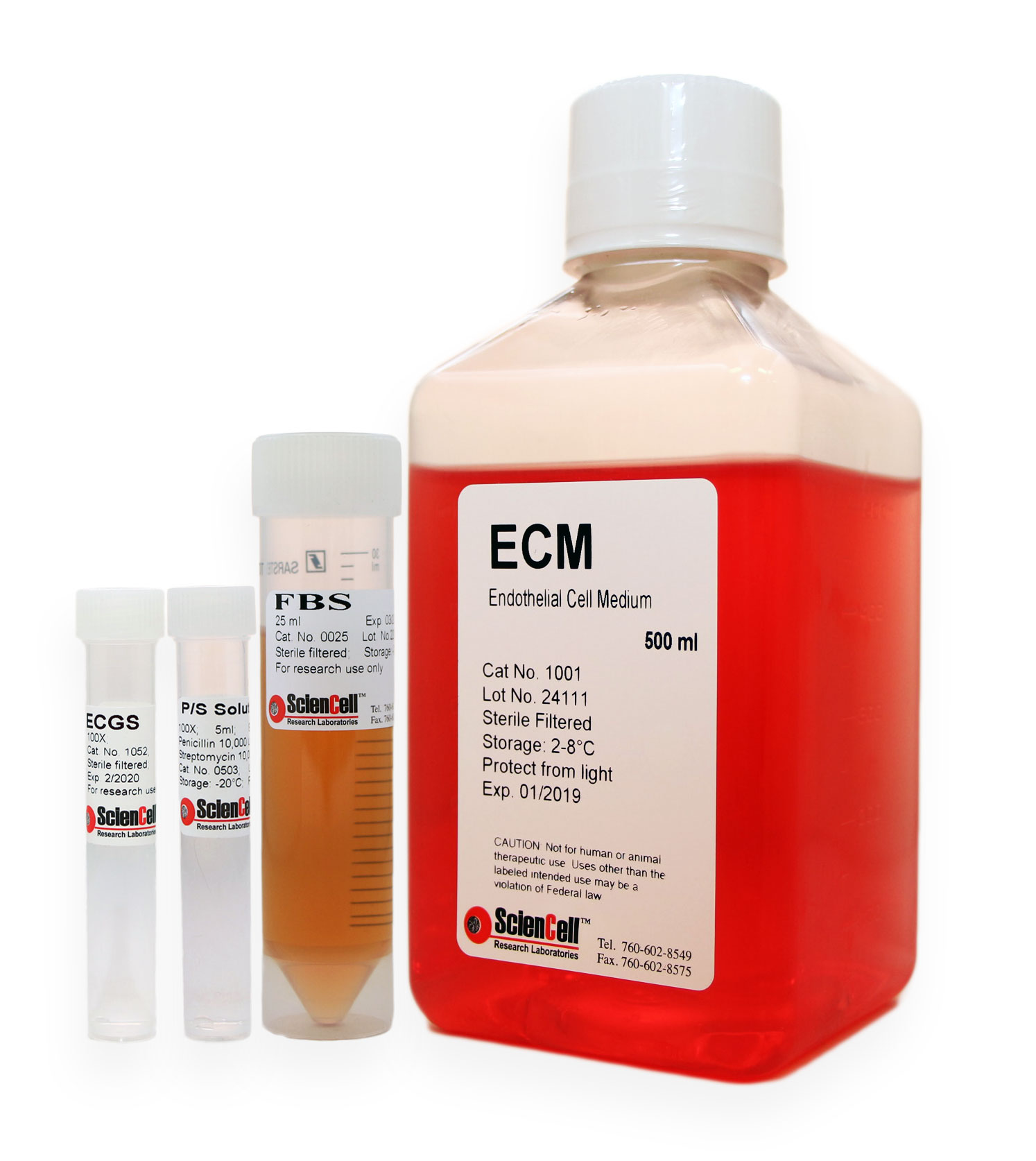
Read more »Achieving physiologically relevant in vitro models is paramount for groundbreaking research. The extracellular matrix (ECM) plays a critical role in cell behavior, influencing everything from adhesion and spreading to growth and differentiation.
To provide your cells with the optimal environment,
-
Posted: May 11, 2025
 Read more »
Introducing the New and Improved GoldNStart TaqGreen qPCR Master Mix –
Now with 2-Color Tracking! (Cat. #MB6018DCT)
ScienCell’s trusted GoldNStart TaqGreen qPCR Master Mix just got even better. Our latest upgrade features an integrated 2-color tracking system designed to make your qPCR workflow faster,
-
Posted: May 08, 2025
 Read more »
Primary Human Cells for Hair Growth Research
As the demand for advanced models in regenerative medicine and hair restoration therapies grows, so does the need for reliable, physiologically relevant primary cells.
ScienCell Research Laboratories offers a suite of human hair follicle cell types to
-
Posted: May 06, 2025
 Read more »
Say goodbye to the hassle of designing and validating primers!
ScienCell Research Laboratories offers over 5,400 individual primer sets for human and mouse gene expression analysis, plus our GeneQuery™ qPCR Array Kits for high-throughput studies. Each lyophilized primer set is pre-validated and provides
-
Posted: May 04, 2025Categories: Newsletter
 Read more »
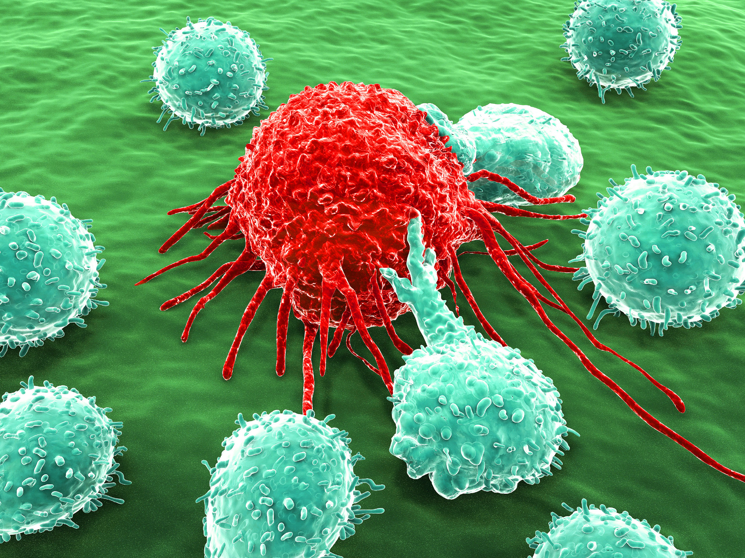
NK Cells
NK cells are a type of cytotoxic lymphocyte that plays a critical role in the innate immune system’s defense against infected and cancerous cells. Unlike T cells, which are part of the adaptive immune response, NK cells do not require prior activation to kill their targets. Instead, they can
-
Posted: May 01, 2025
 Read more »
Advance Regenerative Medicine and Muscle Biology with ScienCell's Primary Human Skeletal Cell Systems
Unlock Deeper Insights into Muscle Physiology
ScienCell Research Laboratories offers a robust foundation for skeletal muscle research with our high-purity Human Skeletal Muscle Cells (HSkMC, Cat. #3500) -
Posted: April 30, 2025
 Read more »
Expand Your Research Horizons with ScienCell's Diverse Dermal Fibroblasts!
Dermal fibroblasts, derived from the embryonic mesoderm are fundamental components of the skin's dermis layer. Their versatility and ease of culture make them extensively used in cellular and molecular studies. Due to their durability,
-
Posted: March 24, 2025
 Read more »
The fourth Tuesday in March is #AmericanDiabetesAlertDay. Today, we highlight this global health concern and emerging standards of care for type 1 and type 2 diabetes mellitus, and how #primarycellmodels might support the ongoing research to combat these diseases.
Though characterized by dysfunction
-
Posted: December 30, 2024Read more »
This kit is a valuable tool for researchers studying the monkeypox virus, and we are proud to offer it to the scientific community.
What is the Monkeypox Virus?
Monkeypox virus (MPXV) is a zoonotic virus that can infect both humans and animals. It belongs to the Orthopoxvirus genus in the Poxviridae
-
Posted: December 19, 2024Categories: Molecular BiologyRead more »
What is DNA/RNA Isolation?
DNA/RNA isolation, also referred as nucleic acid purification, is the process of extracting and purifying DNA or RNA from biological samples like cells, tissues, and body fluids.
The purpose of DNA/RNA isolation is to obtain pure nucleic acid samples free from contaminants
-
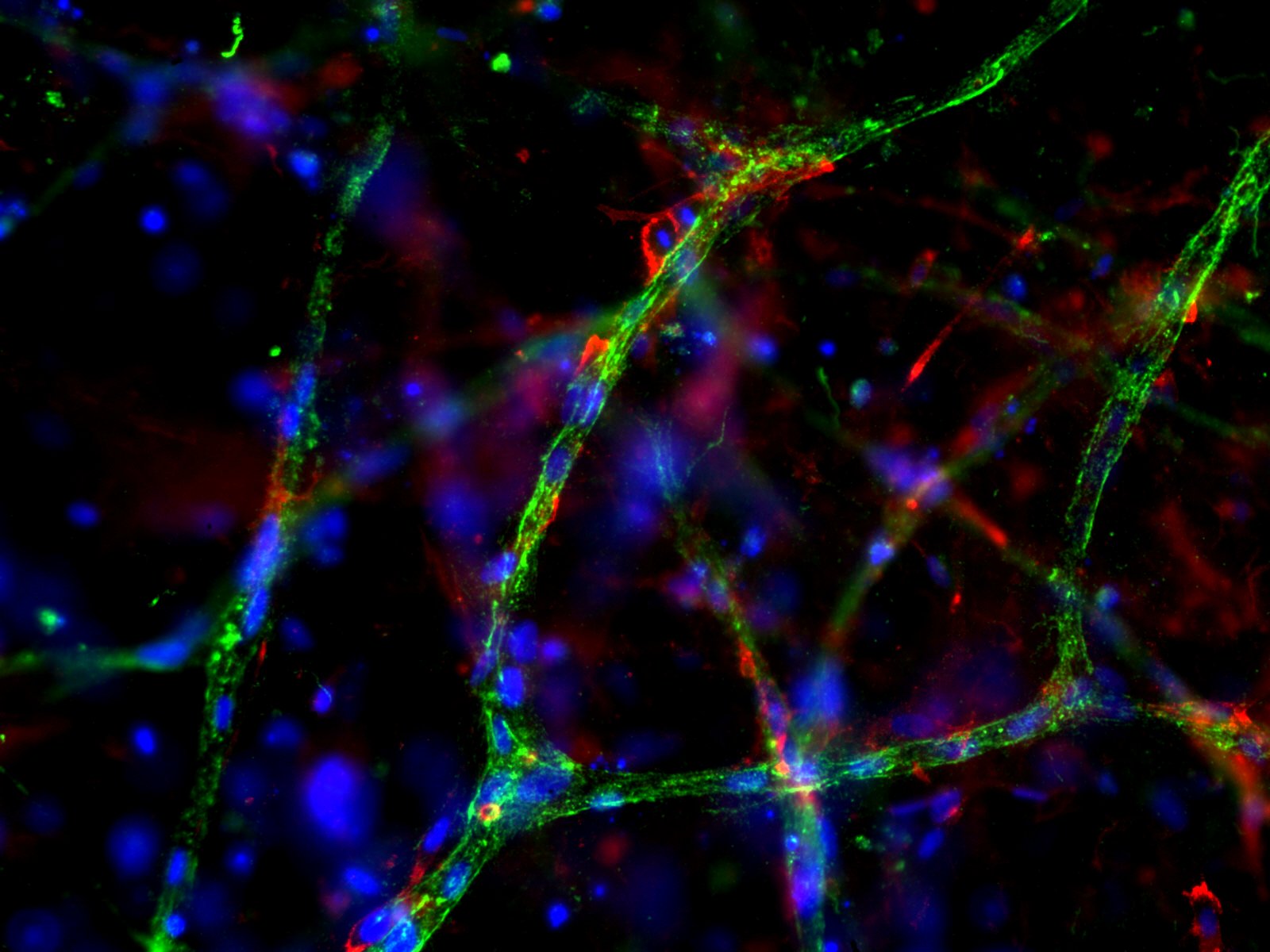
Posted: December 10, 2024Read more »
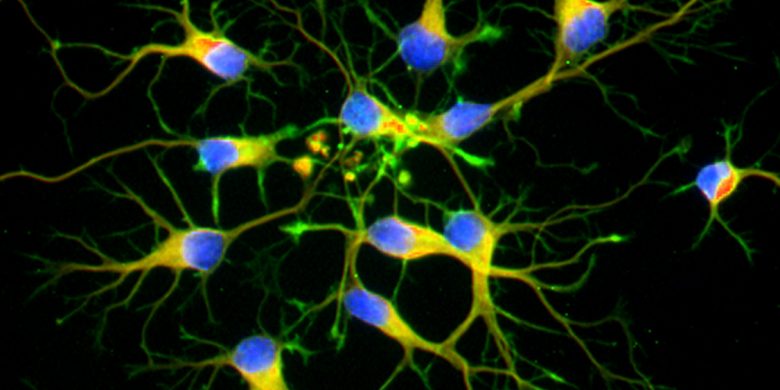
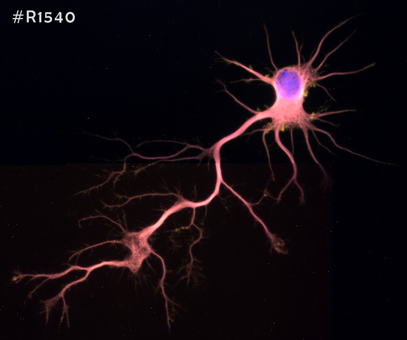
The central nervous system (CNS) is a complex network of neurons and glia that play critical roles in human brain function. Neurons are the primary signaling units of the nervous system, responsible for transmitting information throughout the body.
- A single neuron can contact up to 10,000 other neurons,
-
Posted: October 30, 2024Categories: InformationRead more »
Interleukins (IL) are a group of cytokines initially believed to be expressed solely by leukocytes, but they are now known to be produced by a variety of cells of the body. These proteins are crucial for activating and differentiating immune cells and play roles in cell proliferation, maturation, migration,
-
Posted: January 02, 2024Categories: Primary Cells
 Read more »
The ocular lens is a transparent structure in the eye that is designed to refract and focus light onto the retina. The lens is an avascular unit which includes the lens capsule, lens epithelium, and lens fibers. Lens fiber cells form the bulk of the lens and a monolayer of epithelial cells cover the
-
Posted: October 06, 2022
 Read more »
Telomere “caps” are located at the ends of all chromosomes, including those of the immune system’s T-lymphocytes (T cells), and become shorter with every cell division. Once these chromosomes reach a point where the telomere is too short to provide adequate protection, division ceases and the cell proceeds
-
Posted: November 12, 2020Categories: Information
 Read more »
Isolation of high-quality nucleic acids from biological material is a key factor in successful molecular biology research. While approaches vary, most methods aim to do the following: (1) disrupt the cellular structure, (2) denature and inactivate other macromolecules (e.g., proteins, enzymes, etc.),
-
Posted: September 28, 2020Categories: Primary Cells
 Read more »
Microglia play an essential role in brain homeostasis, neuroinflammation, neurodegenerative diseases, and brain infections. Microglia are integral components of the neuro-glial cell network and are the resident immune cells of the brain. Recent studies indicate that microglia are derived from the yolk
-
Posted: April 30, 2020
 Read more »
Hepatic sinusoidal endothelial cells (HSEC) are fascinating cells that are uniquely adapted to their location in the liver. HSEC are found lining micro-vessels in the liver and are extremely specialized endothelial cells. Structurally and functionally they have distinctive features which include: open
-
Posted: April 20, 2020
 Read more »
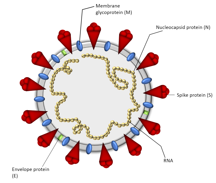
SARS-CoV-2 is the seventh known coronavirus that causes the human disease known as COVID-19. The virus can grow in cells lining the conducting airways and in alveolar epithelial cells. First, the virus generally enters the body through the nose or mouth. From there, the virus travels down into the alveoli
-
Posted: April 03, 2020
 Read more »
Epithelial cells are the most numerous cells in the lungs and contribute to innate and adaptive immunity. Airway epithelial cells are located in the lower respiratory tract which includes the trachea, bronchi, small airways (bronchioles), and alveoli. Due to their location, airway epithelial cells are
-
Posted: February 09, 2020
 Read more »
As of April 10, 2020, the number of U.S. SARS-CoV-2 coronavirus cases surpassed 500,000 with a death toll near 19,000. For over a century, coronaviruses were thought to only cause mild illnesses such as the common cold. With the ou or over a century, coronaviruses were thought to only cause mild illnesses
-
Posted: September 05, 2019Categories: Primary Cells
 Read more »
Migraines and headaches can be extremely debilitating with a diverse set of triggers and symptoms. Although the pathophysiology for headaches is still unclear, evidence suggests that factors such as neurogenic inflammation and meningeal sensory innervation play important roles in the development of
-
Posted: July 22, 2019Comments: 2
 Read more »
Traditional 2D cultures have been used widely over the past decades to study cell biology, molecular biology and conduct translation research such as drug discovery. Cells in 2D culture, however, are forced to adopt a planar morphology and maintain cellular interactions only in lateral directions, altering
-
Posted: June 09, 2019
 Read more »
Hepatic stellate cells have recently gained a great deal of attention regarding their contribution to the progression of diseases such as liver fibrosis, non-alcoholic steatohepatitis (NASH), and hepatocellular carcinoma. They are mesenchymal cells that are located between sinusoidal endothelial cells
-
Posted: February 11, 2019
 Read more »
ScienCell’s wide assortment of cell culture media is in liquid form and includes Specialty, Classical & Supplement varieties. Each product is designed for optimal nutrition and growth of primary cells. ScienCell’s cell culture media is manufactured and tested to ensure a high standard of quality and
-
Posted: August 28, 2018
 Read more »
qPCR is a powerful tool for quantification of gene expression levels and copy number variation. Despite the advances in next-generation sequencing (NGS), qPCR still serves as the "gold standard" for gene expression analysis. Due to poor reproducibility and vast lab-to-lab variation, all NGS data requires
-
Posted: August 19, 2018Categories: Cell Based Assays
 Read more »
Cell-based assays are widely used in basic and translational research as cost-effective and accessible models to mimic in vivo responses. To obtain reliable data, assessing the health of cultured cells prior to any assays is highly recommended. Furthermore, many cell-based assays require quantification
-
Posted: May 30, 2018
 Read more »
When working with primary cells, it is important to remember that they are not cell lines and should be treated with care. At ScienCell, we specialize in primary cell culture and we are very familiar with the common problems researchers encounter when culturing them. We have compiled a list with 13
-
Posted: April 03, 2018Comments: 1
 Read more »
Cancer immunotherapy is one promising cancer treatment option whereby the host's own immune system is used to treat cancer. The therapy works by either stimulating certain immune activities, or counteracting cancer cell signals that suppress immune responses. Cancer immunotherapy has progressed significantly
-
Posted: March 13, 2018
 Read more »Mesenchymal Stem Cell Medium-animal component free (Cat. #7521)
For decades, Fetal Bovine Serum (FBS) has been a vital supplement for the successful culture of a diverse range of cell types. FBS is an undefined, complex source of growth factors, hormones, lipids, attachment factors, and trace elements.
-
 Read more »
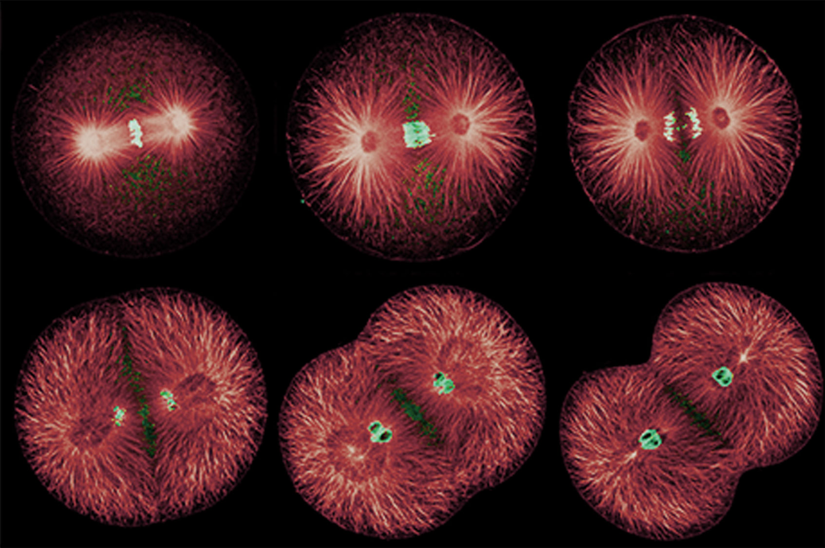
Aging and Telomere Length Quantification by qPCR
Aging is a time-dependent decline of the body’s functional capabilities and is an inevitable course of life (as shown in the image below, extracted from the Wall Street Journal). The rate of aging though is highly variable among individuals. When evaluating
-
Posted: November 29, 2017
 Read more »
Primary cells, which are isolated directly from tissue, show normal cell morphology and maintain many of the important markers and functions seen in vivo. Primary cells, though, have a finite lifespan and limited expansion capacity, so it is critical to use low passage primary cells for your research.
-
Posted: October 24, 2017Comments: 3
 Read more »
Neuronal cell lines are commonly used for in vitro neurobiology studies because they are more easily transfected compared to primary neurons and they proliferate, whereas primary neurons do not. Neuronal cell lines can be induced to differentiate into neuron-like cells, where they express neuronal markers
-
Posted: October 22, 2017Comments: 2
 Read more »
The 2017 Nobel Prize in Physiology or Medicine was awarded to Jeffrey C Hall, Michael Rosbash, and Michael W Young for research that established key mechanistic principles on how circadian rhythms are regulated. Circadian rhythms are endogenous oscillations adjusted to changing external cues and driven
-
Posted: September 20, 2017
 Read more »
Next generation sequencing (NGS), such as Whole Exome Sequencing (WES) and RNA-Sequencing (RNA-Seq), provides a high throughput approach for DNA sequencing and gene expression analysis. It is rapidly advancing our knowledge on almost all aspects of genetic research and shedding light on how individual
-
Posted: July 04, 2017Categories: Cell Based Assays
 Read more »
Cell culturing is a widely used technique to grow cells outside of their natural environment using artificial environments and controlled conditions that has become indispensable to scientific research. The applications of cell culturing are innumerable and there are many ways to differentiate types
-
Posted: May 03, 2017Categories: Gene Expression Profiling
 Read more »
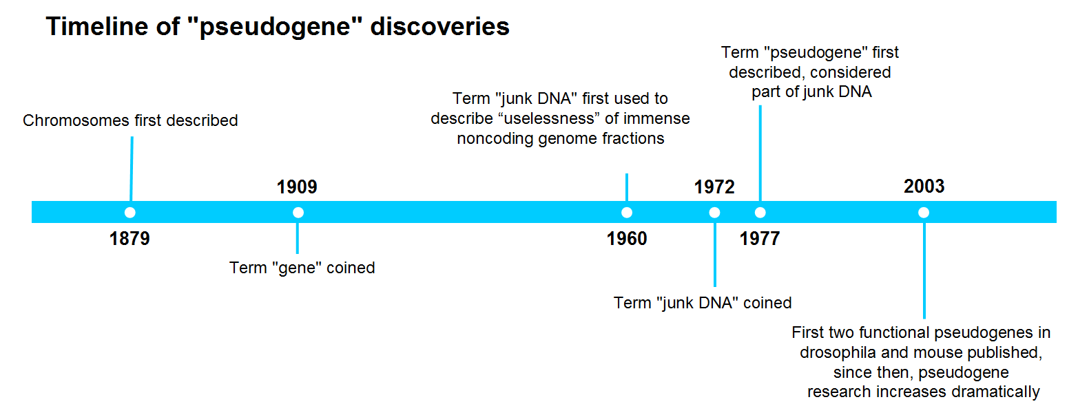
3 Reasons Why You Should Take "Pseudogenes" Seriously in Your Research
Protein-coding DNA, or “functional” genes, only accounts for up to 2% in the human genome. Until recently, the remaining 98% have been referred to as “junk DNA” for their putative noncoding feature. The name itself implies that junk
-
Posted: March 26, 2017Categories: Company, Information, Gene Expression Profiling, Cell Culture Media, Primary Cells, Cell Based Assays, News
-
 Read more »
Hello fellow cell enthusiasts! We at team ScienCell just got back from Baltimore, MD, where we attended ToxExpo 2017, the Society of Toxicology’s annual conference. While the cold weather was quite a shock to us Californians, it was such a treat getting to experience what Baltimore had to offer. After
-
Posted: February 14, 2017
 Read more »
CARLSBAD, Calif. -- ScienCell Research Laboratories, Inc. is a unique supplier of primary Human Schwann Cells to the scientific research community. Schwann cells are glial cells that protect and insulate nerves in the peripheral nervous system.
"There are numerous cell types, such as Schwann cells,
-
Posted: February 14, 2017
 Read more »
SAN DIEGO, Calif. -- ScienCell Research Laboratories, Inc. recently announced it would be expanding its cell culture products to the gene expression profiling market. ScienCell, who prides itself on supplying unique primary cell types for nearly 20 years, will now supply qPCR array kits and individual
-
Posted: February 14, 2017
 Read more »
Online webinar hosted by sciencell to learn tips for successful qPCR, a technique for gene expression profiling
CARLSBAD, CALIFORNIA, USA, -- Sciencell Research Laboratories, Inc. , a global provider of primary cells, cell culture media and reagents for life science industry, announced today that it
-
Posted: December 06, 2016
 Read more »
Cell-based assays are widely used in basic and translational research as cost-effective and accessible models to mimic in vivo responses. To obtain reliable data, assessing the health of cultured cells prior to any assays is essential. Furthermore, many cell-based assays require quantification of cell
-
Posted: October 26, 2016Categories: Primary Cells
 Read more »
ScienCell's Rat Hippocampal Neurons Used in Recent Neurological Disease Therapy Research Published in Scientific Reports Journal
Neurons are dynamically polarized cells responsible for electrochemically transmitting information throughout the nervous system. Scientists have characterized
-
Posted: September 28, 2016Categories: Primary Cells
 Read more »
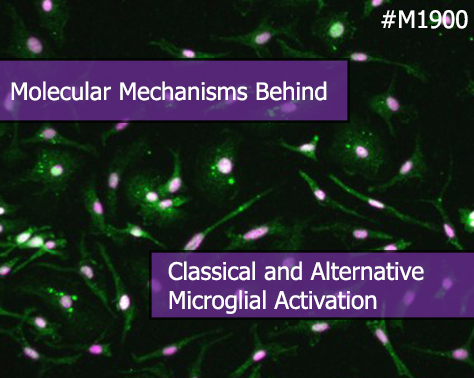
ScienCell's Mouse Microglia Used in Recent Microglial Activation Research Published in Molecular & Cellular Proteomics Journal
Microglia are a type of macrophage-like glial cell in the central nervous system (CNS) and they function as the brain's primary immune defenders. Upon activation,
-
 Read more »
When it comes to primary cell culture, a picture is worth a thousand words. A quality image can convey information on cell morphology, health, culture purity and density. Different types of 2-dimensional microscopy techniques such as phase contrast, relief contrast, and fluorescence microcopy can
-
Posted: August 23, 2016Categories: Gene Expression Profiling
 Read more »
In this final installment on how to improve your qPCR gene expression profiling (see 6 Tips to Improve qPCR: part 1 and part 2 for previous discussion), we will discuss pipetting tips and the benefits of a separate reverse transcription step in qPCR template preparation.
Tip #5. Good Laboratory Practice
-
Posted: August 17, 2016
 Read more »
After watching displays of astounding athletic prowess in the 2016 Olympics, I was inspired to take a closer look at the science behind exercise training, recovery, and injury with a focus on the importance of blood vessels during exercise.
Let’s start with some basic training: Why are blood vessels
-
Posted: August 01, 2016Categories: Primary Cells
 Read more »
Many primary cells have difficulty adhering to uncoated glass and plastic surfaces, especially in low serum or serum-free conditions. For primary cells, it is necessary to use a substrate coating to enhance cell attachment. There are many different substrates to choose from and each type can have dramatically
-
 Read more »
Recently, bone marrow mononuclear cells (BMMC) have been in the spotlight for having the potential to treat a variety of diseases including neurological disorders. Why are BMMC so exciting and do they really show treatment potential? BMMC have regenerative capabilities and are a heterogeneous population
-
 Read more »
Despite being essential for any experiment involving DNA replication, people rarely give primers a second thought. To coincide with the launch of our new qPCR gene reference tool, we’ll be giving them a little more of the praise they deserve.
Dr Kary Mullis received the 1993 Nobel Prize in Chemistry -
Posted: April 26, 2016Categories: Gene Expression Profiling
Read more »
In this next installment on how to improve your qPCR gene profiling (see 6 Tips to Improve qPCR for previous discussion), we would like to discuss the limitations of melting curve analysis and PCR inhibitors.
Tip #3. Melting curve analysis is NOT sufficient for assessing qPCR product specificity.
In
-
Posted: March 10, 2016Categories: Cell Culture Media
 Read more »
Choosing which media to use with your primary cells can be daunting. Whether you are replicating a formula from a publication, optimizing your own supplemented classical or nutrient media, or purchasing commercially available specialty media, it is important to understand how this choice will affect
-
Posted: February 09, 2016Categories: Information
 Read more »
American Heart Month Sponsor: The American Heart Association (http://www.heart.org/HEARTORG/)
Heart disease is the leading cause of death for men and women in the United States. Every year, 1 in 4 deaths are caused by heart disease.
The good news? Heart disease can often be prevented when people
-
Posted: February 03, 2016Categories: Gene Expression Profiling
 Read more »
Gene expression analysis is critical for understanding the transcriptome profiles of primary cells and how they directly influence the cells’ functionality. In addition, the transcriptome profile can affect cell proliferation, differentiation, senescence, or apoptosis. Traditional reporter-gene assays
-
Posted: February 03, 2016Categories: Gene Expression Profiling
Read more »
qPCR array provides a quick, powerful and sensitive approach for gene expression profiling. To help our customers acquire accurate and consistent results, we are happy to share some tips for better qPCR.
Tip #1. Good primer design is the key to a successful qPCR array.
The success of a qPCR array depends
-
Posted: January 21, 2016Categories: Primary Cells
 Read more »
Cell culture studies provide a valuable complement to in vivo experiments, allowing for a more controlled manipulation of cellular functions and processes. For decades, cell lines have played a critical role in scientific advancements, yet researchers have become increasingly cautious when interpreting
-
 Read more »
When working with primary cells, it is important to remember that they are not cell lines and cannot be treated the same. Because ScienCell has extensively working with primary cells, we are very familiar with the common problems researchers encounter when culturing them. Here are six common mistakes
-
Posted: December 27, 2015Categories: Primary Cells
 Read more »
CD-1 IGS mice are outbred mice derived from a group of outbred Swiss mice developed at the Anti-Cancer Center in Lausanne, Switzerland. They were imported to the US in 1926 and to Charles River in 1959. The CD-1 IGS mice are generally used for genetics, toxicology, pharmacology, and aging research.
-
 Read more »
Cell culture studies provide a valuable complement to in vivo experiments, allowing for a more controlled manipulation of cellular functions and processes. For decades, cell lines have played a critical role in scientific advancements, yet researchers have become increasingly cautious when interpreting
-
Posted: November 01, 2015Categories: Information
 Read more »
Alzheimer’s disease (AD) affects almost 44 million people worldwide. The disease typically begins with memory difficulties followed by trouble thinking, reasoning and processing emotions. The cause of AD is not well understood, but there appear to be two forms: 90% of patients have a late-onset sporadic
-
Posted: October 07, 2015Categories: Gene Expression Profiling
 Read more »
Dye-based qPCR assays, such as SYBR® Green and EvaGreen®, have many advantages over probe-based qPCR assays. Dye-based systems are desirable because they are highly adaptable, cost effective, and time efficient. qPCR specificity, however, is a vital factor to consider when using dye-based qPCR assays.
-
Posted: July 15, 2015Categories: Cell Culture Media
 Read more »
For decades, researchers have been culturing cells to study normal and diseased biological processes. Many of these studies use immortalized cell lines which proliferate indefinitely in culture. Immortalized cells thrive in classical media formulated with the minimum requirements for cell growth, including
-
Posted: July 05, 2015Categories: Company
 Read more »
For over a decade, ScienCell Research Laboratories has been a leading provider of human and animal primary cells, cell-derived DNA, RNA and protein, as well as other biological materials. For our business practices, we strictly comply with the following policies:
- We are committed to operating under the


.jpg)
.jpg)
.jpg)
.jpg)