COVID-19
-
- May 01, 2020
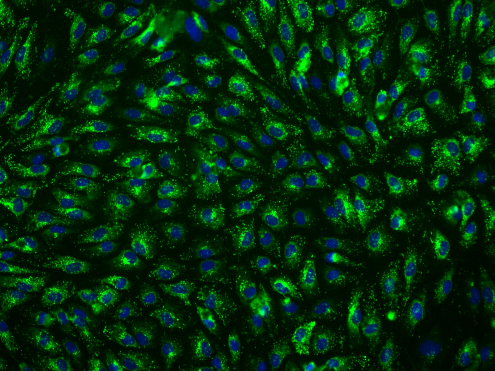
Hepatic sinusoidal endothelial cells (HSEC) are fascinating cells that are uniquely adapted to their location in the liver. HSEC are found lining micro-vessels in the liver and are extremely specialized endothelial cells. Structurally and functionally they have distinctive features which include: open pores known as fenestra which form sieve plates, a lack of an organized basement membrane, expression of scavenger receptors, and performing endocytic activity. Notably, HSEC are highly permeable and play a critical role in removing bloodborne waste. To perform the endocytic function, HSEC express a vast array of scavenger receptors as well as the mannose receptor, which allows them to collect molecules from the bloodstream and transport them to the hepatocytes.
HSEC also play a pivotal role in the innate immunity by their ability to bind viruses and other pathogens through their endocytic receptors. By way of the portal vein, the liver is continuously being exposed to antigens and microbes
-
- April 21, 2020
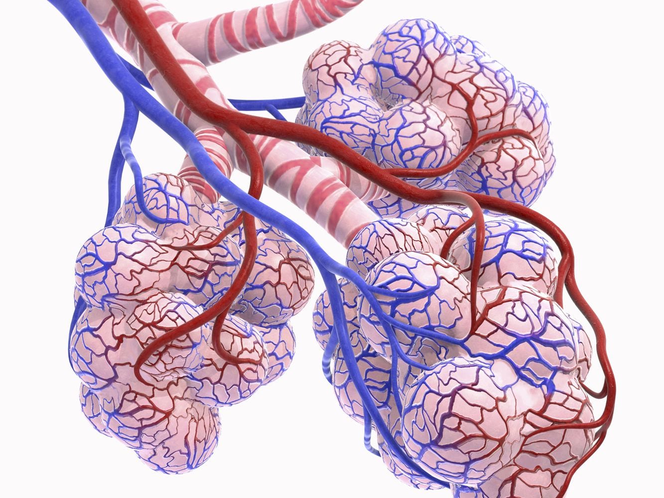
SARS-CoV-2 is the seventh known coronavirus that causes the human disease known as COVID-19. The virus can grow in cells lining the conducting airways and in alveolar epithelial cells. First, the virus generally enters the body through the nose or mouth. From there, the virus travels down into the alveoli which are located in the lungs. Once in the alveoli, the virus “hijacks” cells to make new copies of the virus. The infected cell is then killed, releasing new viruses to infect neighboring cells in the alveolus. Each sac of air, or alveolus, is wrapped with capillaries where red blood cells release carbon dioxide (CO2) and pick up oxygen (O2). Two alveolar epithelial cells (type I and II) facilitate gas exchange. Type I cells are squamous alveolar cells with thin membranes that perform gas exchange. Type II cells are known as progenitor cells in the alveoli and proliferate and differentiate into type I cells. In addition, Type II cells secrete the pulmonary surfactant that lines
-
- April 04, 2020
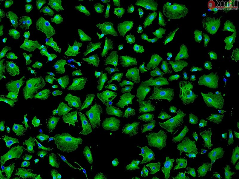
Epithelial cells are the most numerous cells in the lungs and contribute to innate and adaptive immunity. Airway epithelial cells are located in the lower respiratory tract which includes the trachea, bronchi, small airways (bronchioles), and alveoli. Due to their location, airway epithelial cells are constantly exposed to microbes, particles, and pollutants and are essentially the first line of defense against invading pathogens. Airway epithelium acts as a physical barrier and either directly remove pathogens or interact with immune cells which initiate the clearance of pathogens. Epithelial cells also play an important role in reducing inflammation and maintaining homeostasis in the lungs. During an infection, epithelial cell dysfunction can contribute to the development of inflammation of the airways and lungs. Additionally, patients with chronic pulmonary disease are more susceptible to respiratory infections due to defects in epithelial barrier structure and function.
Human Bronchial -
- February 10, 2020
As of April 10, 2020, the number of U.S. SARS-CoV-2 coronavirus cases surpassed 500,000 with a death toll near 19,000. For over a century, coronaviruses were thought to only cause mild illnesses such as the common cold. With the ou or over a century, coronaviruses were thought to only cause mild illnesses such as the common cold. With the outbreak of the 2002-03 SARS (severe acute respiratory syndrome) that was caused by SARS-CoV coronavirus, this concept was rapidly overturned, and as a result coronavirus research has geared up for the fast lane.
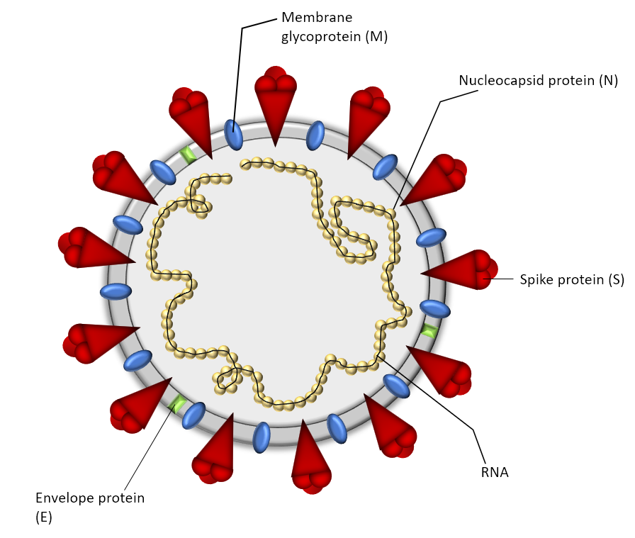
Coronaviruses are a family of large, single-stranded RNA viruses with size ranging from 26 to 32 kb. Like other viruses, coronaviruses proliferate by invading cells, manipulating the cells into making many copies of the virus, and infecting more cells. As an RNA virus, the coronavirus lacks error-repairing mechanisms during replication, and therefore, has a relatively high mutation rate resulting in rapid evolution.
At the 3′-end of the viral