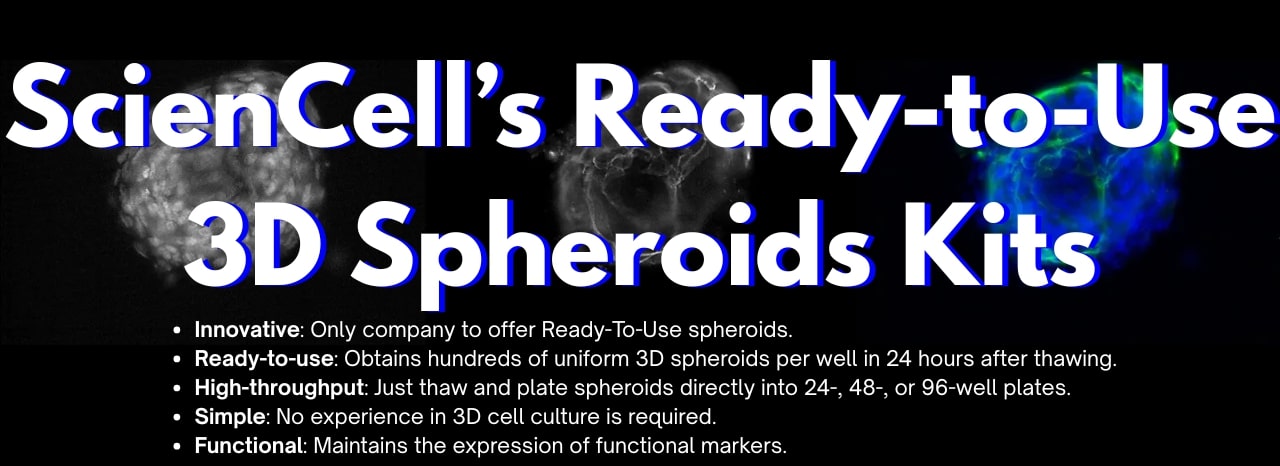
Building and maintaining consistent 3D spheroid models can traditionally involve long and complex workflows sometimes described as tedious and rate limiting.
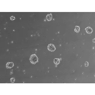
Our featured ready-to-use 3D spheroid kits address this challenge. These innovative products allow researchers to obtain highly homogenous and functional 3D spheroids quickly after thawing. With these kits, the cryopreserved spheroids are thawed and plated directly into ultra-low binding culture plates. Functional and uniform spheroids are typically recovered and ready for experiments within 24-48 hours post-thawing.
We offer a variety of ready-to-use 3D spheroid models, each specifically designed to provide a more relevant in vitro system for distinct research areas:
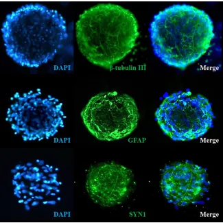
3D Human Cortical Spheroids (SP3D-HCS, Cat. No. SP3D-1520)
- ≥ 1×10⁴ spheroids per vial
- Comprised of primary human neurons and astrocytes at a 1:7 ratio. These spheroids maintain direct cell-cell interactions and form functional synapses throughout the structure
- Networked with quiescent astrocytes, they closely resemble the in vivo brain phenotype
- Ideal for studying CNS functions, diseases, and therapeutics
Ready-to-use 3D Human Blood Brain Barrier Spheroid Kit (SP3D-HBBBS-HBMEC, Cat. No. SP3D-8768)
- ≥ 1x10⁴ spheroids per vial
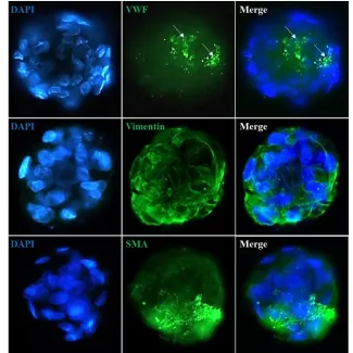
- Comprised of human brain microvascular endothelial cells, brain vascular pericytes, and astrocytes at a 1:1:1 ratio effectively recapitulating the intracellular interactions at the BBB
- This model exhibits the tight junction marker ZO160 and is highly relevant for studying BBB integrity, transport mechanisms, and the delivery of therapeutic molecules to the brain
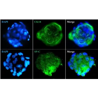
3D Human Retinal Pigment Epithelial Spheroids (SP3D-HRPEpiS, Cat. No. SP3D-6540)
- ≥ 1×10⁴ spheroids per vial
- These spheroids form and maintain a well-differentiated epithelium in 3D
- They exhibit the epithelial marker CK18 and deposit apolipoprotein ApoE, a key constituent of drusen, making this an ideal model for studying drusen formation in vitro and the pathogenesis of Age-related Macular Degeneration (AMD)
3D Hepatic Stellate-Endothelial Cell Spheroids (SP3D-HSteECS, Cat. No. SP3D-5000)
- ≥ 1×10⁴ spheroids per vial
- Co-culture spheroids of hepatic stellate cells and sinusoidal endothelial cells
- This model is designed to investigate the signaling crosstalk between liver fibrogenesis and angiogenesis
3D Human Renal Proximal Tubular Epithelial Spheroids (SP3D-HRPTEpiS, Cat. No. SP3D-4100)
- ≥ 4×10³ spheroids per vial
- Generated by co-culturing human renal proximal tubular epithelial cells with human renal mesangial cells at a 4:1 ratio
- Provides a physiological model for kidney function and immune response studies
Ready-to-use 3D Human Ovarian Fibroblast Spheroids (SP3D-HOFS, Cat. No. SP3D-7330)
- ≥ 1×10⁴ spheroids per vial
- Comprised of primary HOF. These cells are cultured in three-dimensional (3D) architecture and are embedded in their own ECM, which better reflects the native ovarian tissue architecture
- Excellent in vitro model for studying HOF function and its contribution to diseases such as ovarian cancer and ovarian fibrosis
Ready-to-use 3D Human Mesenchymal Stem Cell Spheroids (SP3D-HMSCS, Cat. No. SP3D-7500)
- ≥ 4 × 10³ spheroids per vial
- Can be thawed and plated directly in multiwell plates
- Great model for examining the functions of mesenchymal stem cells in cell therapy and tissue regeneration
3D Human Chondrocyte-articular Spheroid Kit (SP3D-HCaS, Cat. No. SP3D-4650)
- ≥ 1×10⁴ spheroids per vial
- Contains human chondrocyte spheroids that maintain functional markers such as type II collagen, aggrecan, and Sox9 in 3D culture, unlike their tendency to dedifferentiate in monolayer
- Offers an excellent in vitro model for studying normal chondrocyte physiology, degenerative joint diseases, and cartilage repair
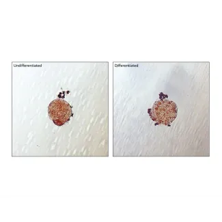
3D Human Preadipocyte Spheroids (SP3D-HPA, Cat. No. SP3D-7220)
- ≥ 1x10⁴ spheroids per vial
- Ready-to-use preadipocyte spheroids that can be easily differentiated into mature adipocytes in culture. Upon differentiation, they accumulate large lipid droplets, confirmed by Oil Red O staining
- This model serves as an excellent tool for drug discovery and the study of adipocyte biology
Ready-to-use 3D Osteogenesis-Angiogenesis Coupling Kit (SP3D-OAC, Cat. No. SP3D-8748)
- ≥ 1×10⁴ spheroids per vial
- Contains co-culture spheroids of human osteoblasts and human dermal microvascular endothelial cells
- This kit offers a superior model for analyzing the complex cellular interactions present in bone tissue, allowing for the study of the crucial molecular crosstalk between angiogenesis and osteogenesis, which are tightly coupled processes in bone formation and repair
Our ready-to-use 3D spheroids are highly suitable for a wide range of research applications, including:
- Studying tissue functions and development
- Investigating disease mechanisms, such as CNS diseases, degenerative joint diseases, ovarian cancer and fibrosis, AMD, and metabolic diseases like type II diabetes
- Drug discovery and toxicity testing, including nephrotoxicity
- Modeling complex interactions, such as neurovascular coupling, liver fibrogenesis and angiogenesis, angiogenesis and osteogenesis coupling and the blood-brain barrier
- Tissue repair and engineering
- Immunology, such as the immune response of kidney cells to viruses and the immune-suppressive properties of MSCs
Elevate your research with more relevant and reliable in vitro models. Explore the possibilities with our unique ready-to-use 3D spheroid kits!