hPSC-Derived Sensory Neurons Transforming Peripheral Neuropathy Research?
Pain, numbness, tingling, hypersensitivity and loss of sensation are common features of disorders affecting the peripheral nervous system. Yet studying the human sensory neurons responsible for these symptoms has traditionally been difficult. Primary human dorsal root ganglia are rarely available for research, immortalized cell lines do not reproduce many specialized properties of sensory neurons, and animal models cannot always capture human-specific ion-channel physiology, genetic variation or responses to therapeutic compounds.
Human pluripotent stem cell-derived sensory neurons offer an increasingly powerful solution. By providing a renewable source of human peripheral neurons, hPSC-derived sensory neurons make it possible to investigate disease mechanisms directly in the cell type responsible for detecting pain, temperature and other somatosensory signals. Reviews of the field now highlight these cells as valuable platforms for studying neuropathic pain, inherited sensory disorders and drug-induced neurotoxicity, as well as for therapeutic screening and personalized medicine.
Why sensory neurons are difficult—and important—to model
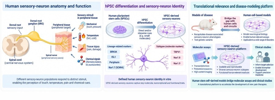
Peripheral sensory neurons reside primarily within dorsal root ganglia and extend remarkably long axons between peripheral tissues and the spinal cord. Different sensory-neuron populations respond to mechanical stimuli, temperature, tissue injury and chemical signals through specialized receptors and ion channels.
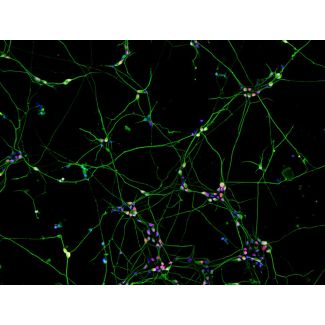
Nociceptive sensory neurons, for example, express channels such as TRPV1 and voltage-gated sodium channels including NaV1.7 and NaV1.8. Alterations in these pathways can profoundly change neuronal excitability and pain perception. Modern hPSC differentiation protocols can generate sensory neurons expressing markers such as BRN3A, ISL1, peripherin and neurofilaments together with disease-relevant sensory channels. Recent protocols have reported substantial populations expressing TRPV1, NaV1.7 and NaV1.8, illustrating the increasingly defined sensory identity achievable in vitro.
The ability to reproduce these features in human cells is important because some key pain-signaling mechanisms differ between species. A therapeutic candidate that appears effective in a rodent system may therefore behave differently in human sensory neurons. Human stem cell-derived models provide an intermediate experimental platform between simplified molecular assays and clinical studies.
From human genotype to measurable pain phenotypes
One of the clearest demonstrations of the power of iPSC-derived sensory neurons comes from inherited erythromelalgia, a severe pain disorder frequently caused by gain-of-function mutations in SCN9A, the gene encoding NaV1.7.
Researchers generated iPSCs from affected patients and differentiated them into sensory neurons. The resulting neurons reproduced disease-associated hyperexcitability and abnormal responses to heat. Importantly, pharmacological inhibition of NaV1.7 reduced the abnormal phenotype in vitro, providing a direct connection between patient genotype, sensory-neuron physiology and therapeutic response.
Subsequent studies using patient-derived sensory neurons have further clarified how NaV1.7 mutations alter action-potential threshold and excitability. Such findings illustrate an important advantage of hPSC models: disease-associated variants can be studied within the endogenous human neuronal environment rather than in a non-neuronal cell engineered to express a single ion channel.
This approach can be extended through genome editing. Disease-associated mutations can be introduced into control hPSCs or corrected in patient-derived iPSCs, producing isogenic experimental pairs. Differences in neuronal excitability, morphology or survival can then be linked more directly to the variant of interest.
A human model for acquired peripheral neuropathy
Sensory-neuron models are equally relevant for acquired diseases. Chemotherapy-induced peripheral neuropathy, or CIPN, is a particularly important example. Drugs including paclitaxel, vincristine, bortezomib and platinum compounds can damage peripheral sensory neurons, producing painful or disabling neuropathy that may limit cancer treatment.
Human iPSC-derived sensory neurons exposed to neurotoxic chemotherapy reproduce several clinically relevant cellular phenotypes. Studies have reported axonal blebbing, neurite retraction or fragmentation, impaired electrophysiological function and reduced viability after exposure to neuropathy-associated chemotherapeutics. Importantly, compounds not normally associated with peripheral neurotoxicity produced substantially weaker effects in the same systems.
This creates opportunities that extend beyond descriptive disease modeling. Morphological endpoints such as neurite length, axonal fragmentation and neuronal survival can be quantified at scale, making hPSC-derived sensory neurons attractive for neurotoxicity screening and identification of neuroprotective compounds.
The translational potential is becoming increasingly tangible. A 2026 study developed an iPSC-derived sensory-neuron platform specifically for high-throughput discovery of protective agents against CIPN, illustrating how these cultures can move from small mechanistic experiments toward scalable drug-discovery workflows.
Why sensory neurons alone do not tell the entire story
Although neuron monocultures are valuable for studying neuronal excitability, axonal degeneration and cell-autonomous disease mechanisms, sensory neurons normally function in close association with Schwann cells.
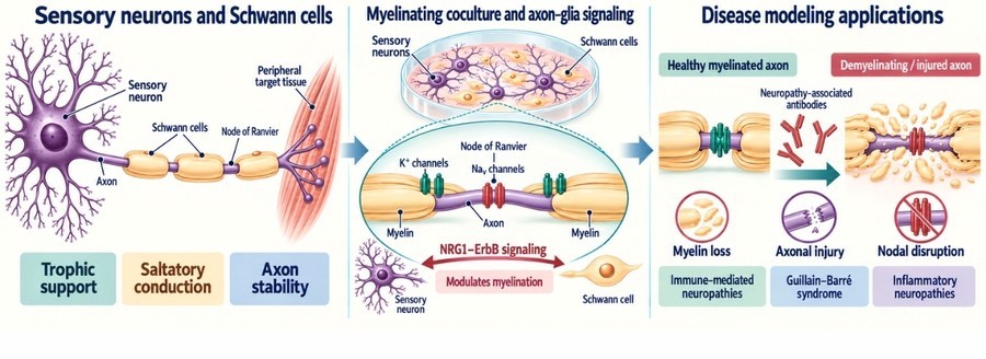
Schwann cells provide trophic support, organize peripheral axons and, for larger sensory fibers, form the myelin sheath required for efficient action-potential conduction. Reciprocal signaling between sensory axons and Schwann cells determines whether an axon becomes myelinated and regulates the organization of specialized structures including nodes of Ranvier.
This biology makes sensory-neuron–Schwann-cell coculture particularly attractive for studying diseases in which both axonal and glial mechanisms contribute to pathology.
A landmark study demonstrated that human iPSC-derived sensory neurons could form stable myelinating cultures with Schwann cells. The cultures developed molecularly organized nodes of Ranvier, including clustered voltage-gated sodium channels and juxtaparanodal potassium channels. Manipulating the NRG1–ErbB signaling pathway altered myelination, demonstrating that the system could reveal mechanisms controlling human sensory axon–Schwann-cell communication.
The same model also showed that neuropathy-associated antiganglioside antibodies could interfere with myelination, induce axonal injury and promote demyelination. This expands the experimental possibilities beyond neuron-autonomous disorders to immune-mediated peripheral neuropathies such as Guillain–Barré syndrome and related inflammatory neuropathies.
Revealing disease phenotypes through axon–glia interactions
Perhaps one of the strongest examples comes from hereditary sensory neuropathy type 1, or HSN1.
Patient-derived iPSC sensory neurons carrying SPTLC1 mutations generated abnormal deoxysphingolipids, showed disrupted ganglioside metabolism, impaired neurotrophic signaling and reduced neurite growth. However, additional pathology became apparent when the neurons were studied in a myelinating coculture.
After several weeks, the system developed abnormalities in nodal and paranodal proteins. With prolonged culture, these defects progressed to extensive myelin breakdown. L-serine treatment ameliorated several disease-associated phenotypes.
This illustrates why multicellular disease models can reveal biology that neuron monocultures may miss. A mutation originating in the neuron can progressively destabilize the axon–Schwann-cell unit, creating a phenotype that only becomes evident after extended cell–cell interaction.
Toward fully human peripheral nervous system models
An especially promising direction is replacing xenogeneic components with human stem cell-derived Schwann cells.
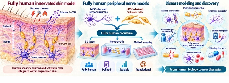
Proof-of-concept studies have already demonstrated that sensory neurons and Schwann cells differentiated from human iPSCs can coexist within complex tissue models. In one fully human innervated skin system, iPSC-derived sensory neurons expressed nociceptive markers and released Substance P and CGRP after stimulation. When Schwann cells were added, neuronal processes extended throughout the engineered tissue toward the epidermal compartment.
Such results point toward a future in which defined hPSC-derived sensory neurons and Schwann cells can be combined to create scalable, human-specific models of peripheral nerve biology.
These systems could be applied to inherited neuropathies, demyelinating disorders, small-fiber neuropathy, diabetic neuropathy, chemotherapy-induced neurotoxicity, nerve injury, neuroinflammation and pain-drug discovery.
A versatile platform for translational neuroscience
hPSC-derived sensory neurons therefore provide more than an alternative neuronal culture. They offer direct access to human peripheral neuronal biology while remaining compatible with imaging, electrophysiology, molecular analysis and pharmacological screening.
Researchers can examine endpoints including neuronal survival, neurite growth, axonal degeneration, calcium responses, action-potential firing, NaV and TRP-channel function, transcriptomic changes and responses to neurotoxic or analgesic compounds.
Adding hPSC-derived Schwann cells can expand those applications further, allowing investigators to examine axon–glia communication, Schwann-cell alignment, myelination, nodal organization and long-term axonal stability.
No in vitro model reproduces the complete complexity of the human peripheral nervous system. However, increasing biological complexity does not always require sacrificing experimental control. A defined sensory-neuron culture provides a tractable human neuronal model, while a sensory-neuron–Schwann-cell coculture adds a physiologically important component precisely when axoglial biology matters.
Together, these approaches provide researchers with a flexible framework: begin with a controlled human sensory-neuron model for neuronal mechanisms and screening, then incorporate Schwann cells when the scientific question requires myelination, glial support or axon–glia interactions.
As neurological research moves toward more human-relevant and mechanistically informative models, hPSC-derived sensory neurons—and increasingly their coculture with hPSC-derived Schwann cells—offer a compelling platform for investigating peripheral neurological disease and accelerating the development of new therapies.