3D Models
-
- 十二月 11, 2024
The central nervous system (CNS) is a complex network of neurons and glia that play critical roles in human brain function. Neurons are the primary signaling units of the nervous system, responsible for transmitting information throughout the body.
- A single neuron can contact up to 10,000 other neurons, highlighting the intricate connectivity of the brain.
- The cerebral cortex, the outermost layer of the brain, is responsible for higher-level functions such as perception, language, decision-making, and memory.
Studying the CNS can be challenging, but in vitro models like primary neurons and 3D human cortical spheroids offer valuable tools for research.
Primary Neurons
ScienCell Research Laboratories provides a variety of primary neurons isolated from different species:
- Human Neurons (HN, Cat. #1520): Isolated from human brain tissue.
- Mouse Neurons-cortical (MN-c): Isolated from embryonic day 18 mouse cerebrum, available from both CD-1 ( Cat. #M1520) and C57BL/6 (Cat. #M1520-57) mice.
- Rat
-
- 可能 01, 2020
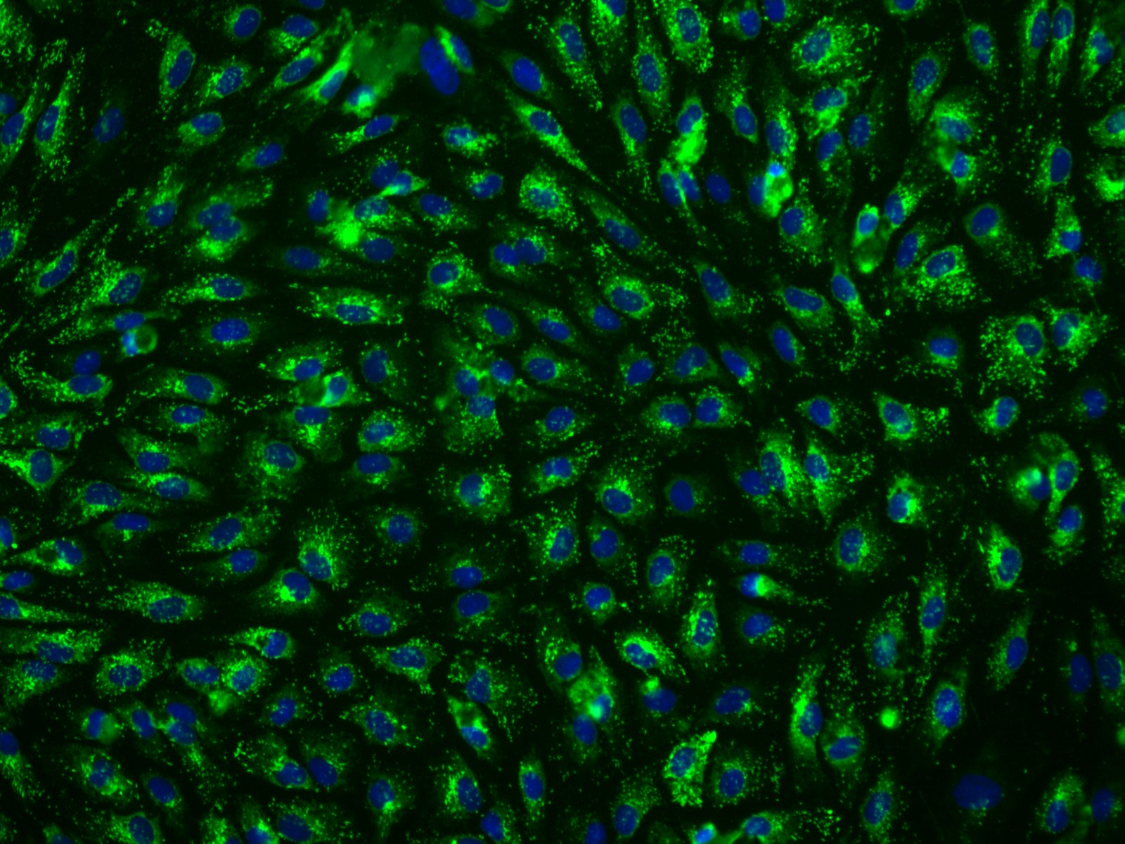
Hepatic sinusoidal endothelial cells (HSEC) are fascinating cells that are uniquely adapted to their location in the liver. HSEC are found lining micro-vessels in the liver and are extremely specialized endothelial cells. Structurally and functionally they have distinctive features which include: open pores known as fenestra which form sieve plates, a lack of an organized basement membrane, expression of scavenger receptors, and performing endocytic activity. Notably, HSEC are highly permeable and play a critical role in removing bloodborne waste. To perform the endocytic function, HSEC express a vast array of scavenger receptors as well as the mannose receptor, which allows them to collect molecules from the bloodstream and transport them to the hepatocytes.
HSEC also play a pivotal role in the innate immunity by their ability to bind viruses and other pathogens through their endocytic receptors. By way of the portal vein, the liver is continuously being exposed to antigens and microbes
-
- 四月 21, 2020
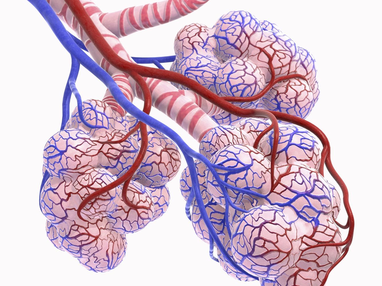
SARS-CoV-2 is the seventh known coronavirus that causes the human disease known as COVID-19. The virus can grow in cells lining the conducting airways and in alveolar epithelial cells. First, the virus generally enters the body through the nose or mouth. From there, the virus travels down into the alveoli which are located in the lungs. Once in the alveoli, the virus “hijacks” cells to make new copies of the virus. The infected cell is then killed, releasing new viruses to infect neighboring cells in the alveolus. Each sac of air, or alveolus, is wrapped with capillaries where red blood cells release carbon dioxide (CO2) and pick up oxygen (O2). Two alveolar epithelial cells (type I and II) facilitate gas exchange. Type I cells are squamous alveolar cells with thin membranes that perform gas exchange. Type II cells are known as progenitor cells in the alveoli and proliferate and differentiate into type I cells. In addition, Type II cells secrete the pulmonary surfactant that lines
-
- 七月 23, 2019
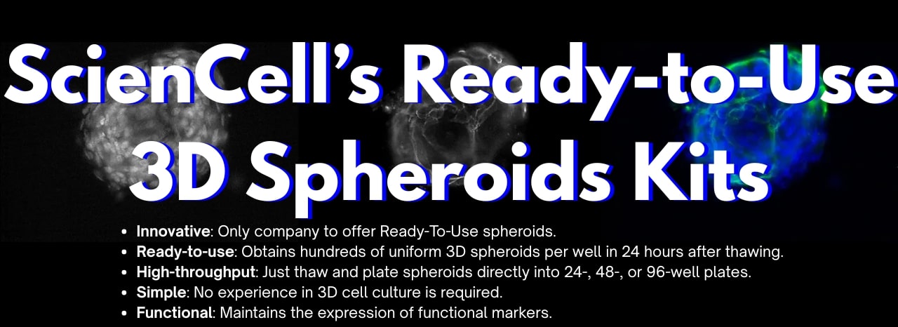
Traditional 2D cultures have been used widely over the past decades to study cell biology, molecular biology and conduct translation research such as drug discovery. Cells in 2D culture, however, are forced to adopt a planar morphology and maintain cellular interactions only in lateral directions, altering gene transcription, protein translation, and functional phenotypes. As a result, there is a shift towards using 3D in vitro models in the last several years as cell morphology and physiology more closely represent cells in vivo.
There are 2 main types of 3D culture systems known as scaffold-based and scaffold-free. In Table 1 below, the advantages and disadvantages of the different 3D cell culture techniques are listed to help researchers determine the most appropriate 3D culture method for their research.
Table 1: Advantages and Disadvantages of Different 3D Cell Culture Techniques.
Due to their novelties and complexities, 3D cell culture technologies may be daunting to some researchers.