
Watching Living Cells Instead of Static Snapshots
Understanding how cells behave over time is fundamental to modern cell biology. Traditional endpoint assays provide valuable information but often capture only a single moment in a dynamic biological process. In contrast, live-cell imaging allows researchers to continuously observe cellular behavior, providing insights into migration, proliferation, morphology, and cell-cell interactions as they occur.
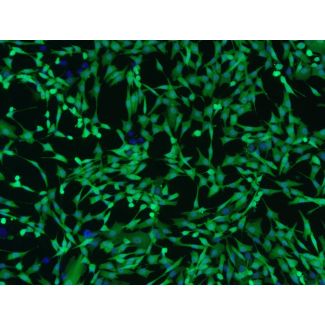
One of the most widely used approaches for live-cell imaging is the expression of Green Fluorescent Protein (GFP). By producing a bright fluorescent signal inside living cells, GFP enables researchers to visualize and monitor cells throughout an experiment without repeated fluorescent labeling (Figure 1).
When combined with primary human cells, GFP labeling offers an effective platform for studying biologically relevant cellular processes under in vitro conditions.
 Figure 1. Representative fluorescence images of GFP-expressing primary human neonatal dermal fibroblasts (HDF-GFP/2310-GFP). GFP fluorescence enables visualization of living cells while preserving normal cell morphology. A. Phase contrast. B. GFP fluorescence. C. Hoechst. D. Merge.
Why GFP?
Since its introduction into molecular and cellular biology, GFP has become one of the most widely used fluorescent reporters. Since GFP is genetically encoded, cells continuously produce the fluorescent protein following successful gene delivery, eliminating the need for repeated staining during live-cell experiments (Figure 2).
Researchers use GFP because it offers several advantages:
- Enables visualization of living cells in real time
- Eliminates repeated fluorescent labeling
Compatible with standard fluorescence microscopes using FITC filter sets
Supports long-term observation during routine cell culture
Easily combined with additional fluorescent probes such as nuclear stains
These characteristics have made GFP an essential tool in cell biology, regenerative medicine research, cancer biology, tissue engineering, and many other research fields.
 Figure 2. Lentiviral transduction enables constitutive GFP expression, allowing visualization of living cells during in vitro experiments.
Why Use Primary Cells?
Immortalized cell lines have become indispensable laboratory models because they are easy to culture and expand. Continuous immortalization, however, may alter important biological characteristics compared to native human tissues.
Primary cells provide an alternative model that more closely resembles normal human physiology. Because they are directly derived from human tissue and have a finite lifespan in culture; primary cells often retain cellular characteristics that are more representative of their tissue of origin.
For many in vitro applications, GFP-expressing primary cells combine the physiological relevance of primary cultures with the convenience of fluorescence-based imaging.
Advantages of GFP-Expressing Primary Cells
Introducing GFP into primary cells expands the range of experiments that can be performed without significantly changing standard cell culture workflows. Common advantages include:
- Live-Cell Imaging: Monitor living cells continuously while preserving cell viability throughout the experiment.
- Cell Tracking: Follow individual cells or cell populations over time during routine culture or image-based assays.
- Morphological Analysis: Observe changes in cell shape, spreading, and confluency using fluorescence microscopy.
- Migration Studies: Visualize cell movement during wound-healing (scratch) assays and other migration experiments.
- Co-Culture Experiments: Differentiate GFP-expressing cells from neighboring unlabeled cell populations in mixed cultures.
- Image-Based Assay Development: Generate clear fluorescent signals for automated imaging platforms and high-content analysis workflows.
The Importance of Quality Control
The quality of fluorescent primary cells depends on more than GFP expression alone. Comprehensive quality control helps verify that cell populations maintain characteristics appropriate for research applications. Typical quality assessments may include:
- Verification of GFP fluorescence
- Cell morphology evaluation
- Confirmation of cell type-specific marker expression
- Assessment of cell quality prior to cryopreservation
- Monitoring of GFP-positive cell populations during routine cell expansion
These evaluations help provide researchers with well-characterized cell populations suitable for reproducible experimental workflows.
Featured Product: Human Dermal Fibroblasts-GFP
HDF-GFP (Cat. No. 2310-GFP) consists of GFP-expressing Primary Human Neonatal Dermal Fibroblasts developed for fluorescence-based in vitro research applications. Supplied as ready-to-use cells with stable GFP expression, HDF-GFP eliminates the need for end users to generate fluorescent cells before starting their experiments.
Following lentiviral transduction during manufacturing, HDF-GFP cells undergo rigorous quality control to verify GFP fluorescence, fibroblast marker expression, cell morphology, and overall cell quality prior to cryopreservation (Figure 3).
HDF-GFP cells are suitable for a variety of fluorescence-based applications, including live-cell imaging, cell tracking, migration studies, image-based assay development, and co-culture experiments.

Figure 3. Immunofluorescence characterization of HDF-GFP/2310-GFP primary human neonatal dermal fibroblasts. Fibronectin staining demonstrates the characteristic extracellular matrix produced by primary dermal fibroblasts.
A. Relief contrast. B. Merged image (GFP fluorescence and Hoechst). C. Fibronectin immunofluorescence.
GFP remains one of the most versatile fluorescent reporters available for cell biology research. When combined with primary human cells, it provides researchers with a practical approach for monitoring cellular behavior in real time while preserving the biological relevance of primary cell culture models.
As fluorescence imaging continues to expand across basic and translational research, GFP-expressing primary cells will remain valuable tools for investigators seeking reliable in vitro models for microscopy-based studies.