Parkinson’s disease research has long centered on one highly vulnerable cell type: the midbrain dopaminergic neuron. These neurons control key aspects of movement, reward, and motivation, and their progressive degeneration in the substantia nigra is one of the defining features of Parkinson’s disease. Yet Parkinson’s disease is not only a dopaminergic neuron problem. Increasing evidence suggests that surrounding glial cells, especially astrocytes, can strongly influence dopaminergic neuron health, stress response, and degeneration.
This is why human stem cell-derived dopaminergic neuron and astrocyte systems are becoming increasingly valuable for disease modeling. By combining disease-relevant neurons with human astrocytes, researchers can move beyond simple monoculture assays and begin to model the cellular interactions that shape neuronal survival, vulnerability, and therapeutic response.
ScienCell’s neural disease modeling products are designed to support this type of research. Human pluripotent stem cell-derived dopaminergic neurons provide a human dopaminergic cell model for Parkinson’s disease-related studies, while human pluripotent stem cell-derived astrocytes provide a biologically relevant glial partner for studying neuron–astrocyte interaction. For researchers who want a streamlined co-culture workflow, ScienCell also offers the Human Pluripotent Stem Cell-derived Midbrain/Dopaminergic Neuron Modeling Kit, which combines dopaminergic neurons, astrocytes, and corresponding culture medium components in one cost-effective bundle.

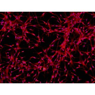
Dopaminergic neurons are a specialized neuronal population with unique developmental identity, neurotransmitter biology, electrophysiological properties, and metabolic demands. In Parkinson’s disease, midbrain dopaminergic neurons are especially vulnerable to cellular stressors such as mitochondrial dysfunction, oxidative stress, impaired proteostasis, lysosomal dysfunction, and alpha-synuclein accumulation.
Because of this selective vulnerability, dopaminergic neurons are a critical model system for studying Parkinson’s disease mechanisms. A generic neuronal cell line cannot fully capture the biology of a human dopaminergic neuron. Human pluripotent stem cell-derived dopaminergic neurons offer a more relevant platform for studying disease-associated phenotypes in the cell type most directly affected in Parkinson’s disease.
Published iPSC-based studies have shown why this matters. LRRK2 mutant iPSC-derived dopaminergic neurons have been reported to show increased oxidative stress sensitivity and elevated alpha-synuclein-associated phenotypes. Other patient-derived Parkinson’s disease models have revealed disease-relevant changes in mitochondrial function, neurite morphology, autophagy, lysosomal pathways, and neuronal survival. These findings demonstrate that human stem cell-derived dopaminergic neurons can capture biologically meaningful disease features that are difficult to study in non-human or non-neuronal systems.
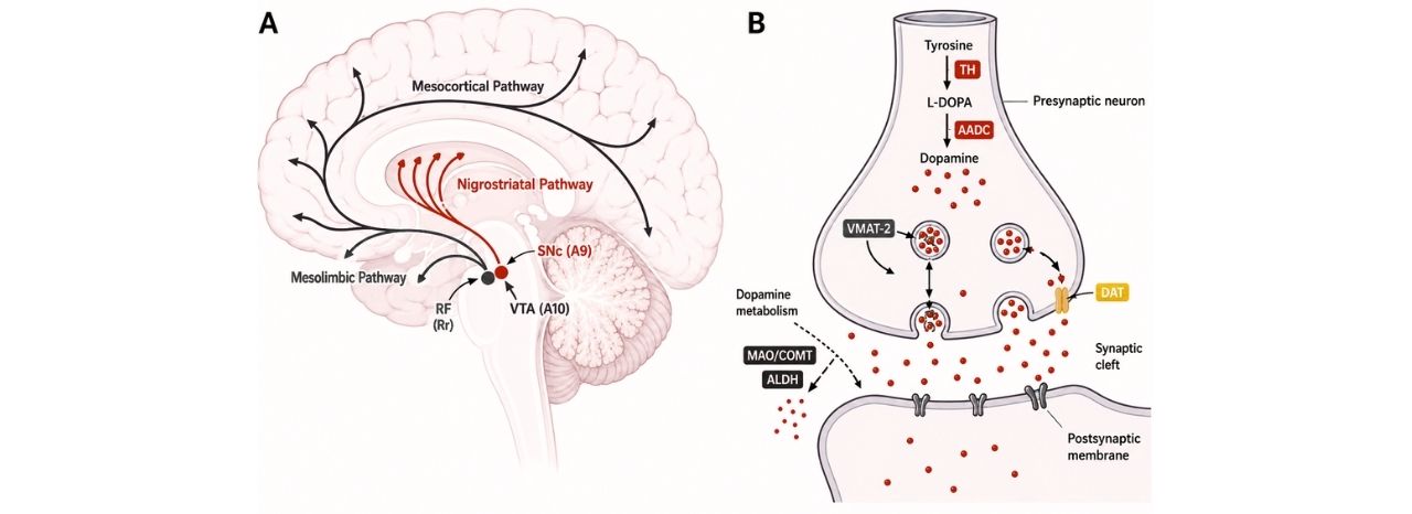
(A) Major dopaminergic pathways in the brain. Midbrain dopaminergic (mDA) neurons are primarily found within three nuclei: the retrorubral field (RrF; A8), substantia nigra pars compacta (SNc; A9), and ventral tegmental area (VTA; A10). Neurons originating in the SNc project to the dorsal striatum through the nigrostriatal pathway, while neurons from the VTA and RrF extend to the ventral striatum and prefrontal cortex, forming the mesolimbic and mesocortical dopaminergic circuits.
(B) Overview of dopamine neurotransmission, illustrating the synthesis, release, reuptake, and metabolic breakdown of dopamine at the synapse.

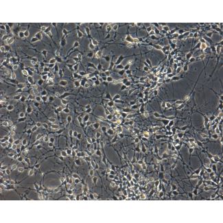
Astrocytes are often described as support cells, but this label understates their importance. Astrocytes regulate extracellular ions, provide metabolic support, help maintain redox balance, modulate synapse formation, participate in glutamate handling, secrete trophic factors, and respond to inflammatory stimuli. In neural disease models, astrocytes can protect neurons under some conditions and contribute to neuronal stress under others.
In Parkinson’s disease research, astrocytes are increasingly recognized as active contributors to disease biology. Parkinson’s disease-associated genes and pathways are not limited to neurons. Several PD-linked proteins have functional roles in astrocytes, and astrocyte dysfunction has been proposed to contribute to neuroinflammation, impaired protein clearance, oxidative stress, and altered neuron–glia communication.
One important example comes from patient-specific iPSC-derived astrocyte studies. In a Parkinson’s disease model using LRRK2 G2019S patient-derived cells, researchers found that PD astrocytes could contribute to non-cell-autonomous dopaminergic neurodegeneration. In co-culture, control ventral midbrain dopaminergic neurons grown on PD astrocytes showed neurodegenerative changes and abnormal astrocyte-derived alpha-synuclein accumulation. This type of result highlights why astrocytes should not be left out of Parkinson’s disease modeling.
For Parkinson’s disease research, astrocytes are particularly useful when the question is not simply “what happens inside the dopaminergic neuron?” but also “how does the surrounding glial environment influence dopaminergic neuron survival?”

Neuron monoculture systems are useful for studying cell-autonomous phenotypes. For example, researchers can test whether dopaminergic neurons are sensitive to oxidative stress, mitochondrial toxins, proteostasis stress, or candidate therapeutic compounds. However, in the brain, neurons do not function in isolation. They constantly interact with astrocytes and other neighboring cells.
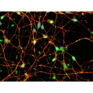
Adding astrocytes to dopaminergic neuron cultures creates a more informative model. A dopaminergic neuron–astrocyte co-culture can help researchers examine both neuronal vulnerability and glial contribution to disease progression. In this type of system, astrocytes may influence neuronal attachment, survival, neurite extension, maturation, inflammatory signaling, oxidative stress response, and alpha-synuclein-associated pathology.
A human dopaminergic neuron–astrocyte model can be used across multiple Parkinson’s disease-related workflows.
Oxidative stress assays
Dopaminergic neurons are highly sensitive to oxidative stress. Researchers can expose cultures to oxidative stressors and evaluate neuronal survival, morphology, neurite integrity, and stress-response readouts. Adding astrocytes allows researchers to test whether glial cells buffer stress or contribute to stress propagation.
Mitochondrial dysfunction studies
Mitochondrial impairment is a major theme in Parkinson’s disease biology. Dopaminergic neuron–astrocyte systems can be used to study mitochondrial stress, energy metabolism, and neuroprotective compounds that may improve cellular resilience.
Alpha-synuclein-related studies
Alpha-synuclein accumulation and propagation are central to Parkinson’s disease and related synucleinopathies. Co-culture systems can help researchers investigate how astrocytes interact with alpha-synuclein-associated pathology and whether neuron–astrocyte communication influences accumulation, clearance, or toxicity.
Neuroinflammation modeling
Astrocytes respond strongly to inflammatory stimuli and can influence neuronal health through cytokines, trophic factors, and stress-related signaling pathways. Dopaminergic neuron–astrocyte co-cultures are useful for modeling how inflammatory environments affect dopaminergic neuron survival.
Compound screening
In drug discovery, a neuron-only assay may miss glia-dependent effects. A neuron–astrocyte co-culture system can reveal whether a compound protects dopaminergic neurons directly, improves astrocyte support function, reduces glial stress responses, or modifies neuron–glia crosstalk.
Neurite degeneration and recovery
Dopaminergic neuron morphology is an important readout in disease modeling. Researchers can quantify neurite length, branching, degeneration, and recovery after stress or compound treatment. Astrocyte-supported cultures may improve the ability to monitor these changes over time.

Parkinson’s disease is a complex disorder involving neuronal vulnerability, glial dysfunction, oxidative stress, mitochondrial impairment, protein aggregation, inflammation, and altered cellular communication. No single in vitro model can capture every aspect of the disease. However, human PSC-derived dopaminergic neuron and astrocyte systems provide a practical and biologically meaningful step toward more predictive disease models.
By combining dopaminergic neurons with astrocytes, researchers can study not only what happens inside vulnerable neurons, but also how the surrounding cellular environment shapes disease progression and therapeutic response.