
Bringing Gene Expression into Focus
Cells continuously fine‑tune their gene expression in response to extracellular signals, environmental stress, and developmental cues. These dynamic changes drive core processes such as proliferation, differentiation, inflammation, and survival. Knowing when and how strongly a signaling pathway is activated is crucial for understanding cellular physiology, disease mechanisms, and responses to therapeutic compounds.
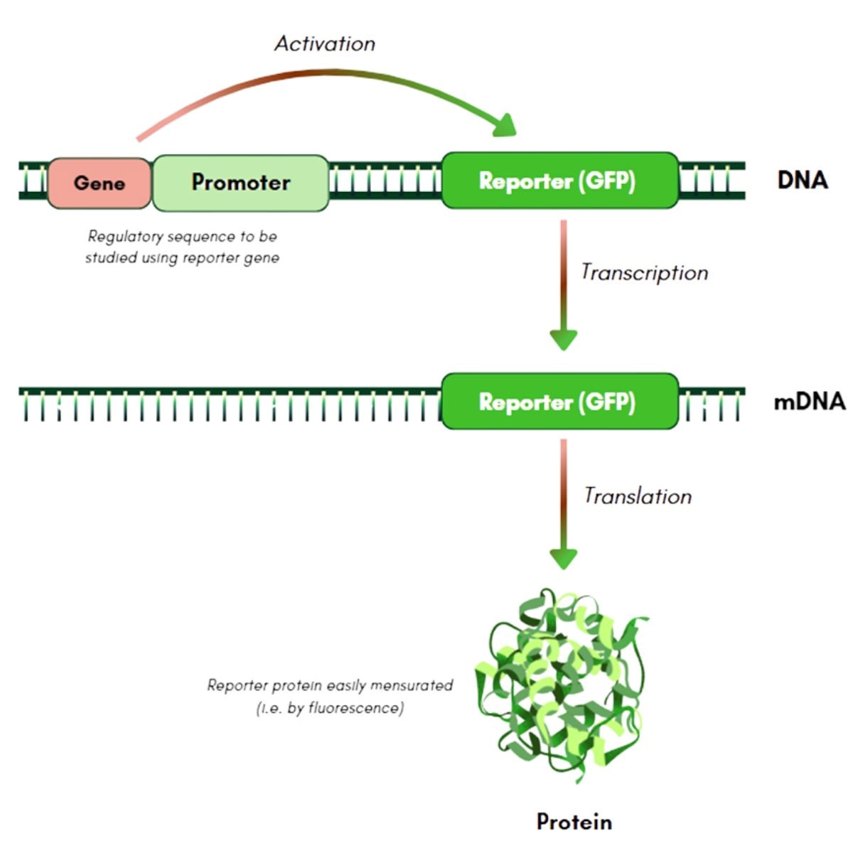
GFP reporter plasmid vectors provide a direct, fluorescence-based window into pathway-specific transcriptional activity in living cells. By placing the green fluorescent protein (GFP) gene under the control of regulatory elements responsive to a given signaling cascade, pathway activation triggers GFP expression, generating a measurable fluorescent signal in viable cells following transient transfection. This signal can be monitored over time using standard fluorescence instrumentation in cultured mammalian cells.
Because the readout arises from living cells rather than a fixed endpoint, researchers can track pathway activation kinetics, reversibility, and cell‑to‑cell variability over time—information often missed by traditional endpoint assays.
Why Use GFP Reporter Plasmids?
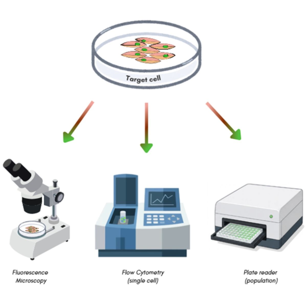
GFP reporters are versatile and compatible with standard instrumentation:
- Fluorescence microscopy for spatial and morphological context.
- Flow cytometry for high‑throughput, single‑cell analysis.
- Fluorescence plate readers for rapid, bulk measurements across many conditions.
- High‑content imaging systems for automated, multiparametric readouts combining intensity, localization, and additional markers.
Beyond overall pathway activity, GFP-positive cells can be visualized individually, enabling assessment of heterogeneity, quantification of responding subpopulations, and isolation via FACS for downstream RNA-seq, proteomics, or functional assays.
Pathway-Specific Reporter Plasmids
ScienCell's GFP Reporter Vectors are engineered to deliver pathway‑specific fluorescent readouts for several widely studied signaling cascades. Each vector incorporates:
- A dedicated response element (promoter/enhancer) recognized by pathway‑specific transcription factors.
- A minimal basal promoter to reduce background expression and improve signal-to-noise.
- A GFP reporter gene whose expression correlates with pathway activation in transfected mammalian cells.
This design allows researchers to connect upstream signaling events, such as receptor activation or kinase activity, to downstream transcriptional responses in a simple, fluorescence‑based assay that fits easily into existing cell-based workflows.
Introducing ScienCell’s GFP Reporter Vectors
ScienCell is pleased to introduce its new GFP Reporter Vectors, designed to provide a direct fluorescent readout of pathway-specific transcriptional activity in living cells. Our GFP Reporter Vector portfolio now includes tools for monitoring Wnt/β-catenin and hypoxia signaling, providing researchers with convenient approaches to visualize pathway activation in real time.
TCF-LEF/Wnt GFP Reporter Vector (TL-GFP, Cat. No. VT007)
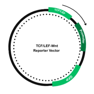
The TL-GFP Reporter Vector is designed to monitor canonical Wnt/β-catenin signaling. The vector contains multiple TCF/LEF response elements upstream of GFP. When Wnt signaling is activated, stabilized β-catenin accumulates in the nucleus and activates TCF/LEF-dependent transcription, resulting in GFP expression that enables visualization of Wnt-responsive cells.
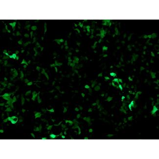
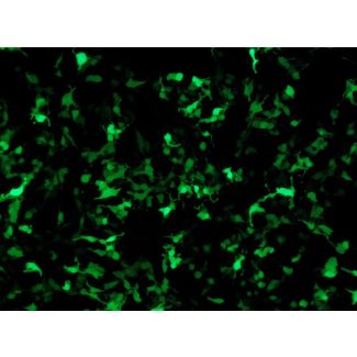
Image on the left: GFP expression in HEK293T cells transfected with the TCF/LEF GFP Reporter Vector after pathway activation (100x)
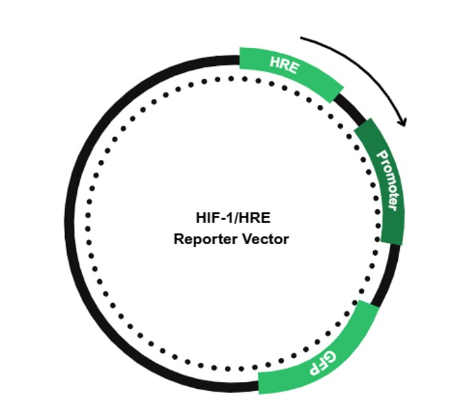
HIF-1/HRE-GFP Reporter Vector (HR-GFP, Cat. No. VT008)
The HRE-GFP Reporter Vector provides a fluorescent readout of cellular responses to hypoxia. Under hypoxic conditions, stabilized HIF-1 accumulate in the nucleus and bind to hypoxia response elements (HREs), activating GFP transcription and enabling visualization of HIF-mediated pathway activation.
Image on the right: GFP expression in HEK293T cells transfected with the HRE GFP Reporter Vector after pathway activation (100x)
Together, these reporters provide convenient fluorescence-based tools for studying dynamic signaling responses in living cells and can be readily integrated into imaging, flow cytometry, and fluorescence plate-based assays.
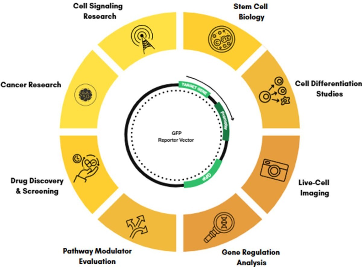
Typical Experimental Workflows
ScienCell’s GFP Reporter Vectors integrate easily into common cell‑based assays:
Live‑cell fluorescence imaging to visualize spatial patterns of pathway activation, track dynamic changes, and correlate signaling with cell morphology or behavior.
Flow cytometry and FACS, enabling quantitative analysis of GFP‑positive cells and isolation of activated subpopulations for downstream molecular profiling.
High‑content imaging, where automated microscopy and image analysis quantify GFP intensity, localization, and co‑markers across thousands of cells per condition.
Drug discovery and compound screening, using reporter activation as a rapid, functional readout of pathway modulation by small molecules or biologics.
Time‑course studies, to map activation, peak response, and signal decay after stimuli such as growth factors, cytokines, hypoxia, or stress.
Evaluation of pathway agonists and inhibitors, benchmarking dose–response relationships and off‑target effects across different signaling cascades in a standardized, fluorescence-based system.
A Simple Window into Complex Signaling
Reporter plasmids have become essential tools for dissecting how cells sense and respond to changes in their environment. By converting transcriptional activation into a visible fluorescent signal, GFP reporters enable researchers to monitor signaling pathways using straightforward, scalable fluorescence‑based assays compatible with a range of instruments and experimental designs.
Whether the focus is Wnt‑mediated differentiation, NF‑κB activation during inflammatory responses, HIF‑1 signaling under hypoxic conditions, or other transcriptional programs, ScienCell’s GFP Reporter Vectors provide a versatile platform for investigating pathway activation in living cells and gaining deeper insights into cellular signaling dynamics.
Stay tuned for more GFP Reporter Vectors coming soon, designed to help you visualize even more cellular signaling pathways.
References:
- Chalfie M, Tu Y, Euskirchen G, Ward WW, Prasher DC. Green fluorescent protein as a marker for gene expression. Science. 1994.
- Albano CR, Randers-Eichhorn L, Bentley WE, Rao G. Green fluorescent protein as a real time quantitative reporter of heterologous protein production. Biotechnol Prog. 1998; 14:351–4.
- Hack NJ, Billups B, Guthrie PB, Rogers JH, Muir EM, Parks TN, Kater SB. Green fluorescent protein as a quantitative tool. J Neurosci Methods. 2000; 95:177–84.
- Heim R, Cubitt AB, Tsien RY. Improved green fluorescence. Nature. 1995; 373:663–4.
- Ni Q, et al. Live-cell imaging of cell signaling using genetically encoded fluorescent reporters. Curr Opin Chem Biol. 2017; 39:108–115.
- Apfel J, Reischmann P, Müller O. A new fluorescence-based reporter gene vector as a tool for analyzing and fishing cells with activated wnt signaling pathway. ISRN Oncol 2013: 603129, 2013.