Page 2 - Company

-
Posted: February 12, 2026
 Read more »
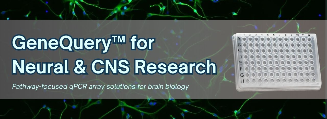
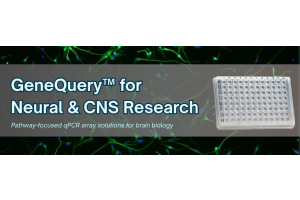
In CNS research, understanding disease mechanisms goes beyond tracking individual biomarkers—it requires insight into how entire biological pathways are regulated. From blood–brain barrier (BBB) integrity to glial activation and neuroinflammatory signaling, meaningful studies depend on high-resolution
-
Posted: February 08, 2026
 Read more »
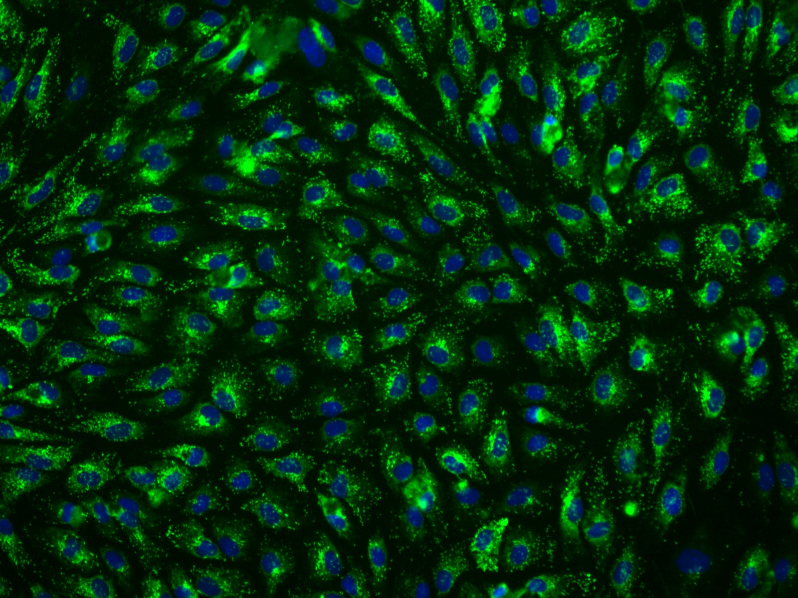
APOE genotype–specific astrocyte biology has emerged as a central driver of Alzheimer’s disease and neurodegenerative risk. Recent studies demonstrate that the APOE4/4 genotype profoundly reshapes astrocyte function across multiple pathways, including lipid metabolism, neuroinflammatory signaling, and
-
Posted: February 04, 2026
 Read more »
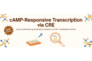
Evaluate cAMP-dependent signaling with ScienCell’s CRE/CREB Dual Reporter Vector (CREVR, Cat. No. VT004). CREB regulates gene expression in response to cAMP/PKA, calcium, and other signaling pathways, controlling metabolism, neuronal activity, and stress responses. Accurate measurement of CRE activity
-
Posted: February 03, 2026
.png) Read more »Heart Health in Focus: Cardiac Primary Cells for Cardiovascular DiscoveryFebruary marks American Heart Month, an observance dedicated to advancing awareness of cardiovascular health and the critical research needed to prevent and treat heart disease. At ScienCell, we recognize the essential role that
Read more »Heart Health in Focus: Cardiac Primary Cells for Cardiovascular DiscoveryFebruary marks American Heart Month, an observance dedicated to advancing awareness of cardiovascular health and the critical research needed to prevent and treat heart disease. At ScienCell, we recognize the essential role that -
Posted: January 06, 2026
 Read more »
Apolipoprotein E (ApoE) is a key regulator of lipid transport and cholesterol homeostasis, and genetic differences in the APOE gene significantly influence susceptibility to neurodegenerative and cardiovascular diseases. Understanding a sample’s APOE genotype provides essential biological context across
-
Posted: October 09, 2025
.jpg) Read more »
Read more »The Wnt pathway plays a central role in cell signaling, development, and disease, yet studying it often poses technical challenges. Its complexity stems from a tightly regulated network of proteins and receptors that drive processes such as stem cell renewal, tissue regeneration, and cancer progression.
-
Posted: October 07, 2025
 Read more »
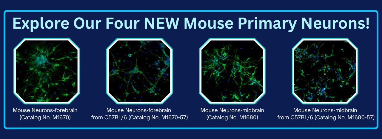
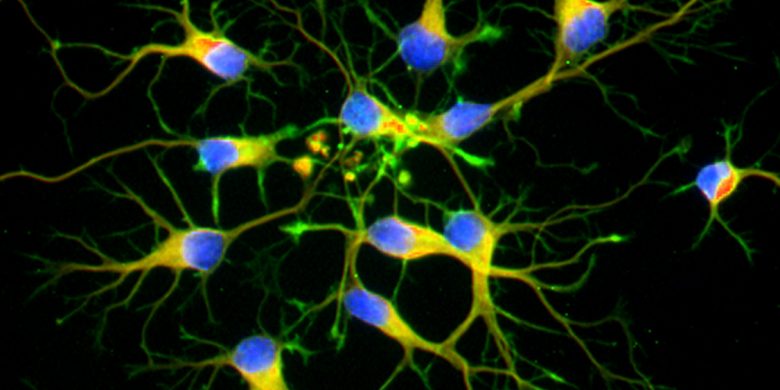
Neurons are the dynamic, anatomically, functionally, and trophically vital units of the brain, responsible for electrochemically transmitting information across the nervous system. While all neurons share common morphological features, their regional specialization is what truly drives brain function.
-
Posted: October 06, 2025
.jpg) Read more »
Read more »We are excited to announce and launch two new products for Wnt/β-catenin signaling research: the TCF/LEF Dual Reporter Vector (TDRV, Catalog No. 9038) and the Luciferase Assay Kit (LAK, Catalog No. 9048).
The TDRV provides the reporter system, and the LAK provides the robust, accurate measurement,
-
Posted: October 06, 2025
 Read more »
Highly bioactive, animal-free growth factors and cytokines for iPSC and ESC cell culture
Induced Pluripotent Stem Cells (iPSC) are a type of pluripotent stem cell derived from adult somatic cells such as skin cells or blood cells, through cellular reprogramming. Embryonic stem cells (ESC) are derived
-
Posted: September 30, 2025
 Read more »
Telomeric repeat–containing RNA (TERRA) plays a vital role in telomere biology, influencing telomerase activity, chromosome stability, and cellular aging. Abnormal TERRA expression has been linked to cancer, aging, and age-related diseases. But detecting TERRA has always been a challenge due to:
-
Posted: September 29, 2025
 Read more »
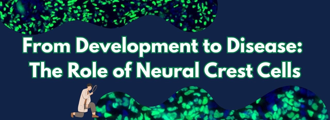
When reliable, high-purity cells are essential, origin and quality matter. ScienCell offers two well-characterized Human Neural Crest Cell (NCC) lines from distinct stem cell sources for precision in disease modeling and tissue regeneration.
-
Posted: August 11, 2025
 Read more »
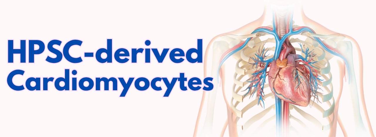
Human primary cardiomyocytes and cardiac tissue are invaluable for heart disease studies, drug discovery, and toxicity testing, often surpassing animal models or immortalized cell lines in relevance. However, obtaining adult human primary cardiomyocytes is challenging due to limited tissue availability,
-
Posted: July 30, 2025
.png) Read more »
Read more »If your research depends on DNA, RNA, or protein lysates from human primary cells, you know how challenging and expensive it can be to isolate these materials yourself. That’s where ScienCell’s Human Cell Pellets come in, offering an opportunity to work with primary cells without the high cost and time
-
Posted: July 28, 2025
.jpg) Read more »
Read more »At Sciencell, we are committed to providing high-quality tools to support groundbreaking research. This month, we are excited to showcase two powerful proteases: Recombinant Yeast SUMO Protease (Catalog No. MB9018) and Recombinant TEV Protease (Catalog No. MB9028). These reagents are designed to streamline
-
Posted: July 06, 2025
.jpg) Read more »
Read more »Unlock precise control over cellular processes with ScienCell's extensive range of recombinant human proteins. Engineered for stability and biological activity, our products are essential tools for studying cell proliferation, differentiation, tissue development, and disease mechanisms.
Explore our
-
Posted: June 10, 2025
.jpg) Read more »
Read more »Unlocking the full potential of human pluripotent stem cells (hPSCs) often hinges on effective cryopreservation techniques.
ScienCell provides StemCryo®, a cutting-edge solution designed to ensure the viability and pluripotency of these invaluable cells for your critical research.
-
Posted: June 09, 2025
.jpg) Read more »
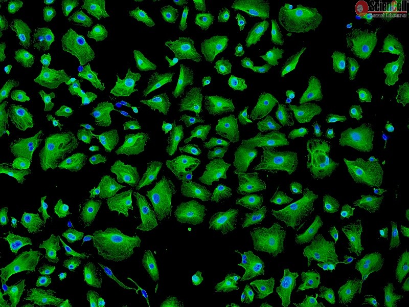
Read more »Fibroblasts, fundamental mesenchymal cells derived from the embryonic mesoderm, are widely utilized in cellular and molecular research due to their ease of culture and robust nature. These versatile cells are key secretors of a non-rigid extracellular matrix, abundant in type I and/or type III collagen,
-
Posted: June 09, 2025
.jpg) Read more »
Read more »Stem cells are multipotent cells with the ability to self-renew, self-replicate, and differentiate into multiple cell types, including mesenchymal stem cells (MSCs), hematopoietic stem cells (HSCs), neural stem cells (NSCs), embryonic stem cells (ESCs), and induced pluripotent stem cells (iPSCs). Remarkable
-
Posted: June 05, 2025
.jpg) Read more »
Read more »Save Time. Reduce Variability. Get Results.
Working with primary cells is essential for biologically relevant data, but sourcing tissues, isolating cells, and extracting high-quality RNA, DNA, or protein can be time-consuming, expensive, and technically demanding.
Our line of cell-derived molecular
-
Posted: May 28, 2025
.jpg) Read more »
Read more »Pseudogenes are DNA segments similar to functional genes but with lost function. They were first described in 1977. Based on origin, they are classified as processed (e.g., human PTENP1), non-processed (e.g., human ARHGAP27P1), or unitary (e.g., human GULOP).
While many may not be functional or transcribed,
-
Posted: May 28, 2025
.jpg) Read more »
Read more »Macrophages are crucial cells in the body, known for their role in removing cellular debris and destroying invading pathogens. Their main functions include phagocytizing invading microorganisms and scavenging dead, damaged cells, and cellular debris.
Given their primary functions involving phagocytosis,
-
Posted: May 27, 2025
.jpg) Read more »
Read more »Several key metabolites play crucial roles in cellular bioenergetic homeostasis. ScienCell has developed sensitive assays to accurately quantify L-lactate, glucose, glutamate, and pyruvate.
L-Lactate is a key intermediate in glucose metabolism. Under hypoxic conditions, lactate dehydrogenase
-
Posted: May 26, 2025
.jpg) Read more »
Read more »Why Choose Our MSCs? Our Mesenchymal Stem Cells are:
- Multipotent—capable of differentiating into a variety of lineages, such as osteoblasts, chondrocytes, endothelial cells, hepatocyte-like cells, and many more.
- Multiple organ systems—derived from adipose, liver, bone, and the umbilical cord, our MSCs
-
Posted: May 21, 2025
.jpg) Read more »
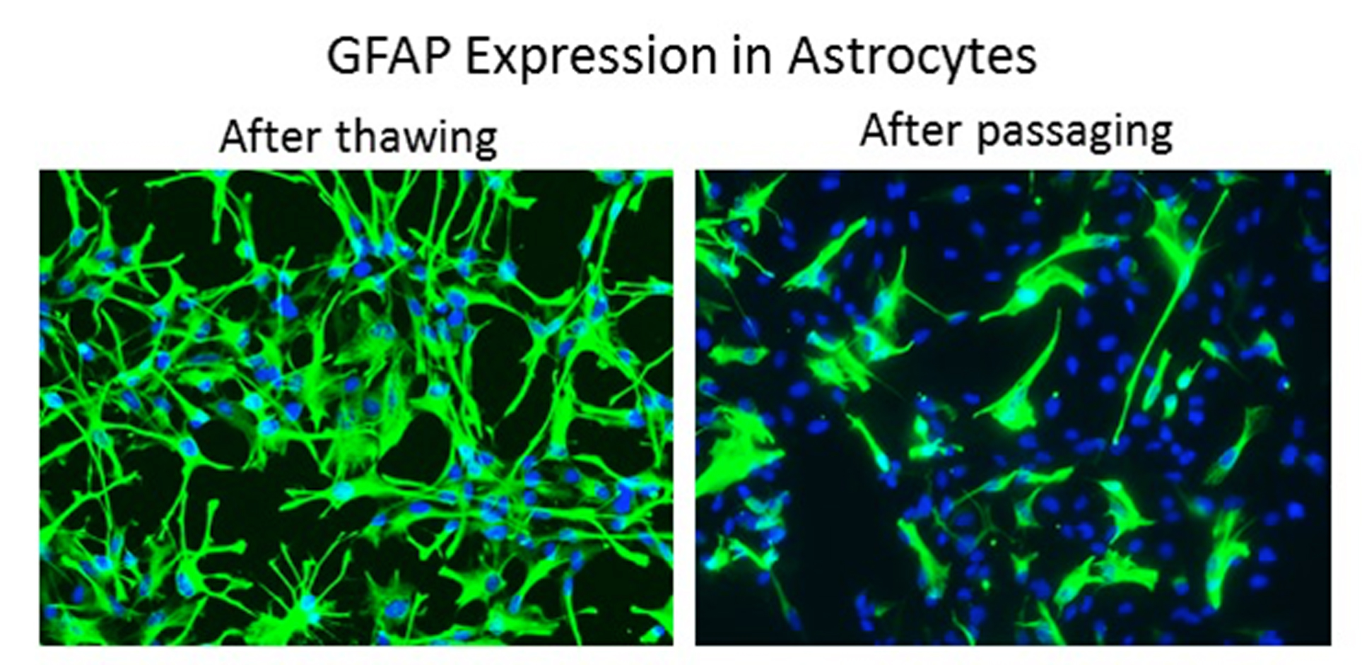
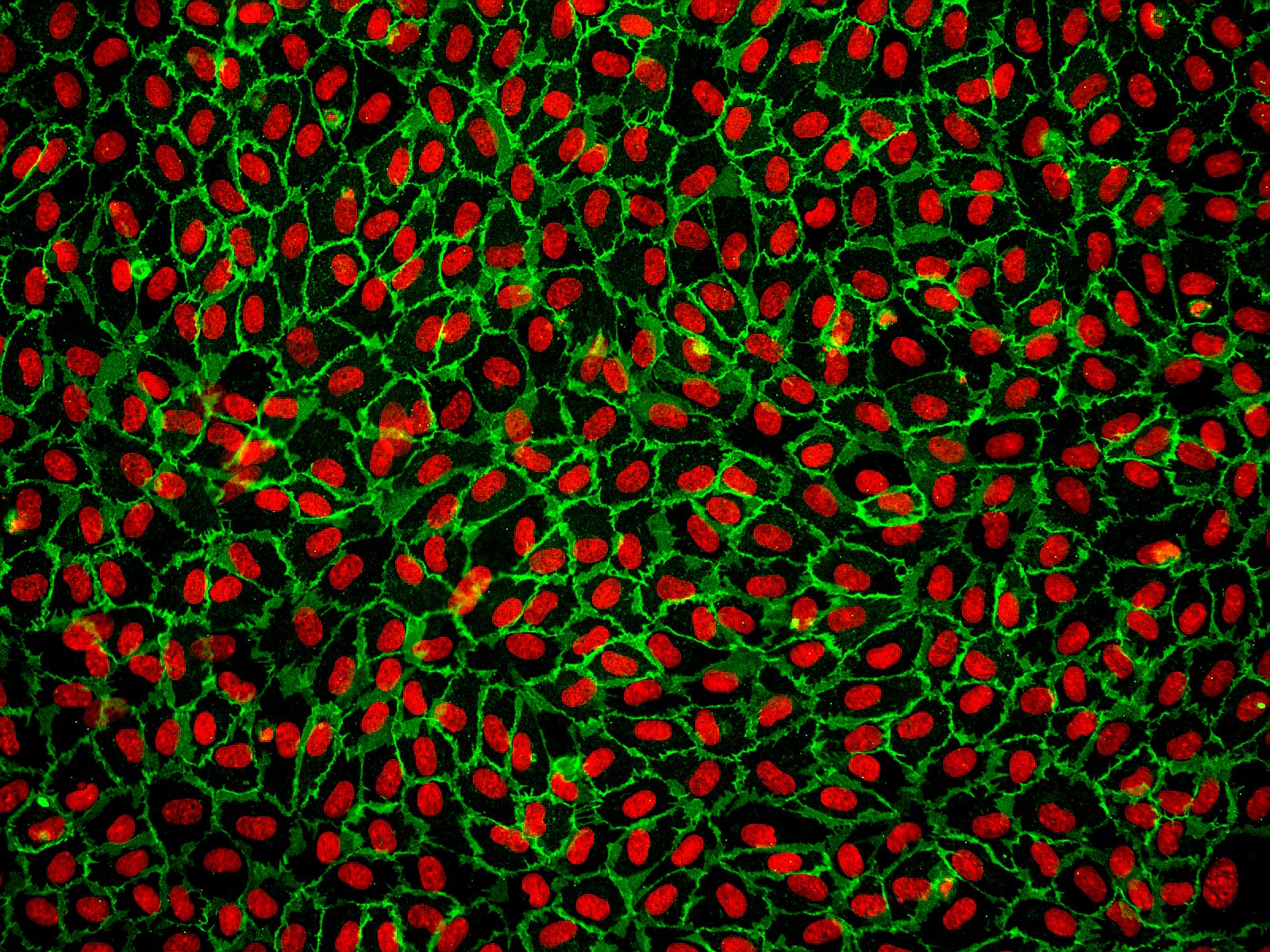
Read more »Astrocytes are the most abundant cell type in the central nervous system (CNS). They serve a variety of vital functions including supporting neuronal transmission and survival, regulating the blood-brain barrier, contributing to anti-inflammatory responses, and aiding in wound repair.
Astrocytes also
-
Posted: May 19, 2025
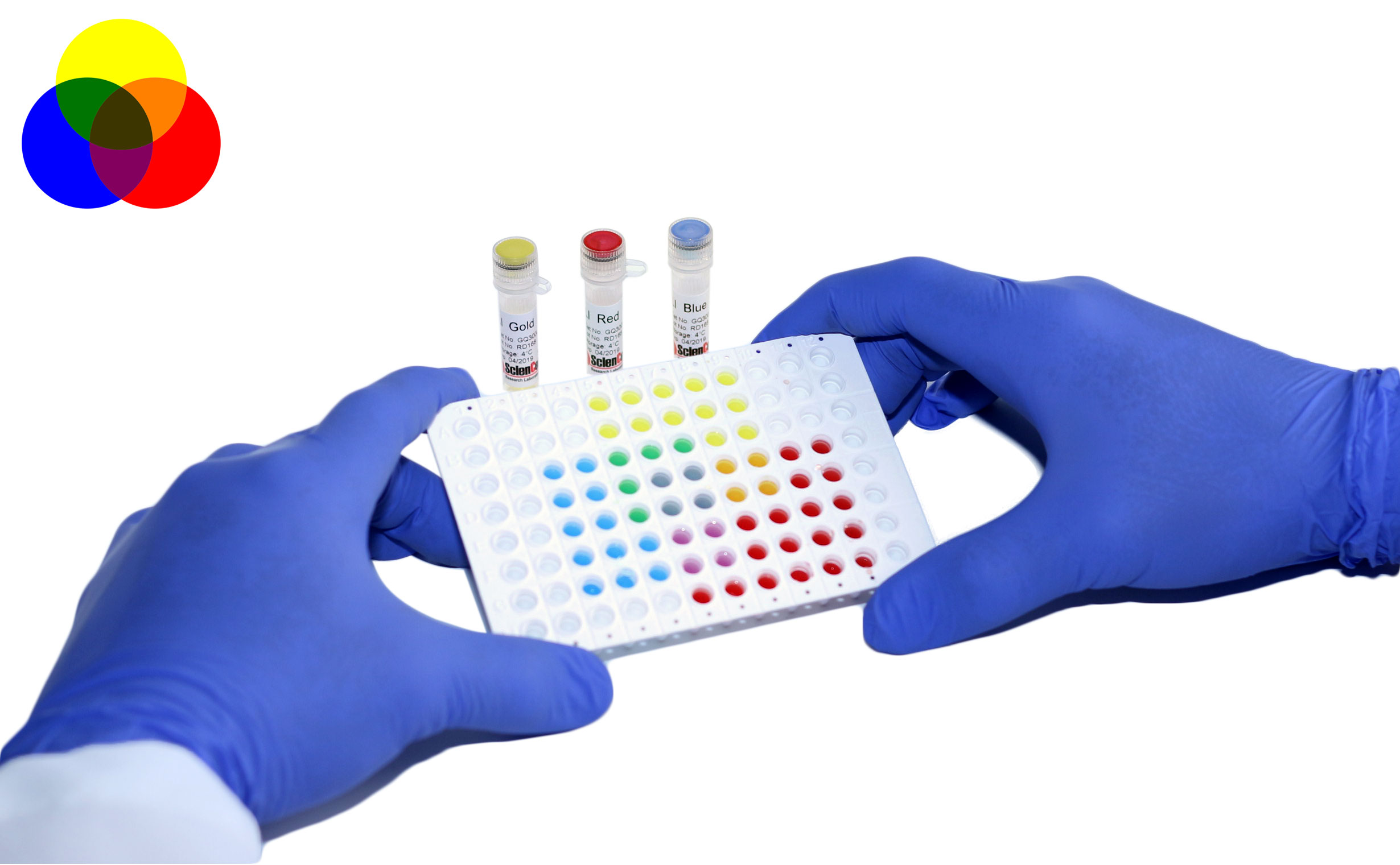
.jpg) Read more »
Read more »GeneQuery™ Disease Models qPCR Array Kits are powerful tools for understanding the molecular mechanisms underlying disease onset and progression. By measuring the activity levels of specific genes, researchers can gain crucial insights into disease pathways, identify potential biomarkers, and evaluate
-
Posted: May 18, 2025
 Read more »
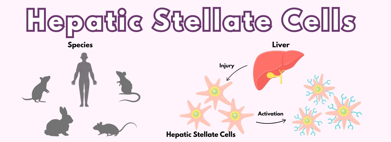
Hepatic stellate cells (HSteC) are essential intralobular connective tissue cells that perform diverse functions in the liver, including extracellular matrix homeostasis, repair, regeneration, fibrosis, and control of retinol metabolism, storage, and release. HSteC are a powerful model for studying liver
-
Posted: May 15, 2025
 Read more »
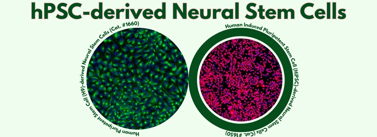
Leading the way in providing advanced tools for life science research, ScienCell Research Laboratories offers high-quality, well-characterized neural stem cell products derived from pluripotent stem cell sources, along with supporting reagents.
These products provide unique advantages for diverse applications,
-
Posted: May 15, 2025
 Read more »
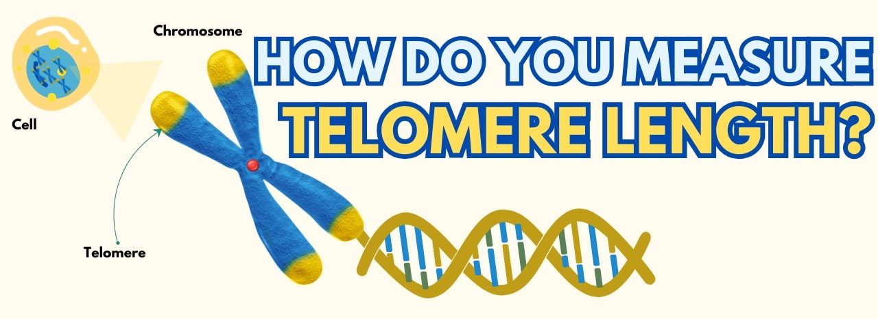
Advance Your Telomere Research Using ScienCell’s Absolute Telomere Length Quantification qPCR Kits!
Understanding Telomeres and Their Role in Genomic Stability
Telomeres are nucleoprotein structures that cap chromosome ends, protecting them from degradation and ensuring genomic stability. Each cell
-
Posted: May 14, 2025
 Read more »
In cellular and molecular research, the quality and reliability of your reagents are paramount to achieving reproducible and meaningful results. ScienCell offers a selection of high-quality
cell culture reagents designed to support critical steps in your experimental workflows!
-
Posted: May 14, 2025
 Read more »
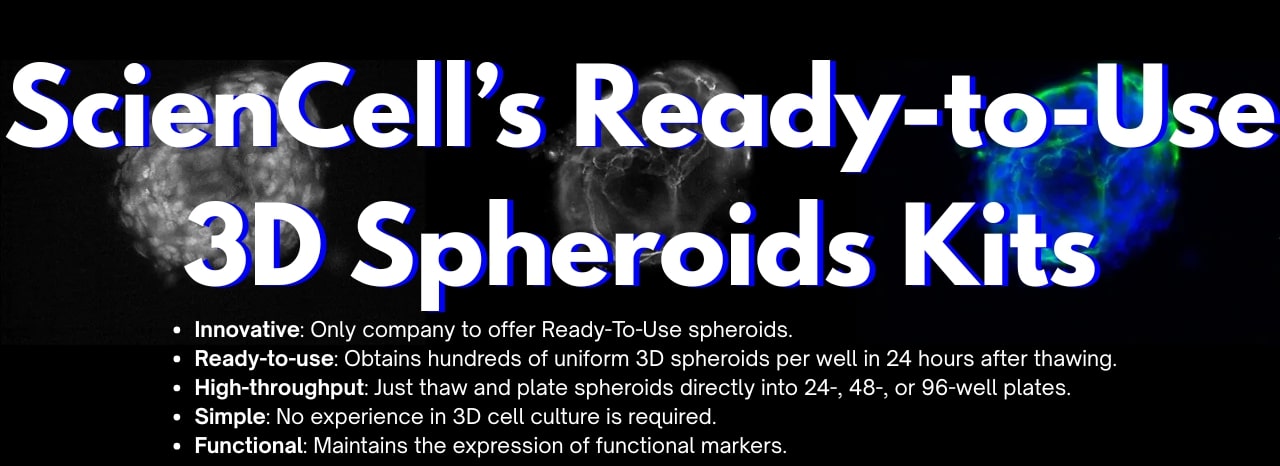
Building and maintaining consistent 3D spheroid models can traditionally involve long and complex workflows sometimes described as tedious and rate limiting.
Our featured ready-to-use 3D spheroid kits address this challenge. These innovative products allow researchers to obtain highly homogenous and
-
Posted: May 12, 2025
.jpg) Read more »
Read more »Achieving physiologically relevant in vitro models is paramount for groundbreaking research. The extracellular matrix (ECM) plays a critical role in cell behavior, influencing everything from adhesion and spreading to growth and differentiation.
To provide your cells with the optimal environment,
-
Posted: May 11, 2025
 Read more »
ScienCell’s trusted GoldNStart TaqGreen qPCR Master Mix just got even better. Our latest upgrade features an integrated 2-color tracking system designed to make your qPCR workflow faster, easier, and more accurate—without sacrificing any of the reliability you know and trust.
-
Posted: May 08, 2025
 Read more »
As the demand for advanced models in regenerative medicine and hair restoration therapies grows, so does the need for reliable, physiologically relevant primary cells.
ScienCell Research Laboratories offers a suite of human hair follicle cell types to empower your research — each isolated from human
-
Posted: May 06, 2025
 Read more »
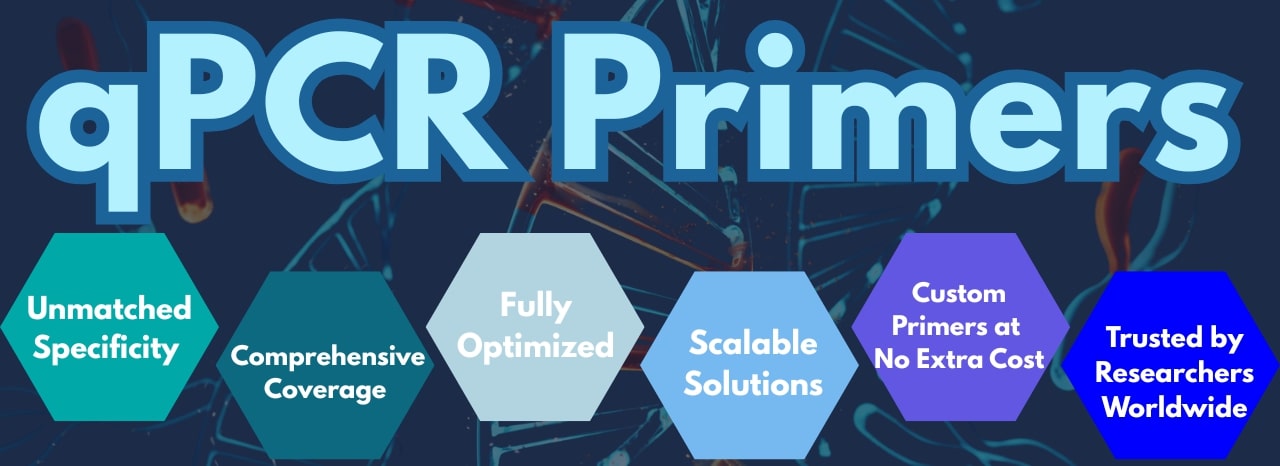
ScienCell Research Laboratories offers over 5,400 individual primer sets for human and mouse gene expression analysis, plus our GeneQuery™ qPCR Array Kits for high-throughput studies. Each lyophilized primer set is pre-validated and provides enough material for 100 x 20µL qPCR reactions—perfect for
-
Posted: May 04, 2025Categories: Newsletter
 Read more »
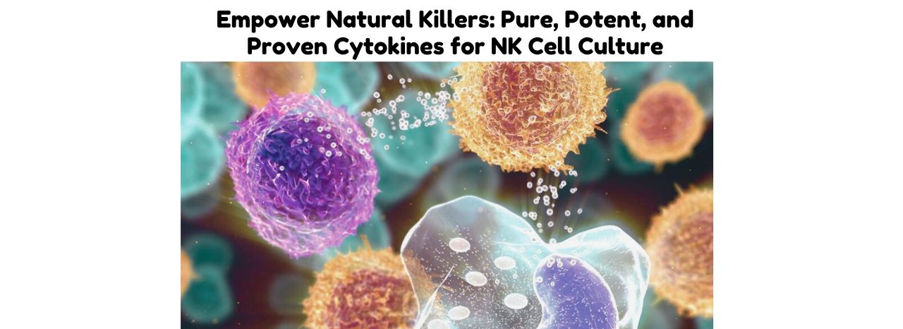
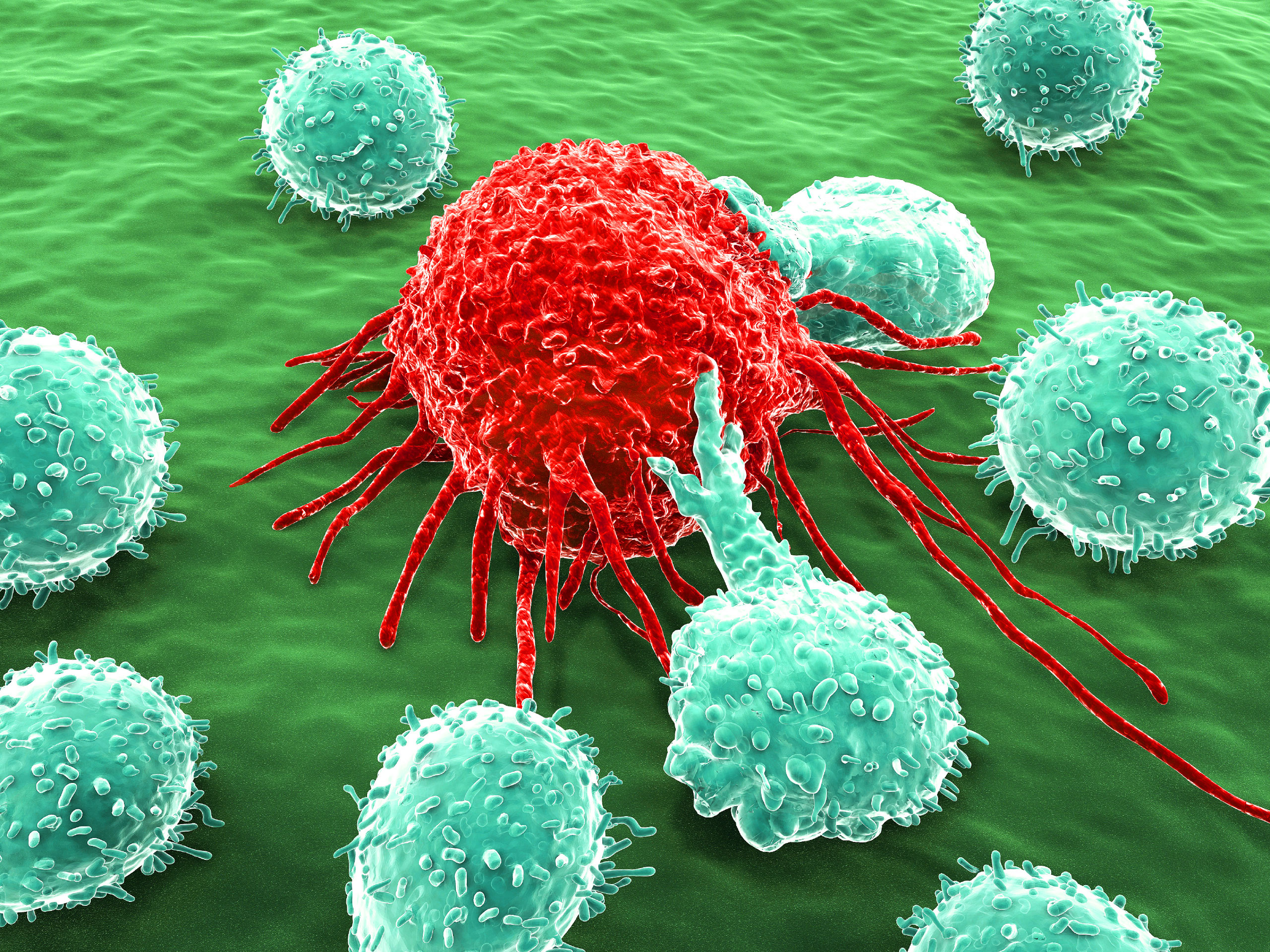
NK Cells
NK cells are a type of cytotoxic lymphocyte that plays a critical role in the innate immune system’s defense against infected and cancerous cells. Unlike T cells, which are part of the adaptive immune response, NK cells do not require prior activation to kill their targets. Instead, they can
-
Posted: May 01, 2025
 Read more »
ScienCell Research Laboratories offers a robust foundation for skeletal muscle research with our high-purity Human Skeletal Muscle Cells (HSkMC, Cat. #3500). These cells are isolated from adult human skeletal muscle and cryopreserved at passage one to ensure optimal viability and physiological relevance. -
Posted: April 30, 2025
 Read more »
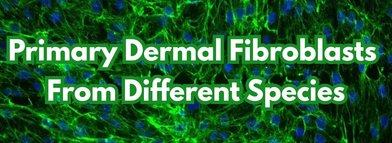
Dermal fibroblasts, derived from the embryonic mesoderm are fundamental components of the skin's dermis layer. Their versatility and ease of culture make them extensively used in cellular and molecular studies. Due to their durability, they are also suitable for a variety of manipulations, such as gene
-
Posted: April 08, 2025
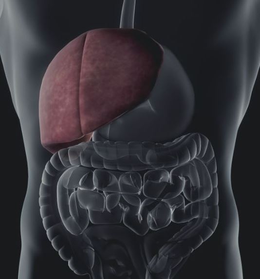
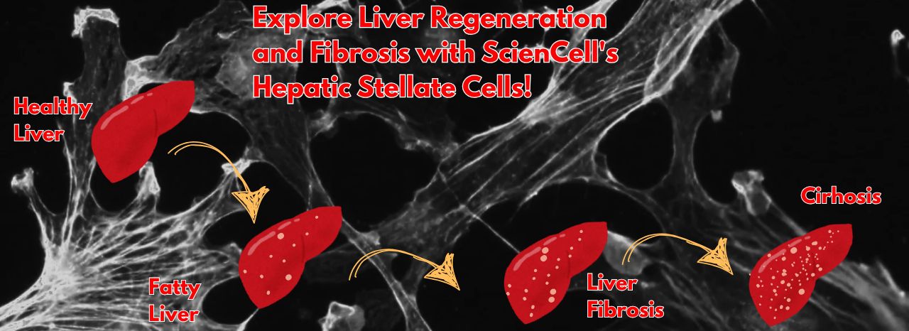 Read more »Hepatic stellate cells (HSCs) are essential to liver function, playing a central role in homeostasis, extracellular matrix maintenance, repair, and regeneration. ScienCell Research Laboratories provides high-quality primary human hepatic stellate cells (HHSteC) to support cutting-edge research into liver
 -
Posted: October 06, 2022
 Read more »
Telomere “caps” are located at the ends of all chromosomes, including those of the immune system’s T-lymphocytes (T cells), and become shorter with every cell division. Once these chromosomes reach a point where the telomere is too short to provide adequate protection, division ceases and the cell proceeds
-
Posted: April 30, 2020
 Read more »
Hepatic sinusoidal endothelial cells (HSEC) are fascinating cells that are uniquely adapted to their location in the liver. HSEC are found lining micro-vessels in the liver and are extremely specialized endothelial cells. Structurally and functionally they have distinctive features which include: open
-
Posted: April 03, 2020
 Read more »
Epithelial cells are the most numerous cells in the lungs and contribute to innate and adaptive immunity. Airway epithelial cells are located in the lower respiratory tract which includes the trachea, bronchi, small airways (bronchioles), and alveoli. Due to their location, airway epithelial cells are
-
Posted: February 09, 2020
 Read more »
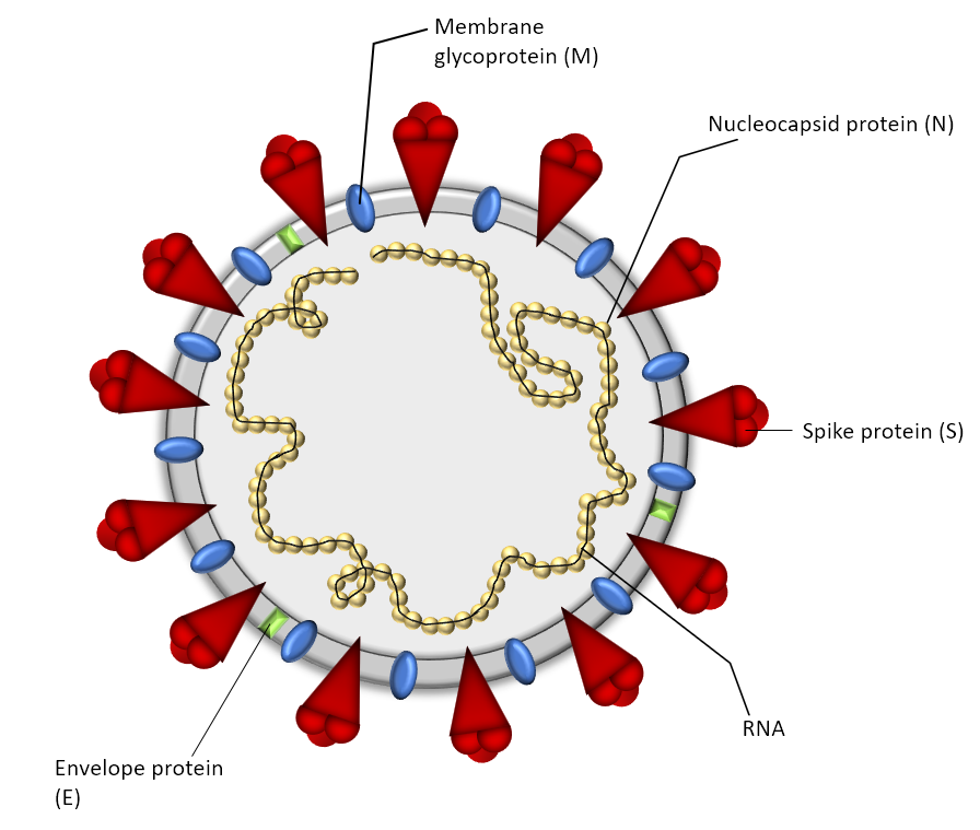
As of April 10, 2020, the number of U.S. SARS-CoV-2 coronavirus cases surpassed 500,000 with a death toll near 19,000. For over a century, coronaviruses were thought to only cause mild illnesses such as the common cold. With the ou or over a century, coronaviruses were thought to only cause mild illnesses
-
Posted: July 22, 2019Comments: 2
 Read more »
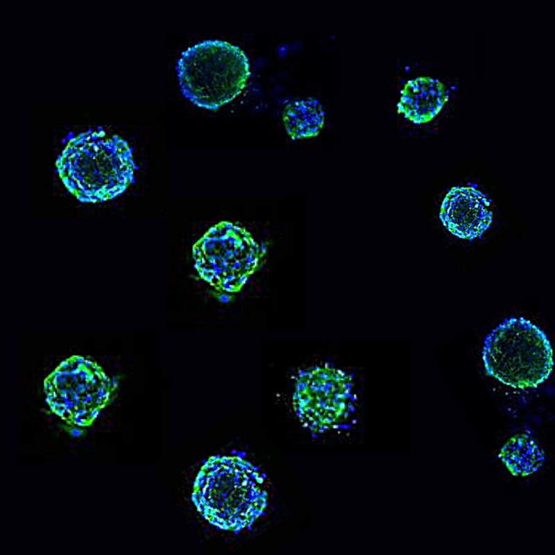
Traditional 2D cultures have been used widely over the past decades to study cell biology, molecular biology and conduct translation research such as drug discovery. Cells in 2D culture, however, are forced to adopt a planar morphology and maintain cellular interactions only in lateral directions, altering
-
Posted: June 09, 2019
 Read more »
Hepatic stellate cells have recently gained a great deal of attention regarding their contribution to the progression of diseases such as liver fibrosis, non-alcoholic steatohepatitis (NASH), and hepatocellular carcinoma. They are mesenchymal cells that are located between sinusoidal endothelial cells
-
Posted: February 11, 2019
 Read more »
ScienCell’s wide assortment of cell culture media is in liquid form and includes Specialty, Classical & Supplement varieties. Each product is designed for optimal nutrition and growth of primary cells. ScienCell’s cell culture media is manufactured and tested to ensure a high standard of quality and
-
Posted: August 28, 2018
 Read more »
qPCR is a powerful tool for quantification of gene expression levels and copy number variation. Despite the advances in next-generation sequencing (NGS), qPCR still serves as the "gold standard" for gene expression analysis. Due to poor reproducibility and vast lab-to-lab variation, all NGS data requires
-
Posted: May 30, 2018
 Read more »
When working with primary cells, it is important to remember that they are not cell lines and should be treated with care. At ScienCell, we specialize in primary cell culture and we are very familiar with the common problems researchers encounter when culturing them. We have compiled a list with 13
-
Posted: April 03, 2018Comments: 1
 Read more »
Cancer immunotherapy is one promising cancer treatment option whereby the host's own immune system is used to treat cancer. The therapy works by either stimulating certain immune activities, or counteracting cancer cell signals that suppress immune responses. Cancer immunotherapy has progressed significantly
-
Posted: October 24, 2017Comments: 3
 Read more »
Neuronal cell lines are commonly used for in vitro neurobiology studies because they are more easily transfected compared to primary neurons and they proliferate, whereas primary neurons do not. Neuronal cell lines can be induced to differentiate into neuron-like cells, where they express neuronal markers
-
Posted: March 26, 2017Categories: Company, Information, Gene Expression Profiling, Cell Culture Media, Primary Cells, Cell Based Assays, News
-
 Read more »
Hello fellow cell enthusiasts! We at team ScienCell just got back from Baltimore, MD, where we attended ToxExpo 2017, the Society of Toxicology’s annual conference. While the cold weather was quite a shock to us Californians, it was such a treat getting to experience what Baltimore had to offer. After
-
Posted: February 14, 2017
 Read more »
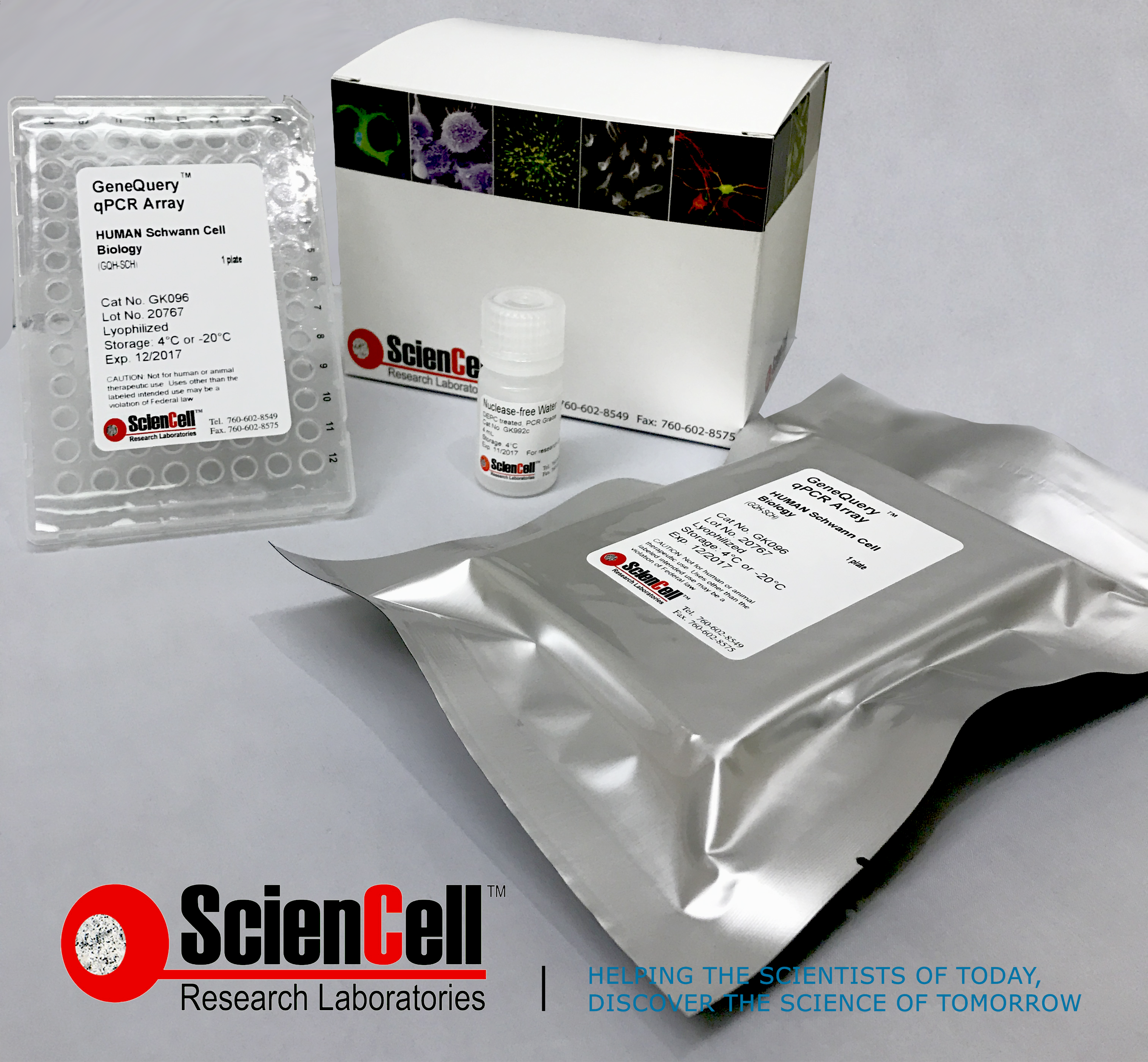
CARLSBAD, Calif. -- ScienCell Research Laboratories, Inc. is a unique supplier of primary Human Schwann Cells to the scientific research community. Schwann cells are glial cells that protect and insulate nerves in the peripheral nervous system.
"There are numerous cell types, such as Schwann cells,
-
Posted: February 14, 2017
 Read more »
SAN DIEGO, Calif. -- ScienCell Research Laboratories, Inc. recently announced it would be expanding its cell culture products to the gene expression profiling market. ScienCell, who prides itself on supplying unique primary cell types for nearly 20 years, will now supply qPCR array kits and individual
-
Posted: February 14, 2017
 Read more »
Online webinar hosted by sciencell to learn tips for successful qPCR, a technique for gene expression profiling
CARLSBAD, CALIFORNIA, USA, -- Sciencell Research Laboratories, Inc. , a global provider of primary cells, cell culture media and reagents for life science industry, announced today that it
-
Posted: July 05, 2015Categories: Company
 Read more »
For over a decade, ScienCell Research Laboratories has been a leading provider of human and animal primary cells, cell-derived DNA, RNA and protein, as well as other biological materials. For our business practices, we strictly comply with the following policies:
- We are committed to operating under the


.png)