Safranin O Staining Kit
SafraninO is for research use only. It is not approved for human or animal use, or for application in clinical or in vitro diagnostic procedures.
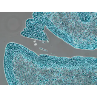
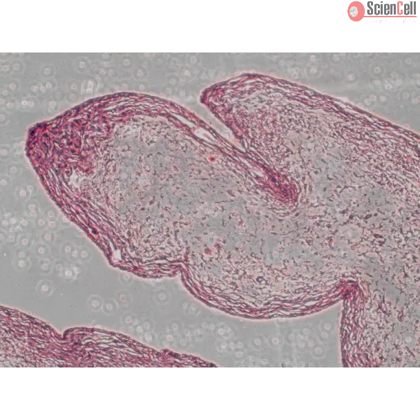
Safranin O, an indicator of cell chondrogenesis, is a cationic dye that stains acidic proteoglycan present in cartilage tissues. The Safranin O Staining Kit contains 0.1g of Safranin O Stain in powder, which can easily be dissolved in deionized water to make the staining solution. Safranin O binds to glycosaminoglycan and shows an orange-red color [1].
| Catalog No. | 8348 |
|---|---|
| Country of Manufacture | United States |
| Product Code | SafraninO |
| Size/Quantity | 100 reactions |
| Product use | This product is for research use only. It is not approved for use in humans, animals, or in vitro diagnostic procedures. |
| Storage | Room temperature |
| Shipping | Ambient temperature. |
We have recently shown that IL-29 was an important proinflammatory cytokine in pathogenesis of rheumatoid arthritis (RA). Inflammation also contributes to the pathogenesi... More
ScienCell Research Laboratories (SRL) takes pride in being a resource for researchers all over the world. The publications listed here are not meant as an endorsement or confirmation of the reliability of the products.
Related Products
Check items to add to the cart or