There has been little research on the seeding of human umbilical cord mesenchymal stem cells (hUCMSCs) in three-dimensional scaffolds for muscle tissue engineering. The o... More
There has been little research on the seeding of human umbilical cord mesenchymal stem cells (hUCMSCs) in three-dimensional scaffolds for muscle tissue engineering. The objectives of this study were: (i) to seed hUCMSCs in a fibrin hydrogel containing fast-degradable microbeads (dMBs) to create macropores to enhance cell viability; and (ii) to investigate the encapsulated cell proliferation and myogenic differentiation for muscle tissue engineering. Mass fractions of 0–80% of dMBs were tested, and 35% of dMBs in fibrin was shown to avoid fibrin shrinkage while creating macropores and promoting cell viability. This construct was referred to as “dMB35”. Fibrin without dMBs was termed “dMB0”. Microbead degradation created macropores in fibrin and improved cell viability. The percentage of live cells in dMB35 reached 91% at 16 days, higher than the 81% in dMB0 (p < 0.05). Live cell density in dMB35 was 1.6-fold that of dMB0 (p < 0.05). The encapsulated hUCMSCs proliferated, increasing the cell density by 2.6 times in dMB35 from 1 to 16 days. MTT activity for dMB35 was substantially higher than that for dMB0 at 16 days (p < 0.05). hUCMSCs in dMB35 had high gene expressions of myotube markers of myosin heavy chain 1 (MYH1) and alpha-actinin 3 (ACTN3). Elongated, multinucleated cells were formed with positive staining of myogenic specific proteins including myogenin, MYH, ACTN and actin alpha 1. Moreover, a significant increase in cell fusion was detected with myogenic induction. In conclusion, hUCMSCs were encapsulated in fibrin with degradable microbeads for the first time, achieving greatly enhanced cell viability and successful myogenic differentiation with formation of multinucleated myotubes. The injectable and macroporous fibrin–dMB–hUCMSC construct may be promising for muscle tissue engineering applications. Keywords: Fibrin hydrogel, Fast-degradable microbeads, Human umbilical cord mesenchymal stem, cells, Injectable cell delivery, Macroporous hydrogel Less
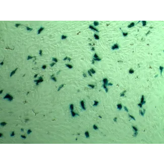
MicroRNAs (miRNAs) have emerged as potential cancer therapeutics; however, their clinical use is hindered by lack of effective delivery mechanisms to tumor sites. Mesench... More
MicroRNAs (miRNAs) have emerged as potential cancer therapeutics; however, their clinical use is hindered by lack of effective delivery mechanisms to tumor sites. Mesenchymal stem cells (MSCs) have been shown to migrate to experimental glioma and to exert anti-tumor effects by delivering cytotoxic compounds. Here, we examined the ability of MSCs derived from bone marrow, adipose tissue, placenta and umbilical cord to deliver synthetic miRNA mimics to glioma cells and glioma stem cells (GSCs). We examined the delivery of miR-124 and miR-145 mimics as glioma cells and GSCs express very low levels of these miRNAs. Using fluorescently labeled miRNA mimics and in situ hybridization, we demonstrated that all the MSCs examined delivered miR-124 and miR-145 mimics to co-cultured glioma cells and GSCs via gap junction- dependent and independent processes. The delivered miR-124 and miR-145 mimics significantly decreased the luciferase activity of their respected reporter target genes, SCP-1 and Sox2, and decreased the migration of glioma cells and the self-renewal of GSCs. Moreover, MSCs delivered Cy3-miR-124 mimic to glioma xenografts when administered intracranially. These results suggest that MSCs can deliver synthetic exogenous miRNA mimics to glioma cells and GSCs and may provide an efficient route of therapeutic miRNA delivery in vivo. Less
Tissue engineering approaches are promising to meet the increasing need for bone regeneration. Calcium phosphate cement (CPC) can be injected and self-set to form a scaff... More
Tissue engineering approaches are promising to meet the increasing need for bone regeneration. Calcium phosphate cement (CPC) can be injected and self-set to form a scaffold with excellent osteoconductivity. The objectives of this study were to develop a macroporous CPC-chitosan-fiber construct containing alginate-fibrin microbeads encapsulating human umbilical cord mesenchymal stem cells (hUCMSCs) and to investigate hUCMSC release from the degrading microbeads and proliferation inside the porous CPC construct. The hUCMSC-encapsulated microbeads were completely wrapped inside the CPC paste, with the gas-foaming porogen creating macropores in CPC to provide for access to culture media. Increasing the porogen content in CPC significantly increased the cell viability, from 49% of live cells in CPC with 0% porogen to 86% of live cells in CPC with 15% porogen. The alginate-fibrin microbeads started to degrade and release the cells inside CPC at 7 days. The released cells started to proliferate inside the macroporous CPC construct. The live cell number inside CPC increased from 270 cells/mm(2) at 1 day to 350 cells/mm(2) at 21 days. The pore volume fraction of CPC increased from 46.8% to 78.4% using the gas-foaming method, with macropore sizes of approximately 100 to 400 μm. The strength of the CPC-chitosan-fiber scaffold at 15% porogen was 3.8 MPa, which approximated the reported 3.5 MPa for cancellous bone. In conclusion, a novel gas-foaming macroporous CPC construct containing degradable alginate-fibrin microbeads was developed that encapsulated hUCMSCs. The cells had good viability while wrapped inside the porous CPC construct. The degradable microbeads in CPC quickly released the cells, which proliferated over time inside the porous CPC. Self-setting, strong CPC with alginate-fibrin microbeads for stem cell delivery is promising for bone tissue engineering applications. Less
Human umbilical cord mesenchymal stem cells (hUCMSCs) are inexhaustible and can be obtained without an invasive surgery. To date, there has been no report on seeding hUCM... More
Human umbilical cord mesenchymal stem cells (hUCMSCs) are inexhaustible and can be obtained without an invasive surgery. To date, there has been no report on seeding hUCMSCs in three-dimensional scaffolds for muscle tissue engineering. The objectives of this study were to (1) investigate hUCMSC seeding in a scaffold for muscle engineering and (2) develop a novel construct consisting of hUCMSC-encapsulating and fast-degradable microbeads inside a hydrogel matrix. The rationale was that the hydrogel matrix would maintain the defect volume, while the microbeads would degrade to release the cells and concomitantly create macropores in the matrix. hUCMSCs were encapsulated in alginate-fibrin microbeads, which were packed in an Arg-Gly-Asp (RGD)-modified alginate matrix (AM). This construct is referred to as hUCMSC-microbead-AM. The control consisted of the usual cell encapsulation in AM without microbeads (referred to as hUCMSC-AM). In the hUCMSC-AM construct, the hUCMSCs showed as round dots with no spreading at 1-14 days. In contrast, cells in the hUCMSC-microbead-AM construct had a healthy spreading and elongated morphology. The microbeads successfully degraded and released the cells at 8 days. Myogenic expressions for hUCMSC-microbead-AM were more than threefold those of hUCMSC-AM (p<0.05). Immunofluorescence for myogenic markers was much stronger for hUCMSC-microbead-AM than hUCMSC-AM. Muscle creatine kinase of hUCMSC-microbead-AM at 14 days was twofold that of hUCMSC-AM (p<0.05). In conclusion, hUCMSC encapsulation in novel fast-degradable microbeads inside a hydrogel matrix was investigated for muscle engineering. Compared to the usual method of seeding cells in a hydrogel matrix, hUCMSC-microbead-AM construct had greatly improved cell viability and myogenic differentiation, and hence, is promising to enhance muscle regeneration. Less
Stem cell-based tissue engineering offers immense promise for bone regeneration. The objective of this study was to develop a self-setting, mannitol-containing calcium ph... More
Stem cell-based tissue engineering offers immense promise for bone regeneration. The objective of this study was to develop a self-setting, mannitol-containing calcium phosphate cement (CPC) encapsulating human umbilical cord mesenchymal stem cells (hUCMSCs) for bone tissue engineering. hUCMSCs could be an inexhaustible and low-cost alternative to the gold-standard bone marrow MSCs, which require an invasive procedure to harvest. hUCMSCs were encapsulated in alginate beads and mixed into the CPC paste. Water-soluble mannitol porogen was incorporated into CPC to create macropores. The porosity was increased from 49% for the hUCMSC-encapsulating CPC to 64% after adding mannitol and absorbable-fibres (p < 0.05). Flexural strength of the construct was increased from 0.3 MPa to 2.0 MPa via fibres. Live cell percentage was > 80% for all constructs. The ALP and OC gene expressions were low at 1 day and greatly increased at 14 days. The constructs that contained mannitol had significantly higher ALP and OC expressions than that without mannitol. ALP activity of hUCMSCs inside CPC with mannitol and fibre was significantly higher than that without mannitol. At 14 days, mineralization by the encapsulated hUCMSCs was eight-fold higher than that at 1 day. In conclusion, a novel mannitol-containing porous CPC-hUCMSC construct was developed for bone tissue engineering. Its advantages include cell delivery inside a load-bearing CPC that has injectable and in situ setting capabilities. hUCMSCs inside CPC had good viability and successfully osteodifferentiated. The self-setting and strong hUCMSC-encapsulating CPC scaffold is promising for bone tissue engineering in a wide range of orthopaedic and craniofacial applications. Less
The need for bone repair has increased as the population ages. Non-rigid calcium phosphate scaffolds could provide compliance for micro-motions within the tissues and yet... More
The need for bone repair has increased as the population ages. Non-rigid calcium phosphate scaffolds could provide compliance for micro-motions within the tissues and yet have load-supporting strength. The objectives of this study were to: (a) develop a non-rigid calcium phosphate cement (CPC) with microbeads and fibre reinforcement; and (b) investigate human umbilical cord mesenchymal stem cell (hUCMSC) proliferation, osteodifferentiation and mineralization on non-rigid CPC for the first time. Non-rigid CPC was fabricated by adding extra tetracalcium phosphate in the traditional CPC and by incorporating chitosan, absorbable fibres and hydrogel microbeads. The non-rigid CPC-microbead scaffold possessed a strain-at-failure of 10.7%, much higher than the traditional CPC's strain of 0.05% which is typical for brittle bioceramics. Flexural strength of non-rigid CPC-microbead was 4-fold that of rigid CPC-microbead scaffold, while work-of-fracture (toughness) was increased by 20-fold. The strength of non-rigid CPC-microbead-fibre scaffold matched that of cancellous bone. hUCMSCs on non-rigid CPC proliferated from 100 cells/mm(2) at 1 day to 600 cells/mm(2) at 8 days. Alkaline phosphatase, osteocalcin and collagen gene expressions of hUCMSCs were greatly increased, and the cells synthesized bone minerals. hUCMSCs on non-rigid CPC-microbead-fibre constructs had higher bone markers and more mineralization than those on rigid CPC controls. In conclusion, this study developed the first non-rigid, in situ-setting calcium phosphate-microbead-fibre scaffold with a strain-at-failure exceeding 10%. hUCMSCs showed excellent proliferation, osteodifferentiation and mineralization on non-rigid CPC scaffold. The novel non-rigid CPC-hUCMSC construct with good strength, high strain-at-failure and toughness, as well as superior stem cell proliferation, osteodifferentiation and mineralization, is promising for load-bearing bone regeneration applications. Keywords: bone tissue engineering; calcium phosphate cement (CPC); human umbilical cord stem cells; non-rigid scaffold; osteogenic differentiation; strength and strain. Less
Stem cell-encapsulating microbeads could be mixed into a paste such as calcium phosphate cement (CPC), where the microbeads could protect the cells from the mixing and in... More
Stem cell-encapsulating microbeads could be mixed into a paste such as calcium phosphate cement (CPC), where the microbeads could protect the cells from the mixing and injection forces. After being placed, the microbeads could quickly degrade to release the cells throughout the scaffold, while creating macropores. The objectives of this study were to (1) construct alginate-fibrin microbeads encapsulating human umbilical cord mesenchymal stem cells (hUCMSCs) embedded in the surface of novel biofunctionalized CPC and (2) investigate microbead degradation, cell release, and osteodifferentiation on CPC. Hydrogel microbeads were fabricated that encapsulated hUCMSCs at 1×10(6) cells/mL. CPC was biofunctionalized with fibronectin (Fn) and Arg-Gly-Asp (RGD). Four scaffolds were tested: CPC control, CPC mixed with Fn, CPC mixed with RGD, and CPC grafted with RGD. The degradable microbeads released hUCMSCs at 7 days, which attached to CPC. Adding Fn or RGD to CPC greatly improved cell attachment. CPC grafted with RGD showed the fastest cell proliferation, with cell density being ninefold that on CPC control. The released hUCMSCs underwent osteodifferentiation. Alkaline phosphatase, osteocalcin, collagen 1, and runt-related transcription factor 2 (Runx2) gene expression increased by 10 to 30 fold at 7-21 days, compared with day 1. The released cells on CPC synthesized bone minerals, with the mineralization amount at 21 days being two orders of magnitude higher than that at 7 days. In conclusion, alginate-fibrin microbeads embedded in CPC surface were able to quickly release the hUCMSCs that attached to biofunctionalized CPC. Incorporating Fn and RGD into CPC greatly improved cell function, and CPC grafted with RGD had the fastest cell proliferation. The released cells on CPC differentiated into the osteogenic lineage and synthesized bone minerals. The new biofunctionalized CPC with hUCMSC-encapsulating microbeads is promising for bone regeneration applications. Less
Human umbilical cord mesenchymal stem cells (hUCMSCs) avoid the invasive procedure required to harvest bone marrow MSCs. The addition of collagen fibers into self-setting... More
Human umbilical cord mesenchymal stem cells (hUCMSCs) avoid the invasive procedure required to harvest bone marrow MSCs. The addition of collagen fibers into self-setting calcium phosphate cement (CPC) may increase the scaffold strength, and enhance cell attachment and differentiation. The objectives of this study were to develop a novel class of collagen-CPC composite scaffolds, and to investigate hUCMSC attachment, proliferation, and osteogenic differentiation on collagen-CPC scaffolds for the first time. Collagen fibers in CPC improved the load-bearing capability. Flow cytometry showed that the hUCMSCs expressed cell surface markers characteristic of MSCs, and were negative for hematopoietic and endothelial cell markers. hUCMSCs proliferated rapidly in all CPC composite scaffolds, with cell number increasing by sevenfold in 8 days. Cellular function was enhanced with collagen fibers in CPC scaffolds. Cell density increased from (645±60) cells/mm(2) on CPC with 0% collagen, to (1056±65) cells/mm(2) on CPC with 8% collagen (p<0.05). The actin stress fibers inside the hUCMSCs were stained, and the fluorescence intensity was doubled when the collagen in CPC was increased by 0% to 8%. RT-PCR showed that hUCMSCs on CPC with collagen had higher osteogenic expression than those on CPC without collagen. Alizarin Red S staining revealed a great increase in mineralization by hUCMSCs on CPC with collagen than that without collagen. In conclusion, hUCMSCs showed excellent proliferation, differentiation, and synthesis of bone minerals in collagen-CPC composite scaffolds for the first time. The novel hUCMSC-seeded collagen-CPC construct with superior cell function and load-bearing capability is promising to enhance bone regeneration in a wide range of orthopedic and craniofacial applications. Less
Intercellular communication through gap junctions (GJIC) plays an essential role in maintaining the functional integrity of vascular endothelium. Despite emerging evidenc... More
Intercellular communication through gap junctions (GJIC) plays an essential role in maintaining the functional integrity of vascular endothelium. Despite emerging evidence suggests that (-)-Epigallocatechin gallate (EGCG) may improve endothelial function. However, its effect on Cx43 gap junction in endothelial cells remains unexplored. Here we investigated the effect of EGCG on connexin43 (Cx43) gap junction in endothelial cells. The levels of Cx43 protein in human umbilical vein endothelial cells (HUVECs) cultured under serum-deprivation 48 h decreased about 50%, accompanied by decreased GJIC. This reduction can be reversed by treatments with EGCG. In addition, EGCG activated ERK, P38, and JNK mitogen-activated protein kinases (MAPKs), which were supposed to participate in the regulation of Cx43. A MEK inhibitor PD98059, but not SB203580 (a p38 kinase inhibitor) or SP600125 (a JNK kinase inhibitor), abolished the effects of EGCG on Cx43 expression and GJIC. Moreover, although both Akt and eNOS phosphorylation were time-dependently augmented by EGCG, neither PI3K inhibitor LY294002 nor eNOS inhibitor L-NAME blocked the effects of EGCG on Cx43 gap junctions. Thus, EGCG attenuated Cx43 down-regulation and impaired GJIC induced by serum deprivation, ERK MAPK Signal transduction pathway appears to be involved in these processes. Less
Background It is of growing interest to develop novel approaches to initiate differentiation of mesenchymal stem cells (MSCs) into cardiomyocytes. The purpose of this inv... More
Background It is of growing interest to develop novel approaches to initiate differentiation of mesenchymal stem cells (MSCs) into cardiomyocytes. The purpose of this investigation was to determine if Sphingosine-1-phosphate (S1P), a native circulating bioactive lipid metabolite, plays a role in differentiation of human umbilical cord mesenchymal stem cells (HUMSCs) into cardiomyocytes. We also developed an engineered cell sheet from these HUMSCs derived cardiomyocytes by using a temperature-responsive polymer, poly(N-isopropylacrylamide) (PIPAAm) cell sheet technology. Methods Cardiomyogenic differentiation of HUMSCs was performed by culturing these cells with either designated cardiomyocytes conditioned medium (CMCM) alone, or with 1 μM S1P; or DMEM with 10% FBS + 1 μM S1P. Cardiomyogenic differentiation was determined by immunocytochemical analysis of expression of cardiomyocyte markers and patch clamping recording of the action potential. Results A cardiomyocyte-like morphology and the expression of α-actinin and myosin heavy chain (MHC) proteins can be observed in both CMCM culturing or CMCM+S1P culturing groups after 5 days' culturing, however, only the cells in CMCM+S1P culture condition present cardiomyocyte-like action potential and voltage gated currents. A new approach was used to form PIPAAm based temperature-responsive culture surfaces and this successfully produced cell sheets from HUMSCs derived cardiomyocytes. Conclusions This study for the first time demonstrates that S1P potentiates differentiation of HUMSCs towards functional cardiomyocytes under the designated culture conditions. Our engineered cell sheets may provide a potential for clinically applicable myocardial tissues should promote cardiac tissue engineering research. Less
Calcium phosphate cement (CPC) can fill complex-shaped bone defects and set in situ to form a scaffold with intimate adaptation to neighboring bone. The objectives of thi... More
Calcium phosphate cement (CPC) can fill complex-shaped bone defects and set in situ to form a scaffold with intimate adaptation to neighboring bone. The objectives of this study were to determine (1) the effects of fiber length and alginate microbead volume fraction on CPC mechanical properties, and (2) the effect of cell seeding density of human umbilical cord mesenchymal stem cells (hUCMSCs) on their proliferation and osteodifferentiation on CPC. Adding microbeads to CPC degraded the strength. However, increasing the fiber length improved the mechanical properties. Strength and elastic modulus of CPC-microbead-fiber scaffold matched those reported for cancellous bone. When the cell seeding density was increased from 50k to 300k, the cell viability, osteodifferentiation, and bone mineral synthesis also increased. When the seeding density was further increased to 500k, the osteodifferentiation and mineralization decreased. Hence, the 300k seeding density was optimal for CPC-microbead-fiber under the specified conditions. At day 8, alkaline phosphatase (ALP) gene expression of hUCMSCs with seeding density of 300k was threefold the ALP at 150k, and 200-fold the ALP at 50k. At day 14, osteocalcin and runt-related transcription factor 2 with cell seeding density of 300k was fourfold those at 50k. At day 14, mineralization by hUCMSCs at seeding density of 300k was 5-fold the mineralization at 150k, and 25-fold that at 50k. In conclusion, the effect of stem cell seeding density on CPC was determined for the first time. At low cell densities, cell viability and mineralization increased with seeding density. However, a higher seeding density was not necessarily better, and an optimal seeding density on CPC resulted in the best osteodifferentiation and mineralization. The stem cell-seeded CPC-fiber scaffold with excellent osteodifferentiation and mineralization is promising for orthopedic and craniofacial applications. Less
Stem cell-encapsulating hydrogel microbeads of several hundred microns in size suitable for injection, that could quickly degrade to release the cells, are currently unav... More
Stem cell-encapsulating hydrogel microbeads of several hundred microns in size suitable for injection, that could quickly degrade to release the cells, are currently unavailable. The objectives of this study were to: (1) develop oxidized alginate-fibrin microbeads encapsulating human umbilical cord mesenchymal stem cells (hUCMSCs); (2) investigate microbead degradation, cell release, and osteogenic differentiation of the released cells for the first time. Three types of microbeads were fabricated to encapsulate hUCMSCs: (1) Alginate microbeads; (2) oxidized alginate microbeads; (3) oxidized alginate-fibrin microbeads. Microbeads with sizes of about 100-500 μm were fabricated with 1 × 10(6) hUCMSCs/mL of alginate. For the alginate group, there was little microbead degradation, with very few cells released at 21 d. For oxidized alginate, the microbeads started to slightly degrade at 14 d. In contrast, the oxidized alginate-fibrin microbeads started to degrade at 4 d and released the cells. At 7 d, the number of released cells greatly increased and showed a healthy polygonal morphology. At 21 d, the oxidized alginate-fibrin group had a live cell density that was 4-fold that of the oxidized alginate group, and 15-fold that of the alginate group. The released cells had osteodifferentiation, exhibiting highly elevated bone marker gene expressions of ALP, OC, collagen I, and Runx2. Alizarin staining confirmed the synthesis of bone minerals by hUCMSCs, with the mineral concentration at 21 d being 10-fold that at 7 d. In conclusion, fast-degradable alginate-fibrin microbeads with hUCMSC encapsulation were developed that could start to degrade and release the cells at 4 d. The released hUCMSCs had excellent proliferation, osteodifferentiation, and bone mineral synthesis. The alginate-fibrin microbeads are promising to deliver stem cells inside injectable scaffolds to promote tissue regeneration. Less
Cell-based therapy has achieved promising functional recovery for peripheral nerve repair. Although Schwann cells (SCs) and bone marrow derived mesenchymal stromal cells ... More
Cell-based therapy has achieved promising functional recovery for peripheral nerve repair. Although Schwann cells (SCs) and bone marrow derived mesenchymal stromal cells (BM-MSCs) are the main cell source for nerve tissue engineering, the clinical application is limited because of donor site morbidity, the invasive procedure, and the decreased number of SCs and BM-MSCs. Wharton's jelly-derived mesenchymal stem cells (WJMSCs) could be a promising cell source for nerve tissue engineering because they are easily accessible and their use has no ethical issues. We investigated the phenotypic, molecular and functional characteristics of WJMSCs differentiated along a Schwann-cell lineage. Cultured WJMSCs were isolated from human umbilical cord, and the undifferentiated WJMSCs were confirmed by the detection of MSC-specific cell-surface markers. WJMSCs treated with a mixture of glial growth factors (basic fibroblast growth factor, platelet-derived growth factor and forskolin) adopted a spindle-like morphology similar to SCs. Immunocytochemical staining, RT-PCR analysis, and Western blot analysis revealed that the treated cells expressed the glial markers glial fibrillary acidic protein, p75, S100 and P0 and indicative of differentiation. On co-culture with dorsal root ganglia neurons, the differentiated WJMSCs enhanced the number of sprouting neurites and neurite length in dorsal root ganglia neurons. Furthermore, using enzyme-linked immunosorbent assay and RT-PCR methodology, we found differentiated WJMSCs secrete and express neurotrophic factors, including brain-derived neurotrophic factor (BDNF), nerve growth factor (NGF), and neurotrophin-3 (NT-3). Quantification of neurite outgrowth from PC12 cells grown in differentiated WJMSCs-conditioned media demonstrates that the neurite length is significantly more than control medium and undifferentiated WJMSCs group. WJMSCs can be differentiated into cells that are Schwann-like in terms of morphologic features, phenotype, and function and could be suitable Schwann-cell substitutes for nerve repair in clinical applications. Copyright © 2010 Elsevier Inc. All rights reserved. Less
Calcium phosphate cement (CPC) has in situ-setting ability and bioactivity, but the brittleness and low strength limit CPC to only non-load-bearing bone repairs. Human um... More
Calcium phosphate cement (CPC) has in situ-setting ability and bioactivity, but the brittleness and low strength limit CPC to only non-load-bearing bone repairs. Human umbilical cord mesenchymal stem cells (hUCMSCs) can be harvested without an invasive procedure required for the commonly studied bone marrow MSCs. However, little has been reported on hUCMSC delivery via bioactive scaffolds for bone tissue engineering. The objectives of this study were to develop CPC scaffolds with improved resistance to fatigue and fracture, and to investigate hUCMSC delivery for bone tissue engineering. In fast fracture, CPC with 15% chitosan and 20% polyglactin fibers (CPC-chitosan-fiber scaffold) had flexural strength of 26mPa, higher than 10mPa for CPC control (p<0.05). In cyclic loading, CPC-chitosan-fiber specimens that survived 2x10(6) cycles had the maximum stress of 10MPa, compared to 5MPa of CPC control. CPC-chitosan-fiber specimens that failed after multiple cycles had a mean stress-to-failure of 9MPa, compared to 5.8MPa for CPC control (p<0.05). hUCMSCs showed excellent viability when seeded on CPC and CPC-chitosan-fiber scaffolds. The percentage of live cells reached 96-99%. Cell density was about 300cells/mm(2) at day 1; it proliferated to 700cells/mm(2) at day 4. Wst-1 assay showed that the stronger CPC-chitosan-fiber scaffold had hUCMSC viability that matched the CPC control (p>0.1). In summary, this study showed that chitosan and polyglactin fibers substantially increased the fatigue resistance of CPC, and that hUCMSCs had excellent proliferation and viability on the scaffolds. Less