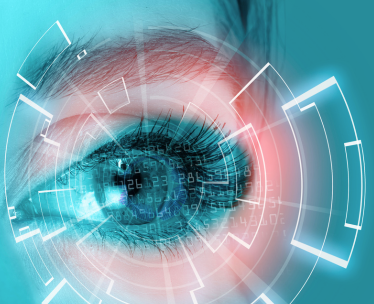
The ocular lens is a transparent structure in the eye that is designed to refract and focus light onto the retina. The lens is an avascular unit which includes the lens capsule, lens epithelium, and lens fibers. Lens fiber cells form the bulk of the lens and a monolayer of epithelial cells cover the anterior surface of the fibers. Lens epithelial cells perform a number of critical functions in the lens including the growth and development of the lens, fluid transport, protecting the lens from environmental and oxidative stress, and maintaining the homeostasis of the lens. Lens epithelial cells are also critical for metabolic activity in the ocular lens, and this activity can be affected by epithelial cell differentiation. During development, lens epithelial cells migrate from the equatorial region to the interior to produce transparent crystallins and develop into lens fiber cells. The crystallins help to maintain the transparency of the lens, but during aging are modified or degraded resulting in decreased lens transparency. The decreased lens transparency leads to the clouding of the lens and development of cataracts.
Due to the location of the lens, it is constantly being exposed to environmental stress from solar radiation, which means it is vulnerable to being damaged. Recent studies have suggested that in order to protect the lens from stress, the lens epithelial cells express molecular chaperones such as aA crystalline, aB crystalline, Heat Shock Protein (Hsp) 25/27, Hsp40, Heat shock cognate (Hsc70), Hsp70, and T Complex Protein (TCP1). During cellular stress molecular chaperones help to reduce unwanted protein aggregation. Other than environmental stress, the lens is also exposed to oxidative stress. Oxidative stress plays a primary role in the formation of cataracts and results in apoptosis of lens epithelial cells. During aging, hydrogen peroxide (H2O2) can build up, resulting in oxidative damage to the lens. Researchers are now focusing on reducing oxidative stress in lens epithelial cells in order to slow cataract progression.
With aging, the lens becomes less transparent leading to the development of cataracts. More recent studies in mice and rats have demonstrated that lens epithelial cells can undergo epithelial-mesenchymal transition (EMT) to produce myofibroblasts. Cytokines or growth factors, such as transforming growth factor b (TGFb), are responsible for initiate the EMT process. The myofibroblasts participate in the development of fibrotic tissue through the deposition of extracellular matrix proteins. The deposition of extracellular matrix proteins by the myofibroblasts can lead to the clouding of the lens or an anterior capsule break injury. Understanding the mechanisms of how these cells coordinate proliferation, survival, and differentiation is key to understanding and maintaining lens transparency.
ScienCell Research Laboratories is excited to add Mouse Lens Epithelial Cells (Cat#M6550) from CD-1 mice to our animal primary cell line up. Mouse lens epithelial cells are a useful model for studying the mechanisms of ocular diseases associated with the lens. We also offer Human Lens Epithelial Cells (Cat#6550), and Rat Lens Epithelial Cells (Cat#R6550).