Engineered mesenchymal stem cells (MSCs) have been investigated extensively for gene delivery and, more recently, for targeted small molecule delivery. While preclinical ... More
Engineered mesenchymal stem cells (MSCs) have been investigated extensively for gene delivery and, more recently, for targeted small molecule delivery. While preclinical studies demonstrate the potential of MSCs for targeted delivery, clinical studies suggest that tumor homing of native MSCs may be inefficient. We report here a surprising finding that loading MSCs with the anticancer drug paclitaxel (PTX) by nanoengineering results in significantly improved tumor homing compared to naïve MSCs. Loading PTX in MSCs results in increased levels of mitochondrial reactive oxygen species (ROS). In response to this oxidative stress, MSCs upregulate two important set of proteins. First were critical antioxidant proteins, most importantly nuclear factor erythroid 2-like 2 (Nrf2), the master regulator of antioxidant responses; upregulation of antioxidant proteins may explain how MSCs protect themselves from drug-induced oxidative stress. The second was CXCR4, a direct target of Nrf2 and a key mediator of tumor homing; upregulation of CXCR4 suggested a mechanism that may underlie the improved tumor homing of nanoengineered MSCs. In addition to demonstrating the potential mechanism of improved tumor targeting of nanoengineered MSCs, our studies reveal that MSCs utilize a novel mechanism of resistance against drug-induced oxidative stress and cell death, explaining how MSCs can deliver therapeutic concentrations of cytotoxic payload while maintaining their viability. Less
Our previous study revealed that miR‑148a, a cyclic adenosine monophosphate‑response element binding protein‑modulated microRNA that promotes adipocyte differentiat... More
Our previous study revealed that miR‑148a, a cyclic adenosine monophosphate‑response element binding protein‑modulated microRNA that promotes adipocyte differentiation by inhibiting Wnt1, is a biomarker of obesity in human subjects and a mouse model. The present study investigated the expression of miR‑148a in human adipose tissue‑derived mesenchymal stem cells (hMSCs‑Ad) in response to inflammatory cytokines and adipokines to clarify its underlying mechanism. miR‑148a expression was detected using reverse transcription‑quantitative polymerase chain reaction analysis and its promoter activity was detected with a luciferase assay. miR‑148a expression levels decreased when differentiated hMSCs‑Ad were exposed to inflammatory cytokines or adipokines, which suggested that miR‑148a may be important in adipocyte metabolism and inflammation. Furthermore, the promoter activity of miR‑148a decreased following treatment of cells with inflammatory cytokines or adipokines. The results of the present study indicated a novel role of miR‑148a in adipocyte inflammation; therefore, miR‑148a may be involved in obesity complications via its own underlying transcriptional mechanism. Less
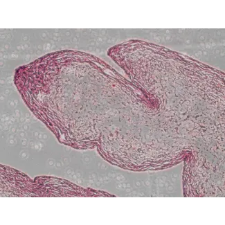
The keratin-associated proteins (KAPs) are the structural proteins of hair fibers and are thought to play an important role in determining the physical properties of hair... More
The keratin-associated proteins (KAPs) are the structural proteins of hair fibers and are thought to play an important role in determining the physical properties of hair fibers. These proteins are activated in a striking sequential and spatial pattern in the keratinocytes of hair fibers. Thus, it is important to elucidate the mechanism that underlies the specific transcriptional activity of these genes. In this study, sheep KRTAP 3–3 and KRTAP11-1 genes were found to be highly expressed in wool follicles in a tissue-specific manner. Subsequently, the promoter regions of the two genes that contained the 5′ flanking/5′ untranslated regions and the coding regions were cloned. Using an in vivo transgenic approach, we found that the promoter regions from the two genes exhibited transcriptional activity in hair fibers. A much stronger and more uniformly expressed green fluorescent signal was observed in the KRTAP11-1-ZsGreen1 transgenic mice. In situ hybridization revealed the symmetrical expression of sheep KRTAP11-1 in the entire wool cortex. Consistently, immunohistochemical analysis demonstrated that the pattern of ZsGreen1 expression in the hair cortex of transgenic mice matches that of the endogenous KRTAP11-1 gene, indicating that the cloned promoter region contains elements that are sufficient to govern the wool cortex-specific transcription of KRTAP11-1. Furthermore, regulatory regions in the 5′ upstream sequence of the sheep KRTAP11-1 gene that may regulate the observed hair keratinocyte specificity were identified using in vivo reporter assays. Less
Background Androgenic alopecia (AGA) is a major type of human scalp hair loss, which is caused by two androgens: testosterone (T) and 5α-dihydrotestosterone (5α-DHT). B... More
Background Androgenic alopecia (AGA) is a major type of human scalp hair loss, which is caused by two androgens: testosterone (T) and 5α-dihydrotestosterone (5α-DHT). Both androgens bind to the androgen receptor (AR) and induce androgen-sensitive genes within the human hair dermal papilla cells (HHDPCs), but 5α-DHT exhibits much higher binding affinity and potency than T does in inducing the involved androgen-sensitive genes. Changes in the induction of androgen-sensitive genes during AGA are caused by the over-production of 5α-DHT by the 5α-reductase (5α-R) enzyme; therefore, one possible method to treat AGA is to inhibit this enzymatic reaction. Methods RT-PCR was used to identify the presence of the 5α-R and AR within HHDPCs. A newly developed AGA-relevant HHDPC-based assay combined with non-radioactive thin layer chromatography (TLC) detection was used for screening crude plant extracts for the identification of new 5α-R inhibitors. Results HHDPCs expressed both 5α-R type 1 isoform of the enzyme (5α-R1) and AR in all of the passages used in this study. Among the thirty tested extracts, Avicennia marina (AM) displayed the highest inhibitory activity at the final concentration of 10 μg/ml, as the production of 5α-DHT decreased by 52 % (IC50 = 9.21 ± 0.38 μg/ml). Conclusions Avicennia marina (AM) was identified as a potential candidate for the treatment of AGA based on its 5α-R1-inhibitory activity. Less
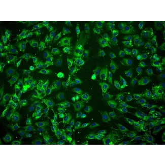
Adipose-derived stem cell (ADSC) is considered as a cell source potentially useful for angiogenesis in tissue engineering and regenerative medicine. This study investigat... More
Adipose-derived stem cell (ADSC) is considered as a cell source potentially useful for angiogenesis in tissue engineering and regenerative medicine. This study investigated the growth and endothelial differentiation of human ADSCs on polyglycolic acid/polylactic acid (PGA/PLA) mesh compared to 2D plastic. Cell adhesion, viability, and distribution of hADSCs on PGA/PLA mesh were observed by CM-Dil labeling, live/dead staining, and SEM examination while endothelial differentiation was evaluated by flow cytometry, Ac-LDL/UEA-1 uptake assay, immunofluorescence stainings, and gene expression analysis of endothelial related markers. Results showed hADSCs gained a mature endothelial phenotype with a positive ratio of 21.4 ± 3.7% for CD31+/CD34- when induced in 3D mesh after 21 days, which was further verified by the expressions of a comprehensive range of endothelial related markers, whereas hADSCs in 2D induced and 2D/3D noninduced groups all failed to differentiate into endothelial cells. Moreover, compared to 2D groups, the expression for α-SMA was markedly suppressed in 3D cultured hADSCs. This study first demonstrated the endothelial differentiation of hADSCs on the PGA/PLA mesh and pointed out the synergistic effect of PGA/PLA 3D culture and growth factors on the acquisition of mature characteristic endothelial phenotype. We believed this study would be the initial step towards the generation of prevascularized tissue engineered constructs. Less
Recovery from tendon injury is based on long periods of rest, which results in sub-optimal repair, often replacing tendon with fibrocartilage scar tissue. Recently, the u... More
Recovery from tendon injury is based on long periods of rest, which results in sub-optimal repair, often replacing tendon with fibrocartilage scar tissue. Recently, the use of stem cells in equine tendon repair has been attempted with variable success. The objective of this work was to determine the expression of scleraxis (scx) and tenascin C (TnC), two markers of tenocytes, in adipose (AdMSC) and umbilical cord blood (UCB) stem cells during culture on various substrata and in response to fibroblast growth factor (FGF) treatment. Equine UCB and AdMSC were cultured on gelatin-coated plasticware, 30 % matrigel or collagen-coated Cytodex beads and treated with 10 ng/ml FGF2, FGF4 or FGF5 prior to measurement of proliferation, kinase activity and tenocyte gene expression. Supplementation with FGF2 or FGF5 activated the ERK1/2 signaling pathway in AdMSC and UCB; no effect of FGF4 was observed in UCB. FGF2 increased proliferation in AdMSC but not UCB. Conversely, FGF5 stimulated proliferation of UCB. Culture in matrigel increased scx expression in both cell populations and increased TnC in AdMSC. In AdMSC grown in matrigel, supplementation with FGF2 or FGF5 increased TnC expression. Thus, culture conditions (substrata and FGF supplementation) impact markers of tenocytes in AdMSC and UCB stem cells, indicating that careful consideration should be given to culture conditions prior to use of UCB or AdMSC as therapeutic aids. Optimal culture conditions may promote early differentiation of these cells, improving their ability to aid tendon regeneration and facilitating more efficient recovery from tendon injury. Less
Primary cicatricial alopecia (PCA) is a group of inflammatory hair disorders that cause scarring and permanent hair loss. Previous studies have implicated PPARγ, a trans... More
Primary cicatricial alopecia (PCA) is a group of inflammatory hair disorders that cause scarring and permanent hair loss. Previous studies have implicated PPARγ, a transcription factor that integrates lipogenic and inflammatory signals, in the pathogenesis of PCA. However, it is unknown what triggers the inflammatory response in these disorders, whether the inflammation is a primary or secondary event in disease pathogenesis, and whether the inflammatory reaction reflects an autoimmune process. In this paper, we show that the cholesterol biosynthetic pathway is impaired in the skin and hair follicles of PCA patients. Treatment of hair follicle cells with BM15766, a cholesterol biosynthesis inhibitor, or 7-dehydrocholesterol (7-DHC), a sterol precursor, stimulates the expression of pro-inflammatory chemokine genes. Painting of mouse skin with 7-DHC or BM15766 inhibits hair growth, causes follicular plugging and induces the infiltration of inflammatory cells into the interfollicular dermis. Our results demonstrate that cholesterologenic changes within hair follicle cells trigger an innate immune response that is associated with the induction of toll-like receptor (TLR) and interferon (IFN) gene expression, and the recruitment of macrophages that surround the hair follicles and initiate their destruction. These findings reveal a previously unsuspected role for cholesterol precursors in PCA pathogenesis and identify a novel link between sterols and inflammation that may prove transformative in the diagnosis and treatment of these disorders. Less
Stromal cells and mesenchymal stem cells (MSCs), 2 important cell populations within the hematopoietic microenvironment, may play an important role in the development of ... More
Stromal cells and mesenchymal stem cells (MSCs), 2 important cell populations within the hematopoietic microenvironment, may play an important role in the development of hematopoietic stem/progenitor cells. We have successfully cultured human umbilical cord blood-derived stromal cells (hUCBDSCs). It has been demonstrated that MSCs also exist in hUCB. However, we have not found any reports on the distinct characteristics of hUCBDSCs and human umbilical cord blood-derived mesenchymal stem cells (hUCBDMSCs). In this study, hUCBDSCs and hUCBDMSCs were isolated from the cord blood of full-term infants using the same density gradient centrifugation and cultured in the appropriate medium. Some biological characteristics and hematopoietic supportive functions were compared in vitro. hUCBDSCs were distinct from hUCBDMSCs in morphology, proliferation, cell cycle, passage, immunophenotype, and the capacity for classical tri-lineage differentiation. Finally, quantitative real-time polymerase chain reaction analysis revealed that granulocyte colony-stimulating factor (G-CSF) gene expression was higher in hUCBDSCs than that in hUCBDMSCs. Enzyme-linked immunosorbent assay revealed that the secretion of G-CSF, thrombopoietin (TPO), and granulocyte macrophage colony-stimulating factor (GM-CSF) by hUCBDSCs was higher than that by hUCBDMSCs. After coculture, the granulocyte/macrophage colony-forming units (CFU-GM) of hematopoietic cells from the hUCBDSC feeder layer was more than that from the hUCBDMSC feeder layer. Flow cytometry was used to detect CD34(+) hematopoietic stem/progenitor cell committed differentiation during 14 days of coculture; the results demonstrated that CD14 and CD33 expression in hUCBDSCs was significantly higher than their expression in hUCBDMSCs. This observation was also true for the granulocyte lineage marker, CD15. This marker was expressed beginning at day 7 in hUCBDSCs. It was expressed earlier and at a higher level in hUCBDSCs compared with hUCBDMSCs. In conclusion, hUCBDSCs are different from hUCBDMSCs. hUCBDSCs are superior to hUCBDMSCs in supporting hematopoiesis stem/progenitor cells differentiation into myeloid lineage cells at an early stage in vitro. Less
Cell-substrate interaction was functionally essential for phenotypic maintenance and multipotency remodeling of stem cells. For bone tissue engineering, electrospinning t... More
Cell-substrate interaction was functionally essential for phenotypic maintenance and multipotency remodeling of stem cells. For bone tissue engineering, electrospinning techniques are useful to create fibrous scaffolds mimicking natural mineralized collagen fibrous structure in bone. In this study, influence of electrospun fiber alignment on MSCs differentiation potential was investigated on PHBHHx electrospun meshes. Compared with randomly-oriented ones, the aligned fiber orientation increased elastic modulus and tension stress of the PHBHHx meshes. Most of the attached MSCs elongated along the aligned fibers. From the transcriptome microarray results, there were a total of 67 differentially expressed genes between aligned and random groups, and most of them were involved in cell adhesion and actin cytoskeleton regulation. In addition, PPAR signaling pathway was reduced on the aligned fibers, which might contribute to the impaired adipogenesis and enhanced osteogenesis. It was further confirmed by RT-PCR and western blotting. The PPARγ downregulation on the aligned fibers was related to phosphorylated activation of ERK, with no effect on total ERK expression. However, the induction of osteogenic by PHBHHx fiber alignment was relatively less significant that it could only support initial adipo-osteogenic switch and would be partially covered up by osteogenic or adipogenic inductive chemicals. Less
Normal cardiomyocytes are highly dependent on the functional expression of ion channels to form action potentials and electrical coupling with other cells. To fully deter... More
Normal cardiomyocytes are highly dependent on the functional expression of ion channels to form action potentials and electrical coupling with other cells. To fully determine the scientific and therapeutic potential of stem cells for cardiovascular-disease treatment, it is necessary to assess comprehensively the regulation of stem-cell electrical properties during stem cell-cardiomyocyte interaction. It has been reported in the literature that contact with native cardiomyocytes induced and regulated stem-cell cardiogenic differentiation. However, in conventional cell-culture models, the importance of cell-cell contact for stem-cell functional coupling with cardiomyocytes has not been elucidated due to insufficient control of the cell-contact mode of individual cells. Using microfabrication and laser-guided cell micropatterning techniques, we created two biochips with contact-promotive and -preventive microenvironments to systematically study the effect of contact on cardiogenic regulation of stem-cell electrical properties. In contact-promotive biochips, connexin 43 expression was upregulated and relocated to the junction area between one stem cell and one cardiomyocyte. Only stem cells in contact with cardiomyocytes were induced by adjacent cardiomyocytes to acquire electrophysiological properties for action-potential formation similar to that of a cardiomyocyte. Less
To understand how stem cells benefit native cardiomyocytes is crucial for cell-based therapies to rescue cardiomyocytes (CMCs) damaged during heart infarction and other c... More
To understand how stem cells benefit native cardiomyocytes is crucial for cell-based therapies to rescue cardiomyocytes (CMCs) damaged during heart infarction and other cardiac diseases. However, the current conclusions on the protective effect of mesenchymal stem cells (MSCs) were obtained by analyzing the overall amount of protein and factor secretion in a conventional co-culture system. These results neglected the heterogeneity of MSC population and failed to determine the importance of cellular contact to the protective effects. To address these issues, we have constructed two biochips by microfabrication methods and laser-guided cell micropatterning technique. Using the biochips, the protective effect of MSCs on CMCs can be quantitatively analyzed at single-cell level with defined cellular contact. The role of cellular contact on protective effect can be clarified according to our statistical results. Less
To assess the regenerative properties and potential therapeutic value of adipose-derived stem cells (ASCs) in the bottlenose dolphin, there is a need to determine whether... More
To assess the regenerative properties and potential therapeutic value of adipose-derived stem cells (ASCs) in the bottlenose dolphin, there is a need to determine whether an adequate adipose depot exists, in addition to the development of a standardized technique for minimally invasive adipose collection. In this study, an ultrasound-guided liposuction technique for adipose collection was assessed for its safety and efficacy. The ultrasound was utilized to identify and measure the postnuchal adipose depot and aid in the guidance of the liposuction cannula during aspiration. Liposuction procedures from 6 dolphins yielded 0.9–12.7 g of adipose. All samples yielded sufficient nucleated cells to initiate primary cell cultures, and at passage 2, were successfully differentiated into adipogenic, chondrogenic, neurogenic, and osteogenic cell lineages. The cultured dolphin cells expressed known stem-cell-associated CD markers, CD44 and CD90. Ultrasound-guided liposuction proved to be a safe and minimally invasive procedure that resulted in the successful isolation of ASCs in bottlenose dolphins. This is the first article that conclusively establishes the presence of stem cells in the dolphin. Less
Recently, the genetic modification of mesenchymal stem cells (MSCs) has led to increased differentiation potential. For the therapeutic application of genetically modifie... More
Recently, the genetic modification of mesenchymal stem cells (MSCs) has led to increased differentiation potential. For the therapeutic application of genetically modified MSCs, it is crucial to evaluate their characteristics and safety. In this study, we investigated the effects of bone morphogenetic protein 2 (BMP2) gene transfer on the characteristics and biodistribution of human MSCs. Lentiviral-mediated BMP2 transduction to MSCs enhanced osteocyte differentiation and decreased adipocyte differentiation. Although there is no significant difference in cell proliferation capacity, MSCs transduced BMP2 proliferate somewhat higher than nontransduced or GFP transduced MSCs. No significant changes were observed in surface antigen expression in genetically modified MSCs. In vivo transplantation of lentiviral-mediated BMP2 gene transferred MSCs to nude mice did not result in tumor formation. To evaluate the biodistribution of genetically modified cells, MSCs carrying BMP2 were injected into the tail vein of femur fractured mice. The introduced MSCs were detected in the spleen, testis and fractured femur 28 days post-implantation. These findings suggest that diverse safety tests for genetically modified MSCs should be considered, particularly when a lentivirus mediated gene transfer method is used. Less
Background It is of growing interest to develop novel approaches to initiate differentiation of mesenchymal stem cells (MSCs) into cardiomyocytes. The purpose of this inv... More
Background It is of growing interest to develop novel approaches to initiate differentiation of mesenchymal stem cells (MSCs) into cardiomyocytes. The purpose of this investigation was to determine if Sphingosine-1-phosphate (S1P), a native circulating bioactive lipid metabolite, plays a role in differentiation of human umbilical cord mesenchymal stem cells (HUMSCs) into cardiomyocytes. We also developed an engineered cell sheet from these HUMSCs derived cardiomyocytes by using a temperature-responsive polymer, poly(N-isopropylacrylamide) (PIPAAm) cell sheet technology. Methods Cardiomyogenic differentiation of HUMSCs was performed by culturing these cells with either designated cardiomyocytes conditioned medium (CMCM) alone, or with 1 μM S1P; or DMEM with 10% FBS + 1 μM S1P. Cardiomyogenic differentiation was determined by immunocytochemical analysis of expression of cardiomyocyte markers and patch clamping recording of the action potential. Results A cardiomyocyte-like morphology and the expression of α-actinin and myosin heavy chain (MHC) proteins can be observed in both CMCM culturing or CMCM+S1P culturing groups after 5 days' culturing, however, only the cells in CMCM+S1P culture condition present cardiomyocyte-like action potential and voltage gated currents. A new approach was used to form PIPAAm based temperature-responsive culture surfaces and this successfully produced cell sheets from HUMSCs derived cardiomyocytes. Conclusions This study for the first time demonstrates that S1P potentiates differentiation of HUMSCs towards functional cardiomyocytes under the designated culture conditions. Our engineered cell sheets may provide a potential for clinically applicable myocardial tissues should promote cardiac tissue engineering research. Less
Introduction: Dogs are commonly used animal models for regenerative endodontics research. Although several studies have used stem cells isolated from dog teeth to investi... More
Introduction: Dogs are commonly used animal models for regenerative endodontics research. Although several studies have used stem cells isolated from dog teeth to investigate the dentin/pulp regeneration in vivo, less attention has been paid for the characterization of these cells. Therefore, this study aimed to characterize the dental pulp stem cells isolated from dog teeth (cDPSCs) in order to further define the dog as an animal model for regenerative endodontics. Less
Human bone marrow-derived mesenchymal stem cells (hMSCs) represent an appealing source of smooth muscle cells (SMCs) for engineering small-diameter vascular grafts due to... More
Human bone marrow-derived mesenchymal stem cells (hMSCs) represent an appealing source of smooth muscle cells (SMCs) for engineering small-diameter vascular grafts due to the limited availability and replicative capacity of somatic SMCs. However, lack of standardization of hMSC culture conditions has limited some progress in hMSC research. Because, at the moment, a chemically defined, serum-free medium without growth factors is not capable of amplifying hMSCs in vitro, the usage of serum (either human serum or fetal bovine serum [FBS]) continues in hMSC research. The emergence of commercial hMSCs and hMSC media opened a series of questions regarding the compatibility of commercial and homemade hMSCs and hMSC media. In this study, two types of commonly used FBS-containing hMSC media-MSCGM (containing 10% FBS) and MesenPro (containing 2% FBS), along with our homemade medium (low-glucose Dulbecco's modified Eagle's medium plus 10% selected lot FBS)-were compared in their ability to support SMC differentiation from hMSCs. The effects of FBS level, medium supplements (ascorbic acid, copper, etc.), and growth factors (transforming growth factor beta1) were also examined for their impact on SMC differentiation. It was discovered that MesenPro and transforming growth factor beta1 are the strongest SMC inducers from hMSCs. In contrast, hMSCs grown in homemade (10% Dulbecco's modified Eagle's medium) and commercial MSCGM media remained undifferentiated. FBS concentration did not affect SMC differentiation when 10% FBS was compared with 2%. Finally, the mechanism underlying SMC differentiation from hMSCs grown in FBS-containing medium was explored by following the expression changes of serum response factor during the establishment of hMSC culture. Less
Mesenchymal stem cells (MSCs) offer promise as therapeutic aids in the repair of tendon, ligament, and bone damage suffered by sport horses. The objective of the study wa... More
Mesenchymal stem cells (MSCs) offer promise as therapeutic aids in the repair of tendon, ligament, and bone damage suffered by sport horses. The objective of the study was to identify and characterize stem-like cells from newborn foal umbilical cord blood (UCB). UCB was collected and MSC isolated using human reagents. The cells exhibit a fibroblast-like morphology and express the stem cell markers Oct4, SSEA-1, Tra1-60 and Tra1-81. Culture of the cells in tissue-specific differentiation media leads to the formation of cell types characteristic of mesodermal and endodermal origins. Chondrogenic differentiation reveals proteoglycan and glycosaminoglycan synthesis as measured histochemically and Sox9 and collagen 2A1 gene transcription. Osteocytes capable of mineral deposition, osteonectin and Runx2 transcription were evident. Hepatogenic cells formed from UCBs express albumin and cytokeratin 18. Multinucleated myofibers that express desmin were observed indicating partial differentiation into mature muscle cells. Interestingly, conventional human protocols for UCB differentiation into adipocytes were unsuccessful in foal UCB and adult horse adipose-derived MSC. These results demonstrate that equine UCB can be induced to form multiple cell types that underlie their value for regenerative medicine in injured horses. In addition, this work suggests that subtle differences exist between equine and human UCB stem cells. Less