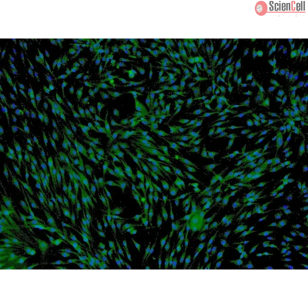
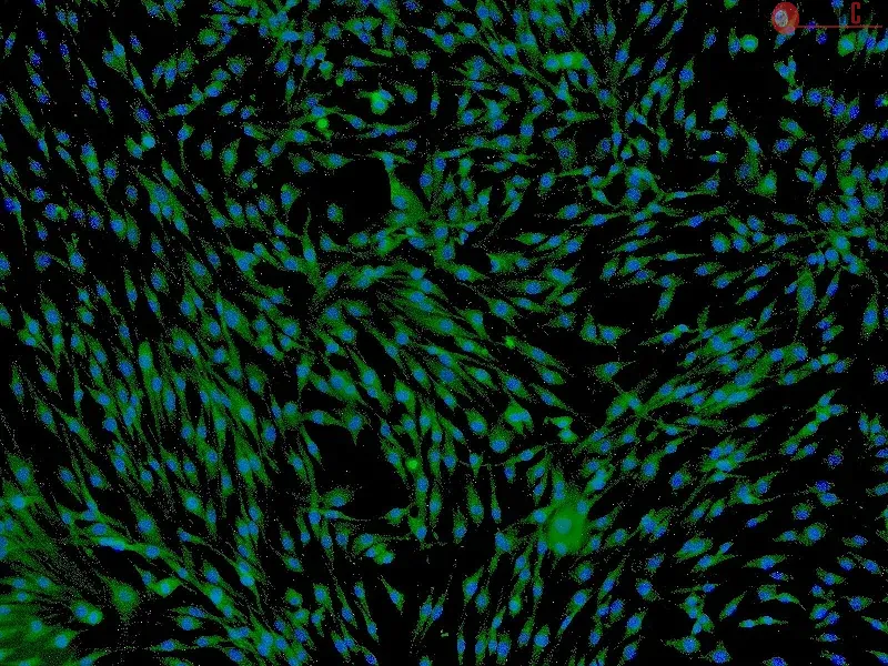
Photobiomodulation (PBM) has recently emerged in cellular therapy as a potent alternative in promoting cell proliferation, migration, and differentiation during tissue re... More
Photobiomodulation (PBM) has recently emerged in cellular therapy as a potent alternative in promoting cell proliferation, migration, and differentiation during tissue regeneration. Herein, a single-cell near-infrared (NIR) laser irradiation system (830 nm) and the image-based approaches were proposed for the investigation of the modulatory effects in mitochondrial membrane potential (ΔΨm), reactive oxygen species (ROS), and vesicle transport in single living human adipose mesenchymal stem cells (hADSCs). The irradiated-hADSCs were then stained with 2′,7′-dichlorodihydrofluorescein diacetate (H2DCFDA) and Rhodamine 123 (Rh123) to represent the ΔΨm and ROS production, respectively, with irradiation in the range of 2.5–10 (J/cm2), where time series of bright-field images were obtained to determine the vesicle transport phenomena. Present results showed that a fluence of 5 J/cm2 of PBM significantly enhanced the ΔΨm, ROS, and vesicle transport phenomena compared to the control group (0 J/cm2) after 30 min PBM treatment. These findings demonstrate the efficacy and use of PBM in regulating ΔΨm, ROS, and vesicle transport, which have potential in cell proliferation, migration, and differentiation in cell-based therapy. Less
Spurred by recent progress in biomaterials and therapeutics, stimulus-responsive strategies that deliver an active substance in temporal-, spatial-, and dose-controlled f... More
Spurred by recent progress in biomaterials and therapeutics, stimulus-responsive strategies that deliver an active substance in temporal-, spatial-, and dose-controlled fashions have become achievable. Implementation of such strategies necessitates the use of bio-safe materials that are sensitive to a specific pathological incitement or that, in response to a precise stimulus, undergo hydrolytic cleavage or a change in biomolecular conformation. An innovative design of polymeric stimulus-responsive systems should controllably release a drug or degrade the drug carrier in response to specific lesion enzymes. Wound healing is a great challenge due to various hidden factors such as pathogenic infections, neurovascular diseases, excessive exudates, lack of an effective therapeutic delivery system, low cell proliferation, and cell migration. In addition, long-term use of antibiotics in chronic wound management can result in side effects and antimicrobial resistance. Novel treatments with antibacterial pharmaceuticals thus vitally need to be developed. Recently, graphene and graphene family members have emerged as shining stars among biomaterials for wound-healing applications due to their excellent bioactive properties, which can overcome limitations of current wound dressings and fulfill wound-healing requirements. Herein, we developed a feasible approach to impregnate graphene oxide (GO) into genipin-crosslinked gelatin (3GO) hydrogels to enzymatically control GO release. The developed hydrogels were characterized by chemical, physical, morphological, and cellular analyses. The results proved that the 3GO1 hydrogel is biocompatible and significantly enhanced the mechanical strength by encapsulating GO. Moreover, the rate of GO release depended on the crosslinking degree and environmental enzyme levels. Enzymatically released GO displayed uniform dispersity, retained its antibacterial activities against Staphylococcus aureus and Pseudomonas aeruginosa through sharp edges and wrapping mechanisms, and promoted human fibroblast migration. This multifunctional hydrogel we developed with antibacterial efficacy is suitable for future application as wound dressings. Less
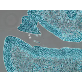
Kaposi's sarcoma (KS), a highly angiogenic and invasive tumor often involving different organ sites, including the oral cavity, is caused by infection with Kaposi's sarco... More
Kaposi's sarcoma (KS), a highly angiogenic and invasive tumor often involving different organ sites, including the oral cavity, is caused by infection with Kaposi's sarcoma-associated herpesvirus (KSHV). Diverse cell markers have been identified on KS tumor cells, but their origin remains an enigma. We previously showed that KSHV could efficiently infect, transform, and reprogram rat primary mesenchymal stem cells (MSCs) into KS-like tumor cells. In this study, we showed that human primary MSCs derived from diverse organs, including bone marrow (MSCbm), adipose tissue (MSCa), dental pulp, gingiva tissue (GMSC), and exfoliated deciduous teeth, were permissive to KSHV infection. We successfully established long-term cultures of KSHV-infected MSCa, MSCbm, and GMSC (LTC-KMSCs). While LTC-KMSCs had lower proliferation rates than the uninfected cells, they expressed mixtures of KS markers and displayed differential angiogenic, invasive, and transforming phenotypes. Genetic analysis identified KSHV-derived microRNAs that mediated KSHV-induced angiogenic activity by activating the AKT pathway. These results indicated that human MSCs could be the KSHV target cells in vivo and established valid models for delineating the mechanism of KSHV infection, replication, and malignant transformation in biologically relevant cell types. Less
Most synthetic polymeric materials currently used for bone tissue engineering lack specific signals through which cells can identify and interact with the surface, result... More
Most synthetic polymeric materials currently used for bone tissue engineering lack specific signals through which cells can identify and interact with the surface, resulting in incompatibility and compromised osteogenic activity. Soluble inductive factors also have issues including a short half-live in vivo. Bone forming peptide-1 is a truncated peptide from the immature form of bone morphogenetic protein-7 (BMP-7) that displays higher osteogenic activity than full-length, mature BMP-7. In this study, we used a mussel-inspired immobilization strategy mediated by polymerization of dopamine to introduce recently discovered stimulators of bone forming peptide-1 (BFP-1) onto the surface of poly-lactic-co-glycolic acid (PLGA) substrate to form a biomaterial that overcomes these challenges. Human adipose-derived stem cells (hASCs), being abundant and easy accessible, were used to test the osteogenic activity of BFP-1 and the novel biomaterial. Under osteoinductive conditions, cells treated with both BFP-1 alone and BFP-1-coated biomaterials displayed elevated expression of the osteogenic markers alkaline phosphatase (ALP), osteocalcin (OC), and RUNX2. Furthermore, hASCs associated with poly-dopamine-assisted BFP-1-immobilized PLGA (pDA-BFP-1-PLGA) scaffolds promoted in vivo bone formation in nude mice. Our novel materials may hold great promise for future bone tissue engineering applications. Less
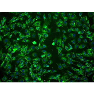
Here, we aimed to investigate osteogenic differentiation of human adipose-derived stem cells (hASCs) in three-dimensional (3D) bioprinted tissue constructs in vitro and i... More
Here, we aimed to investigate osteogenic differentiation of human adipose-derived stem cells (hASCs) in three-dimensional (3D) bioprinted tissue constructs in vitro and in vivo. A 3D Bio-plotter dispensing system was used for building 3D constructs. Cell viability was determined using live/dead cell staining. After 7 and 14 days of culture, real-time quantitative polymerase chain reaction (PCR) was performed to analyze the expression of osteogenesis-related genes (RUNX2, OSX, and OCN). Western blotting for RUNX2 and immunofluorescent staining for OCN and RUNX2 were also performed. At 8 weeks after surgery, osteoids secreted by osteogenically differentiated cells were assessed by hematoxylin-eosin (H&E) staining, Masson trichrome staining, and OCN immunohistochemical staining. Results from live/dead cell staining showed that most of the cells remained alive, with a cell viability of 89%, on day 1 after printing. In vitro osteogenic induction of the 3D construct showed that the expression levels of RUNX2, OSX, and OCN were significantly increased on days 7 and 14 after printing in cells cultured in osteogenic medium (OM) compared with that in normal proliferation medium (PM). Fluorescence microscopy and western blotting showed that the expression of osteogenesis-related proteins was significantly higher in cells cultured in OM than in cells cultured in PM. In vivo studies demonstrated obvious bone matrix formation in the 3D bioprinted constructs. These results indicated that 3D bioprinted constructs consisting of hASCs had the ability to promote mineralized matrix formation and that hASCs could be used in 3D bioprinted constructs for the repair of large
bone tissue defects. Less
MicroRNAs (miRNAs) have been identified as a new class of regulatory molecules that influence many biological functions, including metabolism, adipocyte differentiation. ... More
MicroRNAs (miRNAs) have been identified as a new class of regulatory molecules that influence many biological functions, including metabolism, adipocyte differentiation. To determine the role of adipogenic miRNAs in the adipocyte differentiation process, we used microarray technology to monitor miRNA levels in human adipose-derived mesenchymal stem cells (hMSCs-Ad), human stromal vascular cells (SVCs) and differentiated adipocytes. 79 miRNAs were found to be differentially expressed, most of which are located in obesity related chromosomal regions but have not been previously linked to adipocyte differentiation process. A systematic search was made for relevant studies in academic data bases, involving the Gene Expression Omnibus (GEO) ArrayExpress, Pubmed and Embase database. Eight studies on human adipocyte differentiation or obesity were included in the final analysis. After combining our microarray data with meta-analysis of published microarray data, we detected 42 differently expressed miRNAs (meta-signature miRNAs) in mature adipocytes compared to SVCs or hMSCs-Ad. Our study shows meta-signature miRNAs specific for adipogenesis, several of which are correlated with key gene targets demonstrating functional relationships to pathways in BMP signaling pathway, Cell differentiation, Wnt signaling, insulin receptor signaling pathway, MAPK signaling, Cell cycle and lipid metabolic process. Our study shows that the first evidence of hsa-let-7 family, hsa-miR-15a-5p, hsa-miR-27a-3p, hsa-miR-106b-5p, hsa-miR-148a-3p and hsa-miR-26b-5p got a great weight in adipogenesis. We concluded that meta-signature miRNAs involved in adipocyte differentiation and provided pathophysiological roles and novel insight into obesity and its related metabolic diseases. Less
Our previous study revealed that miR‑148a, a cyclic adenosine monophosphate‑response element binding protein‑modulated microRNA that promotes adipocyte differentiat... More
Our previous study revealed that miR‑148a, a cyclic adenosine monophosphate‑response element binding protein‑modulated microRNA that promotes adipocyte differentiation by inhibiting Wnt1, is a biomarker of obesity in human subjects and a mouse model. The present study investigated the expression of miR‑148a in human adipose tissue‑derived mesenchymal stem cells (hMSCs‑Ad) in response to inflammatory cytokines and adipokines to clarify its underlying mechanism. miR‑148a expression was detected using reverse transcription‑quantitative polymerase chain reaction analysis and its promoter activity was detected with a luciferase assay. miR‑148a expression levels decreased when differentiated hMSCs‑Ad were exposed to inflammatory cytokines or adipokines, which suggested that miR‑148a may be important in adipocyte metabolism and inflammation. Furthermore, the promoter activity of miR‑148a decreased following treatment of cells with inflammatory cytokines or adipokines. The results of the present study indicated a novel role of miR‑148a in adipocyte inflammation; therefore, miR‑148a may be involved in obesity complications via its own underlying transcriptional mechanism. Less
Objective: Roles of bone morphogenetic proteins (BMPs) on osteogenesis of human adipose-derived stem cells (hASCs) remain ambiguous. In this study, we evaluated in vitro ... More
Objective: Roles of bone morphogenetic proteins (BMPs) on osteogenesis of human adipose-derived stem cells (hASCs) remain ambiguous. In this study, we evaluated in vitro and in vivo functional characteristics of BMPs of different dimerization types, with the aim of determining osteoinductive efficiency of heterodimeric BMP-2/7 on osteogenesis of hASCs.
Materials and methods: We explored osteoinductive effects of three different BMPs by using cell DNA assay, alkaline phosphatase (ALP) activity assay, alizarin red staining and mineralization assay, and real-time PCR, in vitro. Also, we examined ectopic bone formation in nude mice by using soft X-ray, histomorphometric and immunohistochemical analyses in vivo.
Results: In our dose-response study, we showed that BMPs with both dimerization types did not induce in vitro osteogenesis of hASCs without osteogenic medium (OM). In the presence of OM, BMPs significantly enhanced hASC osteogenesis in a dose-dependent manner. In in vivo experiments, our analyses indicated that BMPs promoted osteogenesis of hASCs without in vitro osteogenic induction. However, both in vitro and in vivo, there were no significant differences in hASC osteogenic induction between heterodimeric and homodimeric BMPs.
Conclusions: Heterodimeric BMP-2/7 significantly promoted osteogenesis of hASCs in vitro and in vivo. However, BMP-2/7 was not found to be a stronger inducer of osteogenesis compared to homodimeric either BMP-2 or BMP-7. Less
MicroRNAs (miRNAs) have emerged as potential cancer therapeutics; however, their clinical use is hindered by lack of effective delivery mechanisms to tumor sites. Mesench... More
MicroRNAs (miRNAs) have emerged as potential cancer therapeutics; however, their clinical use is hindered by lack of effective delivery mechanisms to tumor sites. Mesenchymal stem cells (MSCs) have been shown to migrate to experimental glioma and to exert anti-tumor effects by delivering cytotoxic compounds. Here, we examined the ability of MSCs derived from bone marrow, adipose tissue, placenta and umbilical cord to deliver synthetic miRNA mimics to glioma cells and glioma stem cells (GSCs). We examined the delivery of miR-124 and miR-145 mimics as glioma cells and GSCs express very low levels of these miRNAs. Using fluorescently labeled miRNA mimics and in situ hybridization, we demonstrated that all the MSCs examined delivered miR-124 and miR-145 mimics to co-cultured glioma cells and GSCs via gap junction- dependent and independent processes. The delivered miR-124 and miR-145 mimics significantly decreased the luciferase activity of their respected reporter target genes, SCP-1 and Sox2, and decreased the migration of glioma cells and the self-renewal of GSCs. Moreover, MSCs delivered Cy3-miR-124 mimic to glioma xenografts when administered intracranially. These results suggest that MSCs can deliver synthetic exogenous miRNA mimics to glioma cells and GSCs and may provide an efficient route of therapeutic miRNA delivery in vivo. Less
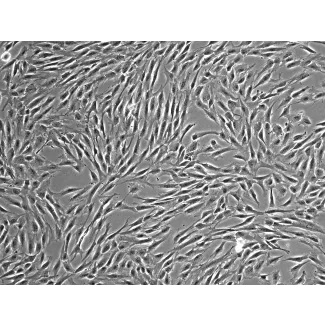
Human umbilical cord-derived mesenchymal stromal cells (UC-MSCs) that are available from cell banks can be induced to differentiate into various cell types, thereby makin... More
Human umbilical cord-derived mesenchymal stromal cells (UC-MSCs) that are available from cell banks can be induced to differentiate into various cell types, thereby making them practical potential sources for cell-based therapies. In injured peripheral nerves, Schwann cells (SCs) contribute to functional recovery by supporting axonal regeneration and myelin reconstruction. Here, we first demonstrate a system toinduce UC-MSCs to differentiate into cells with SC properties (UC-SCs) by treatment with β-mercaptoethanol followed by retinoic acid and a set of specific cytokines. The UC-SCs are morphologically similar to SCs and express SC markers, including P0, as assessed by immunocytochemistry and reverse transcription polymerase chain reaction. Transplantation of UC-SCs into transected sciatic nerves in adult rats enhanced nerve regeneration. The effectiveness of UC-SCs for axonal regeneration was comparable to that of authentic human SCs based on histological criteria and functional recovery. Immunohistochemistry and immunoelectron microscopy also demonstrated myelination of regenerated axons by UC-SCs. These findings indicate that cells with SC properties and with the ability to support axonal regeneration and reconstruct myelin can be successfully induced from UC-MSCs to promote functional recovery after peripheral nerve injury. This system may be applicable for the development of cell-based therapies. Less
Background: Mesenchymal stem (MS) cells are excellent candidates for cell-based therapeutic strategies to regenerate injured tissue. Although human MS cells can be isolat... More
Background: Mesenchymal stem (MS) cells are excellent candidates for cell-based therapeutic strategies to regenerate injured tissue. Although human MS cells can be isolated from bone marrow and directed to differentiate by means of an osteogenic pathway, the regulation of cell-fate determination is not well understood. Recent reports identify critical roles for microRNAs (miRNAs), regulators of gene expression either by inhibiting the translation or by stimulating the degradation of target mRNAs. Methodology/Principal Findings: In this study, we employed a library of miRNA inhibitors to evaluate the role of miRNAs in early osteogenic differentiation of human MS cells. We discovered that miR-148b, -27a and -489 are essential for the regulation of osteogenesis: miR-27a and miR-489 down-regulate while miR-148b up-regulates differentiation. Modulation of these miRNAs induced osteogenesis in the absence of other external differentiation cues and restored osteogenic potential in high passage number human MS cells. Conclusions/Significance: Overall, we have demonstrated the utility of the functional profiling strategy for unraveling complex miRNA pathways. Our findings indicate that miRNAs regulate early osteogenic differentiation in human MS cells: miR-148b, -27a, and -489 were found to play a critical role in osteogenesis. Less