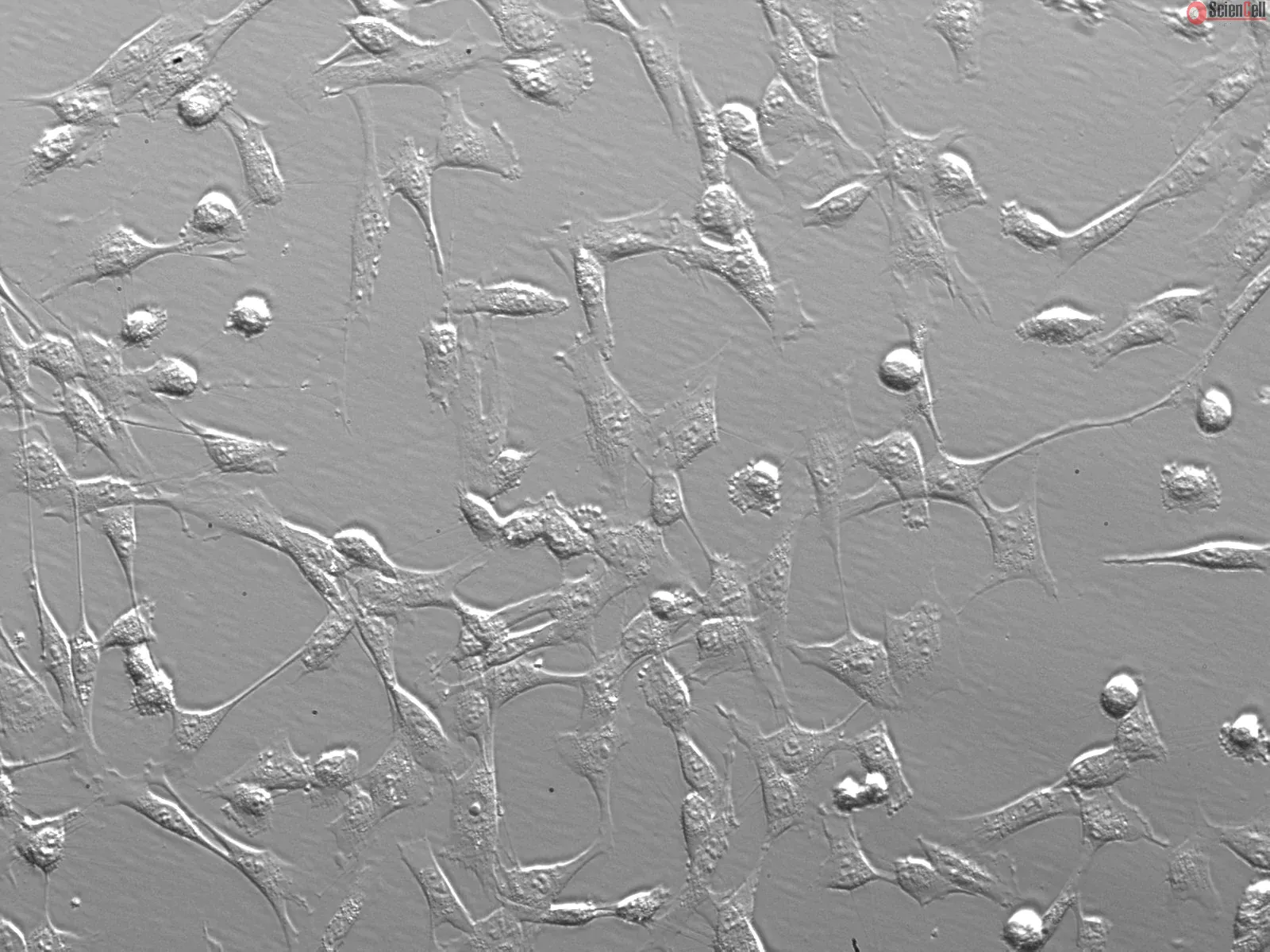
Esophageal fibroblasts, which line the lamina propria of the upper and lower esophagus, are mesenchymal cells derived from the embryonic mesoderm. Fibroblasts secrete a non-rigid extracellular matrix that is both rich in type I and/or type III collagen. They are responsible for much of the synthesis of extracellular matrix in connective tissues and play a central role in wound healing. They have been extensively used for a wide range of cellular and molecular studies, as they are one of the easiest types of cells to grow in culture. In addition, their durability makes them amenable to a variety of manipulations ranging from gene transfection to microinjection. Esophageal fibroblasts have been shown to contribute to the esophageal squamous cell carcinoma-induced angiogenesis through the TGF-β and VEGF signaling pathways.
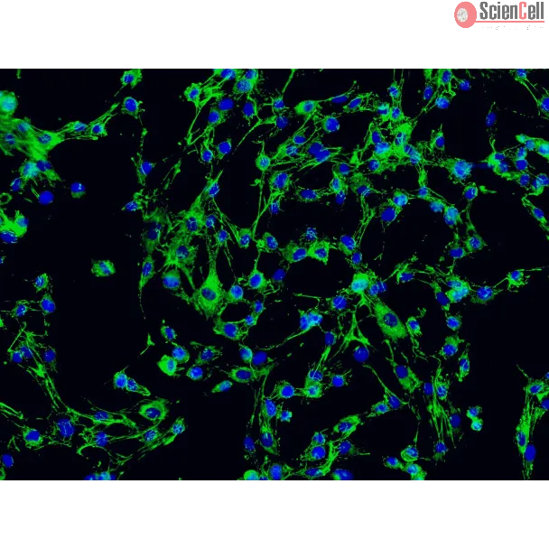
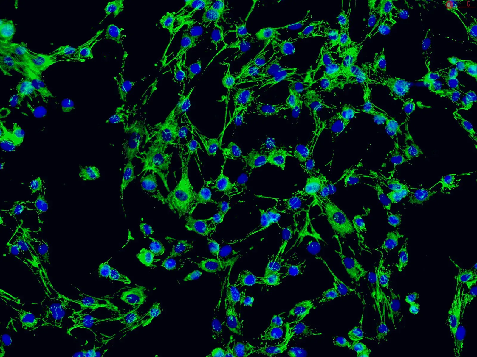
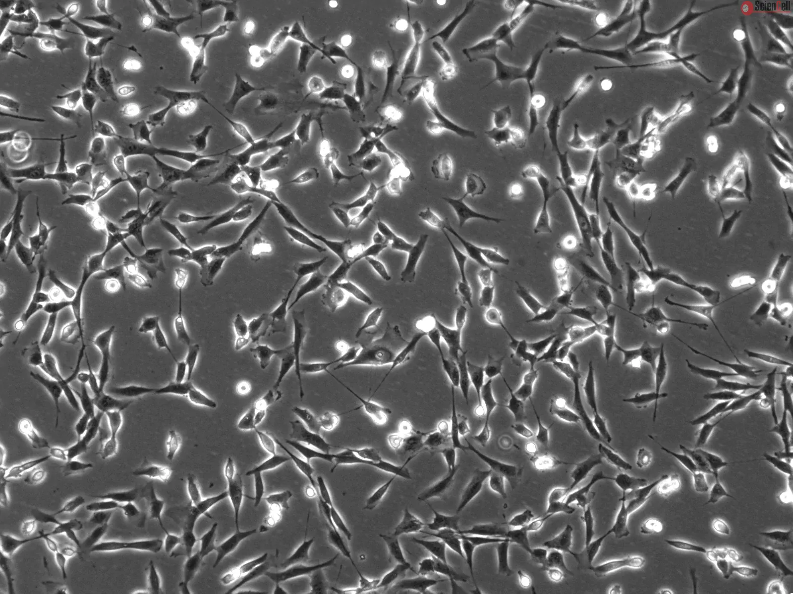
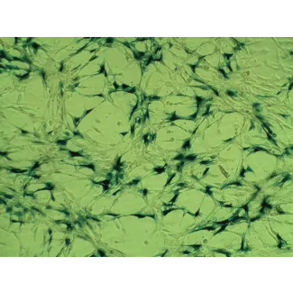
HEsF from ScienCell Research Laboratories are isolated from human esophageal tissue. HEsF are cryopreserved at passage one and delivered frozen. Each vial contains >5 x 10⁵ cells in 1 ml volume. HEsF are characterized by immunofluorescence with antibody specific to fibronectin. HEsF are negative for HIV-1, HBV, HCV, mycoplasma, bacteria, yeast and fungi. HEsF are guaranteed to further expand for 15 population doublings under the conditions provided by ScienCell Research Laboratories.
Recommended Medium
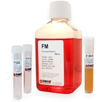
It is recommended to use Fibroblast Medium (FM, Cat. #2301) for culturing HEsF in vitro.
Product Sheets
 2730
2730
Search for a Certificate of Analysis by Lot Number below:
Fibroblast heterogeneity remains undefined in eosinophilic esophagitis (EoE), an allergic inflammatory disorder complicated by fibrosis. We utilized publicly available si... More
Fibroblast heterogeneity remains undefined in eosinophilic esophagitis (EoE), an allergic inflammatory disorder complicated by fibrosis. We utilized publicly available single-cell RNA sequencing data (GSE201153) of EoE esophageal biopsies to identify fibroblast sub-populations, related transcriptomes, disease status-specific pathways and cell–cell interactions. IL13-treated fibroblast cultures were used to model active disease. At least 2 fibroblast populations were identified, F_A and F_B. Several genes including ACTA2 were more enriched in F_A. F_B percentage was greater than F_A and epithelial–mesenchymal transition upregulated in F_B vs. F_A in active and remission EoE. Epithelial–mesenchymal transition was also upregulated in F_B in active vs. remission EoE and TNF-α signaling via NFKB was downregulated in F_A. IL-13 treatment upregulated ECM-related genes more profoundly in ACTA2− fibroblasts than ACTA2+ myofibroblasts. After proliferating epithelial cells, F_B and F_A contributed most to cell–cell communication networks. ECM–Receptor interaction strength was stronger than secreted or cell–cell contact signaling in active vs. remission EoE and significant ligand–receptor pairs were driven mostly by F_B. This unbiased analysis identifies at least 2 fibroblast sub-populations in EoE in vivo, distinguished in part by ACTA2. Fibroblasts play a critical role in cell–cell interactions in EoE, most profoundly via ECM–receptor signaling via the F_B sub-group. Less
Esophageal fibrosis can develop due to caustic or radiation injuries. Umbilical cord–derived mesenchymal stem cells (UC-MSCs) are known to mitigate fibrosis in various ... More
Esophageal fibrosis can develop due to caustic or radiation injuries. Umbilical cord–derived mesenchymal stem cells (UC-MSCs) are known to mitigate fibrosis in various organs. However, the potential effects of UC-MSCs on human esophageal fibrosis remain underexplored. This study investigated the anti-fibrogenic properties and mechanisms of UC-MSC-derived conditioned media (UC-MSC-CM) on human esophageal fibroblasts (HEFs). HEFs were treated with TGF-β1 and then cultured with UC-MSC-CM, and the expression levels of extracellular matrix (ECM) components, RhoA, myocardin related transcription factor A (MRTF-A), serum response factor (SRF), Yes-associated protein (YAP), and transcriptional coactivator with PDZ-binding motif (TAZ) were measured. UC-MSC-CM suppressed TGF-β1-induced fibrogenic activation in HEFs, as evidenced by the downregulation of ECM. UC-MSC-CM diminished the expression of RhoA, MRTF-A, and SRF triggered by TGF-β1. In TGF-β1-stimulated HEFs, UC-MSC-CM decreased the nuclear localization of MRTF-A and YAP. Additionally, UC-MSC-CM diminished the TGF-β1-induced nuclear expressions of YAP and TAZ, while concurrently enhancing the cytoplasmic presence of phosphorylated YAP. Furthermore, UC-MSC-CM reduced TGF-β1-induced phosphorylation of Smad2. These findings suggest that UC-MSC-CM may inhibit TGF-β1-induced fibrogenic activation in HEFs by targeting the Rho-mediated MRTF/SRF and YAP/TAZ pathways, as well as the Smad2 pathway. This indicates its potential as a stem cell therapy for esophageal fibrosis. Less
ScienCell Research Laboratories (SRL) takes pride in being a resource for researchers all over the world. The publications listed here are not meant as an endorsement or confirmation of the reliability of the products.

,-1-mg-ml--2.jpg)