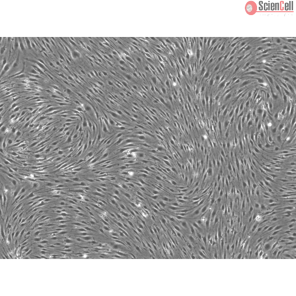
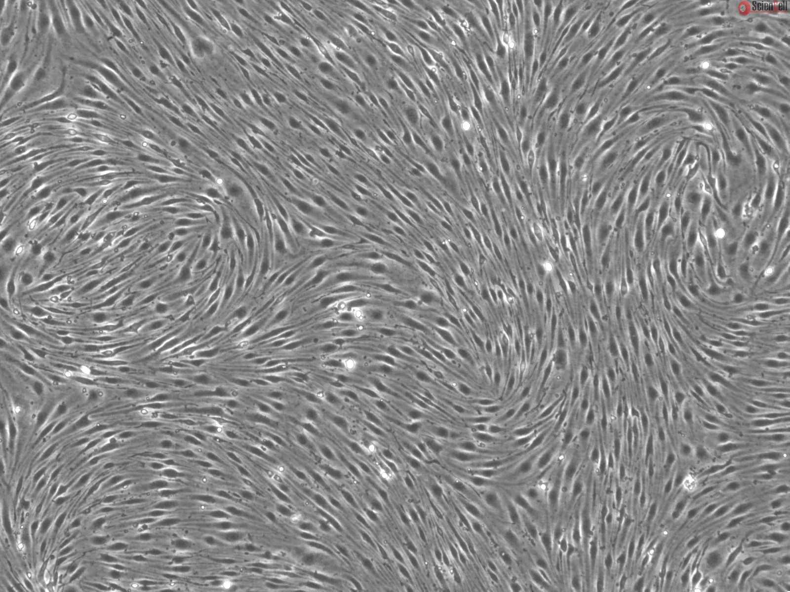
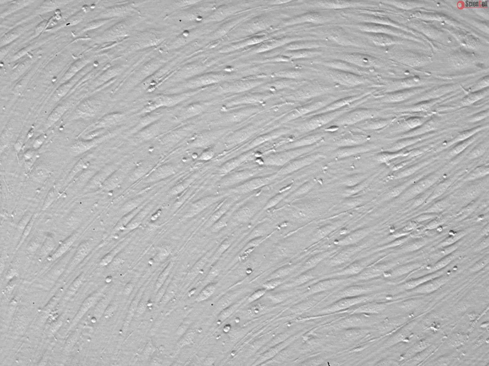
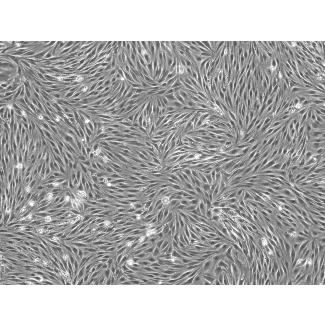
Brain microvascular endothelial cells (BMEC), the major component of the blood-brain barrier, limit the passage of substances, both soluble and cellular, from the blood into the brain. BMEC utilize unique features to distinguish themselves from peripheral endothelial cells, such as 1) intercellular tight junctions that display high electrical resistance and slow paracellular transport, 2) the absence of fenestrae and a reduced level of pinocytic activity, and 3) the expression of specialized pumps that can transport compounds out of the brain via the blood-brain barrier. Similar to peripheral endothelial cells, BMEC express, or can be induced to express, cell adhesion molecules on their surface that regulate the extravasation of leukocytes into the brain. Cultured rat BMEC have been widely used for studying the molecular and cellular properties of blood-brain barrier because of their unique functions. Understanding the molecular mechanisms of blood-brain barrier regulation may help to optimize drug delivery to the CNS and elucidate new therapies for CNS diseases.
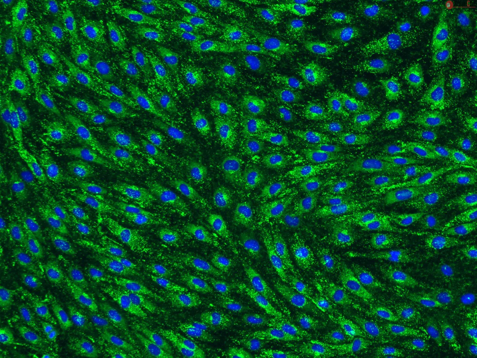
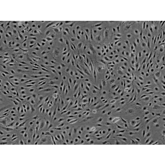
RBMEC from ScienCell Research Laboratories are isolated from adult CD® IGS rat brain. RBMEC are cryopreserved at passage one and delivered frozen. Each vial contains >5 x 10⁵ cells in 1 ml volume. RBMEC are characterized by immunofluorescence with antibodies specific to vWF and/or CD31 (PECAM). RBMEC are negative for mycoplasma, bacteria, yeast, and fungi. RBMEC are guaranteed to further expand for 5 population doublings under the conditions provided by ScienCell Research Laboratories.
Recommended medium:
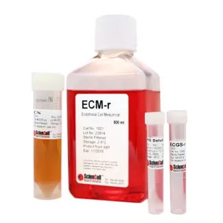
It is recommended to use Endothelial Cell Medium-rat (ECM-r, Cat. #1021) for culturing RBMEC in vitro.
脑微血管内皮细胞(BMEC)是血脑屏障(Blood-Brain Barrier, BBB)的主要组成部分,能够限制可溶性物质及细胞成分由血液进入脑组织。
BMEC 具有区别于外周内皮细胞的独特生物学特性,包括:1)细胞间紧密连接结构,可形成高电阻屏障并减缓细胞旁运输;2)缺乏窗孔结构且胞饮作用水平较低;3)表达多种特异性转运蛋白,可通过血脑屏障将化合物主动转运出脑组织。
与外周内皮细胞类似,BMEC 能够表达或诱导表达多种细胞黏附分子,从而调控白细胞向脑组织的迁移过程。由于具有独特的屏障功能,培养的大鼠 BMEC 已被广泛应用于血脑屏障分子与细胞特性的研究。
深入理解血脑屏障调控的分子机制,将有助于优化中枢神经系统(CNS)药物递送策略,并为中枢神经系统疾病的新型治疗方法开发提供理论基础。
ScienCell Research Laboratories 提供的大鼠脑微血管内皮细胞(RBMEC)分离自成年 CD® IGS 大鼠脑组织。细胞于第一代(Passage 1)完成冻存,并以冷冻状态运输。每瓶产品含有超过 5 × 10⁵ 个细胞,包装体积为 1 ml。
RBMEC 通过针对 vWF 和/或 CD31(PECAM)的特异性抗体进行免疫荧光鉴定。经检测,产品不含支原体、细菌、酵母及真菌污染。在按照 ScienCell Research Laboratories 提供的培养条件操作时,可保证细胞至少完成 5 次群体倍增。
推荐培养基:
建议使用大鼠内皮细胞培养基(Endothelial Cell Medium-rat,ECM-r,产品编号 #1021)进行 RBMEC 的 体外(in vitro) 培养。
Product Sheets
 R1000
R1000
Search for a Certificate of Analysis by Lot Number below:
The endothelial and epithelial cells form biological barriers, the unique anatomical structures that control substance movement between circulation and i.e., organs. Meas... More
The endothelial and epithelial cells form biological barriers, the unique anatomical structures that control substance movement between circulation and i.e., organs. Measuring trans-endothelial electrical resistance (TEER) is a standard method that enables the evaluation of barrier integrity. The capability of a novel measuring system (MS) that combines innovative components, customized electrodes, and user-friendly software tailored to provide accurate and adjustable measurements of endothelial barrier integrity. We demonstrated MS usage in TEER measurement of rat brain microvascular endothelial cell line (RBE-4), primary rat brain microvascular endothelial cells (PBMEC), rat brain microvascular endothelial cells (BMEC), and human intestinal epithelial cell line (HIEC-6). The MS was successfully applied to measure the TEER of cultured cell monolayers, finding that i) the device records stable values; ii) cells treated with ammonium chloride had lower TEER values compared to untreated cells, which was verified with permeability measurements with fluorescein dye and confocal microscopy. In conclusion, we invented a low-cost MS capable of accurate TEER measures in relevant biological ranges. The MS instrument facilitates long-time, real-time monitoring of the cellular barriers, giving vital insights into cell barrier stability, permeability, and the effects of pharmacological substances. The customized measurement duration and electrode configuration meet a broad range of experimental designs. Less
Effective treatment against glioma remains challenging nowadays because the protective blood-brain barrier (BBB) impedes drug penetration into brain and the limited effic... More
Effective treatment against glioma remains challenging nowadays because the protective blood-brain barrier (BBB) impedes drug penetration into brain and the limited efficacy of conventional chemotherapy. While strong positively charged nanoparticles have good permeability through the BBB, they often come with the caveat of cationic toxicity to healthy tissues and organs during blood circulation. Here we show a neutrally charged nanoprobe with a surface decorated with γ-glutamyl moieties that can be cleaved by γ-glutamyl transpeptidase, an enzyme overexpressed on brain capillaries. Upon the cleavage, positively charged primary amines are generated, facilitating the effective crossing of the nanoprobe through BBB via the adsorption-mediated transcytosis pathway, while avoiding the caveat of cationic toxicity. In addition, when reaching the acidic tumor microenvironment, the nanoprobe co-encapsulating sonosensitizer and immune agonist swells, which results in an accelerated drug release under ultrasound irradiation to induce a combined immune response, ultimately leading to a robust anticancer effect. Overall, we report an effective drug delivery nanoplatform across the BBB for an enhanced therapy of glioma. Less
Traumatic brain injury (TBI) is a major cause of morbidity and mortality worldwide, affecting over 10 million people annually, with an estimated cost of $76.5 billion. Al... More
Traumatic brain injury (TBI) is a major cause of morbidity and mortality worldwide, affecting over 10 million people annually, with an estimated cost of $76.5 billion. Although apocynin freely transverses the blood–brain barrier (BBB), its application is limited due to its rapid elimination, low terminal half-life (t1/2 = 6.7 min), narrow dose–response relationship, and cytotoxicity, thereby requiring repeated dosages. With this study, we aimed to develop transferrin-functionalized nanoparticles encapsulating apocynin to treat neuroinflammation for targeted drug delivery to sites of brain injury. As a preliminary approach, we endeavored to optimize the formulation parameters of apocynin-loaded albumin nanoparticles prepared through the desolvation method. The nanoparticles were characterized for their size, polydispersity, surface charge, drug loading and in vitro drug release. In this study, we also investigated the anti-inflammatory and neuroprotective effects of free apocynin and nanoparticle-loaded apocynin in neuronal cells. We show that the developed formulation displayed monodispersed, nanosized particles with higher entrapment efficiency, loading, stability, and sustained release profiles. The permeability of the nanoparticles across HBMECs reached the maximum at 67%. The in vivo evaluation revealed the enhanced uptake of transferrin-anchored nanoparticles in the brain tissues when compared with unmodified nanoparticles after I.V. administration. In vivo nanoparticle localization studies using a blast TBI (bTBI) model and confocal fluorescence microscopy have shown that tf-apoANPs are successful in delivering relatively high amounts of nanoparticles to the brain parenchyma and glial cells compared to non-targeted nanoparticles. We also establish that targeted nanoparticles accumulate in the brain. In conclusion, tf-apoANPs are efficacious carriers for targeted delivery across the blood–brain barrier to potentially treat neuroinflammation in brain injury and other diseases. Keywords: apocynin; nanoparticle; albumin; HPLC; desolvation method; targeted delivery; biodistribution; neuroprotection Less
ScienCell Research Laboratories (SRL) takes pride in being a resource for researchers all over the world. The publications listed here are not meant as an endorsement or confirmation of the reliability of the products.