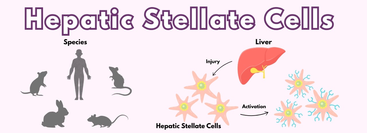
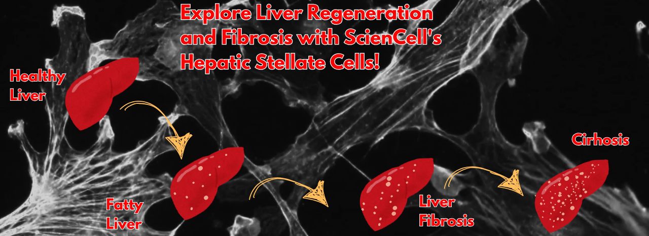
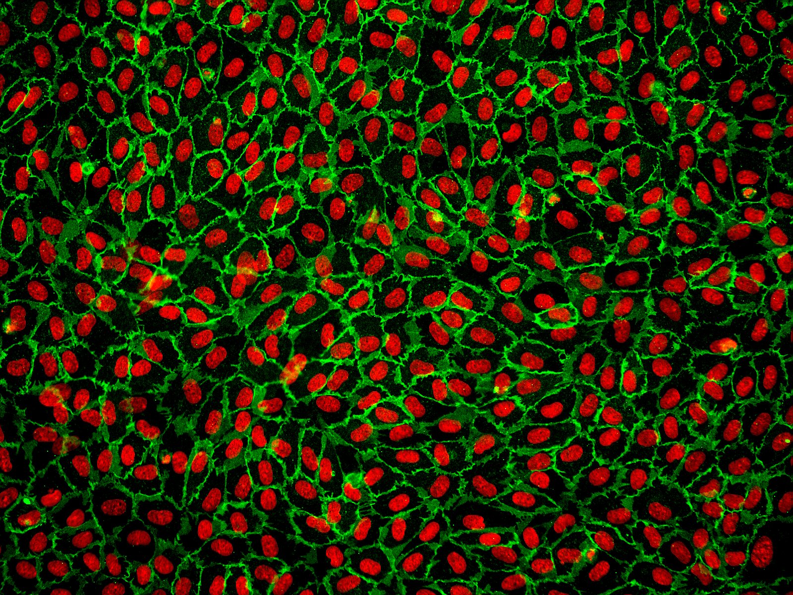
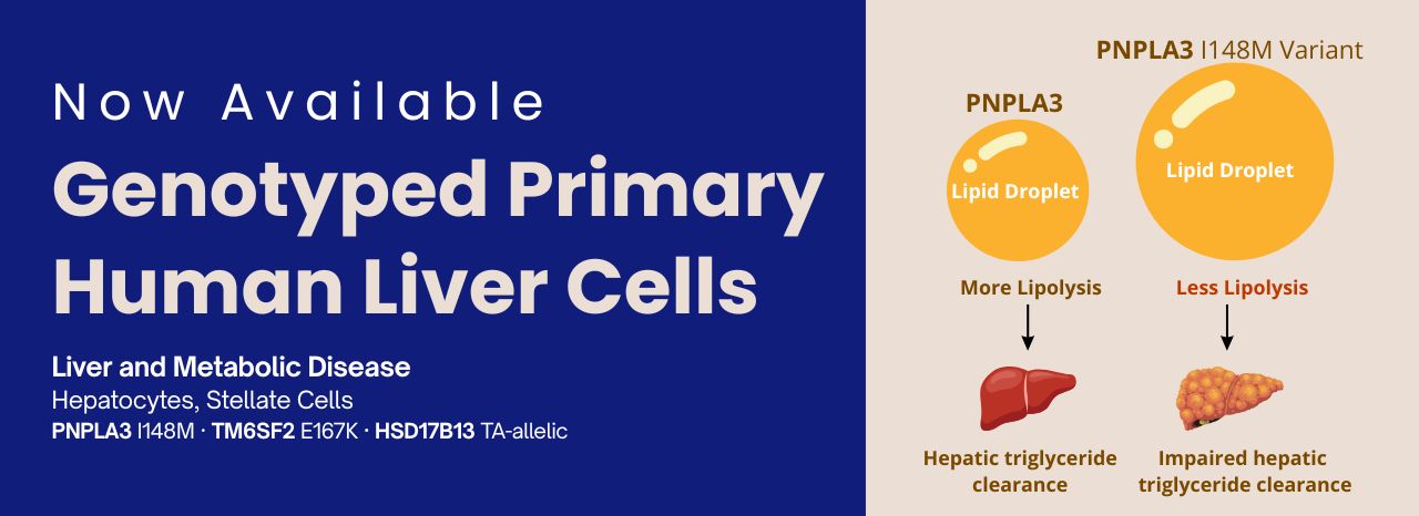
页面 2 - Primary Cells
-
- 十二月 11, 2024
The central nervous system (CNS) is a complex network of neurons and glia that play critical roles in human brain function. Neurons are the primary signaling units of the nervous system, responsible for transmitting information throughout the body.
- A single neuron can contact up to 10,000 other neurons, highlighting the intricate connectivity of the brain.
- The cerebral cortex, the outermost layer of the brain, is responsible for higher-level functions such as perception, language, decision-making, and memory.
Studying the CNS can be challenging, but in vitro models like primary neurons and 3D human cortical spheroids offer valuable tools for research.
Primary Neurons
ScienCell Research Laboratories provides a variety of primary neurons isolated from different species:
- Human Neurons (HN, Cat. #1520): Isolated from human brain tissue.
- Mouse Neurons-cortical (MN-c): Isolated from embryonic day 18 mouse cerebrum, available from both CD-1 ( Cat. #M1520) and C57BL/6 (Cat. #M1520-57) mice.
- Rat
-
- 一月 03, 2024
The ocular lens is a transparent structure in the eye that is designed to refract and focus light onto the retina. The lens is an avascular unit which includes the lens capsule, lens epithelium, and lens fibers. Lens fiber cells form the bulk of the lens and a monolayer of epithelial cells cover the anterior surface of the fibers. Lens epithelial cells perform a number of critical functions in the lens including the growth and development of the lens, fluid transport, protecting the lens from environmental and oxidative stress, and maintaining the homeostasis of the lens. Lens epithelial cells are also critical for metabolic activity in the ocular lens, and this activity can be affected by epithelial cell differentiation. During development, lens epithelial cells migrate from the equatorial region to the interior to produce transparent crystallins and develop into lens fiber cells. The crystallins help to maintain the transparency of the lens, but during aging are modified or degraded resulting
-
- 九月 29, 2020
Microglia play an essential role in brain homeostasis, neuroinflammation, neurodegenerative diseases, and brain infections. Microglia are integral components of the neuro-glial cell network and are the resident immune cells of the brain. Recent studies indicate that microglia are derived from the yolk sac and can self-renew. Microglia are distributed throughout the central nervous system (CNS) to perform brain immune surveillance. Microglia are also the first to respond to injury or infection in the brain and are important for development of the adult brain.
Under homeostatic conditions, the population of microglia is strictly regulated. As microglia are critical cells for the CNS, they perform a diverse array of functions such as scavenging, phagocytic debris removal, antigen presentation, synaptic organization, extracellular signaling and promoting repair. Upon activation, they act as brain macrophages to clear cellular debris, damaged cells, or microbes when programmed cell death occurs
-
- 可能 01, 2020
Hepatic sinusoidal endothelial cells (HSEC) are fascinating cells that are uniquely adapted to their location in the liver. HSEC are found lining micro-vessels in the liver and are extremely specialized endothelial cells. Structurally and functionally they have distinctive features which include: open pores known as fenestra which form sieve plates, a lack of an organized basement membrane, expression of scavenger receptors, and performing endocytic activity. Notably, HSEC are highly permeable and play a critical role in removing bloodborne waste. To perform the endocytic function, HSEC express a vast array of scavenger receptors as well as the mannose receptor, which allows them to collect molecules from the bloodstream and transport them to the hepatocytes.
HSEC also play a pivotal role in the innate immunity by their ability to bind viruses and other pathogens through their endocytic receptors. By way of the portal vein, the liver is continuously being exposed to antigens and microbes
-
- 四月 04, 2020
Epithelial cells are the most numerous cells in the lungs and contribute to innate and adaptive immunity. Airway epithelial cells are located in the lower respiratory tract which includes the trachea, bronchi, small airways (bronchioles), and alveoli. Due to their location, airway epithelial cells are constantly exposed to microbes, particles, and pollutants and are essentially the first line of defense against invading pathogens. Airway epithelium acts as a physical barrier and either directly remove pathogens or interact with immune cells which initiate the clearance of pathogens. Epithelial cells also play an important role in reducing inflammation and maintaining homeostasis in the lungs. During an infection, epithelial cell dysfunction can contribute to the development of inflammation of the airways and lungs. Additionally, patients with chronic pulmonary disease are more susceptible to respiratory infections due to defects in epithelial barrier structure and function.
Human Bronchial -
- 九月 06, 2019
Cells of the dura mater, such as mast cells, macrophages, fibroblasts, and microvascular endothelial cells contribute in varying degrees to headache pathophysiology, but also provide critical normal functions. Dural fibroblasts (DuF), for instance, in the healthy brain produce extracellular matrix proteins such as collagen and fibronectin. Dural endothelial cells regulate blood vessel function
-
- 七月 23, 2019
Traditional 2D cultures have been used widely over the past decades to study cell biology, molecular biology and conduct translation research such as drug discovery. Cells in 2D culture, however, are forced to adopt a planar morphology and maintain cellular interactions only in lateral directions, altering gene transcription, protein translation, and functional phenotypes. As a result, there is a shift towards using 3D in vitro models in the last several years as cell morphology and physiology more closely represent cells in vivo.
There are 2 main types of 3D culture systems known as scaffold-based and scaffold-free. In Table 1 below, the advantages and disadvantages of the different 3D cell culture techniques are listed to help researchers determine the most appropriate 3D culture method for their research.
Table 1: Advantages and Disadvantages of Different 3D Cell Culture Techniques.
Due to their novelties and complexities, 3D cell culture technologies may be daunting to some researchers.
-
- 六月 10, 2019
Hepatic stellate cells have recently gained a great deal of attention regarding their contribution to the progression of diseases such as liver fibrosis, non-alcoholic steatohepatitis (NASH), and hepatocellular carcinoma. They are mesenchymal cells that are located between sinusoidal endothelial cells and hepatocytes in the space of Disse or perisinusoidal space and represent about 5-10% of cells in the liver. Hepatic stellate cells play a critical role in liver homeostasis and perform a diverse set of functions, some of which are poorly understood. In a normal healthy liver, stellate cells are quiescent and store vitamin A droplets. Additionally, stellate cells are involved in vasoregulation, monitoring extracellular matrix deposition, and the production of factors that stimulate hepatocyte regeneration.
In response to liver damage, stellate cells receive signals from hepatocytes, hepatic sinusoidal endothelial cells, and immune cells to activate. Once given the signal to activate,
-
- 可能 31, 2018
When working with primary cells, it is important to remember that they are not cell lines and should be treated with care. At ScienCell, we specialize in primary cell culture and we are very familiar with the common problems researchers encounter when culturing them. We have compiled a list with 13 of the most common problems that researchers encounter when culturing primary cells.
Mistake #1: Being unfamiliar with the primary cell types being cultured.
Correction #1: It is very important to know the morphology of primary cells and to be aware of the morphology of potential contaminating cells.
Mistake #2: Primary cells are 100% pure.
Correction #2: Primary cells are rarely 100% pure so it is essential to pay close attention to cell morphology and to not allow cells to overgrow.
Mistake #3: Thawing a vial of primary cells in a water bath for longer than necessary.
Correction
- 游行 14, 2018For decades, Fetal Bovine Serum (FBS) has been a vital supplement for the successful culture of a diverse range of cell types. FBS is an undefined, complex source of growth factors, hormones, lipids, attachment factors, and trace elements. In addition, it neutralizes harmful substances in the culture media. Despite its utility, many scientists are trying to reduce or replace animal serum in culture media due to ethical concerns, rising costs, batch variability, and concerns over infectious agents. To this end, public and commercial efforts have focused on developing serum replacement products, some of which may be successful for cell lines. For cells with more complex nutritional requirements, such as primary cells, these products cannot yet support cell growth and viability as well as FBS. Attempts to replace serum in primary cell culture continue today.
While researchers typically use classical or nutrient media supplemented
- 十一月 30, 2017Primary cells, which are isolated directly from tissue, show normal cell morphology and
maintain many of the important markers and functions seen in vivo. Primary cells, though, have a finite lifespan and limited expansion capacity, so it is critical to use low passage primary cells for your research.Remember that primary cells are never 100% pure, so if cells at higher passage are used there is greater risk of having contaminating cells outgrow the cells of interest. With each passage, phenotypic and genotypic changes occur as the cells diverge from the original isolated cell population. Additionally, these genetic changes can lead to epigenetic changes. We, therefore, strongly encourage you to use primary cells as early as possible for experiments to prevent genetic drift. Although some primary cell types, such as fibroblasts, can be extremely proliferative and can be passaged many
- 十月 24, 2017Neuronal cell lines are commonly used for in vitro neurobiology studies because they are more easily transfected compared to primary neurons and they proliferate, whereas primary neurons do not. Neuronal cell lines can be induced to differentiate into neuron-like cells, where they express neuronal markers and elaborate processes resembling axons and dendrites. While using these cells may be cost-effective, the results may not be representative of primary neurons.
As with other cell lines, neuronal cell lines are not equivalent to primary cells. By the time the cells are used in experiments, they have likely undergone numerous replications, which can result in mutations and genetic drift. Immortalized cells or those derived from tumors differ biologically from normal, differentiated neurons derived from the nervous system. Indeed, numerous studies have found large differences between neuronal lines and primary neurons. For example, a study found that PC12 cells, a neuronal line derived
- 十月 23, 2017The 2017 Nobel Prize in Physiology or Medicine was awarded to Jeffrey C Hall, Michael Rosbash, and Michael W Young for research that established key mechanistic principles on how circadian rhythms are regulated. Circadian rhythms are endogenous oscillations adjusted to changing external cues and driven by circadian clocks.
All multicellular organisms and almost all tissues in the body regulate circadian rhythms using a similar mechanism as the one elucidated by the 3 Nobel Laureates. Indeed, a majority of the genes expressed in our bodies are regulated by our circadian clock, consequently requiring careful calibration of our physiology with our environment. For example, the cell cycle is regulated by the circadian clock, and disruptions in circadian rhythms can be associated with the sort of aberrant cell cycling associated with tumorigenesis such as breast cancer. Circadian biology as it pertains to overall health, disease, and disease susceptibility has grown into a vast research
- 游行 27, 2017Cancer is a collection of over 200 diseases where the only common denominator is rogue cells1,2. The ways in which a cell can go rogue is so varied that cancer has its own separate biology where order and normalcy are not readily apparent. Cancer does not even have to be solid. Indeed, blood cancers like leukemia and lymphomas account for about 10% of new cancer diagnoses in the US3. Our understanding of cancer is continually being refined, and in preparation for our visit to the 2017 American Association for Cancer Research (AACR) conference, this blog post will give a brief overview of human history with cancer, highlight some accomplishments in cancer research, and discuss two future directions for cancer therapy research.
Fossilized bones and mummies of ancient Egypt provide some of the earliest evidence of cancer, and the first recorded description of cancer dates back to circa 3000 BC characterizing breast tumors as a disease for which “there is no treatment5.”
- 二月 15, 2017CARLSBAD, Calif. -- ScienCell Research Laboratories, Inc. is a unique supplier of primary Human Schwann Cells to the scientific research community. Schwann cells are glial cells that protect and insulate nerves in the peripheral nervous system.
"There are numerous cell types, such as Schwann cells, which were not commercially available to scientists conducting research. Our goal is to "help the scientists of today to discover the science of tomorrow" said James Shen, CEO, ScienCell Research Laboratories, Inc. "The scientists using our primary cells are dedicated to researching and developing potential therapies to significantly improve the quality of life for patients with various diseases."
Schwann Cells Research
The Schwann cell is the principal accessory cell in the peripheral nervous system and has been involved in many important aspects of nerve biology. It interacts with nerve axons to generate myelin. There are many biological and functional questions about Schwann cells which
- 十月 27, 2016ScienCell's Rat Hippocampal Neurons Used in Recent Neurological Disease Therapy Research Published in Scientific Reports Journal
Neurons are dynamically polarized cells responsible for electrochemically transmitting information throughout the nervous system. Scientists have characterized hundreds of neuron types based on location, morphology and gene expression. In general, there are three main types of neurons: motor neurons, sensory neurons, and interneurons. Despite great variability in size and shape, most neurons share common morphological features including a cell body, dendrites, the axon, and the axon terminals.
Researchers at Nanjing University, Jiangsu University and Jiangsu Province Hospital of Chinese Medicine used ScienCell's Rat Hippocampal Neurons in their research on neuroprotection by polynitrogen manganese complexes: regulation of ROS-related pathways. Through this study, they found that due to ROS either inducing cellular oxidative stress or
- 九月 29, 2016ScienCell's Mouse Microglia Used in Recent Microglial Activation Research Published in Molecular & Cellular Proteomics Journal
Microglia are a type of macrophage-like glial cell in the central nervous system (CNS) and they function as the brain's primary immune defenders. Upon activation, microglia may act as scavengers, remove tissue debris, and clear damaged cells during CNS development or injury. There is also evidence that microglia are involved in a variety of physiological and pathological processes in the brain through interactions with neurons, other glial cells, and the production of biologically active substances such as growth factors and cytokines.
Researchers at the University of South Florida (Department of Cell Biology, Microbiology, and Molecular Biology) used ScienCell's mouse microglia in their research on the molecular mechanisms behind classical and alternative microglial activation. Through this study, they found a group of proteins differentially
- 九月 23, 2016When it comes to primary cell culture, a picture is worth a thousand words. A quality image can convey information on cell morphology, health, culture purity and density. Different types of 2-dimensional microscopy techniques such as phase contrast, relief contrast, and fluorescence microcopy can provide invaluable insight into your cultures when used concurrently.
Phase contrast microscopy enhances contrast by translating cell thickness into levels of grayscale. Thicker regions of the cell, such as the nucleus, will appear darker compared to thinner regions of the cell, such as the cytoplasm. This technique is useful for assessing cell morphology and thus the purity of the culture. It can also indicate the health of the cells, as a darkened cytoplasm is sometimes an indication of damaged or dying cells. Figure 1 demonstrates how human endothelial cell, keratinocyte and fibroblast morphology differs using phase contrast microscopy.
Relief contrast microscopy (also known as Hoffman modulation
- 八月 18, 2016After watching displays of astounding athletic prowess in the 2016 Olympics, I was inspired to take a closer look at the science behind exercise training, recovery, and injury with a focus on the importance of blood vessels during exercise.
Let’s start with some basic training: Why are blood vessels important for exercise?
Muscles need oxygen and nutrients to breakdown fats and carbohydrates for energy and the main delivery system to provide these is blood vessels. Under normal conditions, a delicate balance is kept between quiescence and remodeling in blood vasculature to maintain a baseline level of muscle activity, but that balance is upset with physical stress such as exercising due to an increased demand for energy and the components required to make that energy. The “angiogenic switch” is a popular term for the point at which blood vessels change from a quiescent state to an active remodeling state, such as in tumorigenesis [1]. Chemical regulation of angiogenesis is well-researched
- 八月 02, 2016Many primary cells have difficulty adhering to uncoated glass and plastic surfaces, especially in low serum or serum-free conditions. For primary cells, it is necessary to use a substrate coating to enhance cell attachment. There are many different substrates to choose from and each type can have dramatically different effects on cell attachment, morphology, and proliferation. It is important to consider these factors when selecting a substrate to use for a particular cell type.
First, consider the difference between plastic and glass. Tissue culture-treated plastic is modified to be hydrophilic and substrates will coat the surface more thoroughly. Glass coverslips, depending on the manufacturer and how the glass was treated, can be hydrophobic or have residues from manufacturing that hinder substrate binding. Some researchers utilize pre-treatment protocols like acid washing, flaming, and/or autoclaving to enhance substrate binding, though this may not always be necessary. While numerous
- 七月 05, 2016Recently, bone marrow mononuclear cells (BMMC) have been in the spotlight for having the potential to treat a variety of diseases including neurological disorders. Why are BMMC so exciting and do they really show treatment potential? BMMC have regenerative capabilities and are a heterogeneous population of single nucleus cells which include hematopoietic progenitor cells, immature monocytes and lymphocytes, and importantly hematopoietic stem cells. Hematopoietic stem cells have the ability to self-renew and can differentiate into a variety of cell types. Recent studies with animal models show great promise for BMMC in multiple sclerosis, Alzheimer’s disease, ischemic stroke, cardiovascular disease, and type-1 diabetes. BMMC may also be utilized for bone tissue engineering. To date, preliminary human clinical trials indicate treating patients with ischemic vascular disease, peripheral arterial disease, bone nonunion, or pressure ulcers using autologous BMMC appear promising. Encouragingly,
- 一月 22, 2016Cell culture studies provide a valuable complement to in vivo experiments, allowing for a more controlled manipulation of cellular functions and processes. For decades, cell lines have played a critical role in scientific advancements, yet researchers have become increasingly cautious when interpreting data generated from cell lines only. Factors such as misidentified and contaminated cell lines have spurred renewed interest in primary cells [1, 2]. Moreover, cell lines often differ genetically and phenotypically from their tissue origin, whereas primary cells maintain many of the important markers and functions seen in vivo [3, 4]. Endothelial cell lines, for example, lack various functional markers, while primary endothelial cells retain these critical features. Despite these advantages, obtaining a pure population of primary cells can be a difficult and arduous process. At ScienCell Research Laboratories, where we specialize in primary cell culture, we understand the many challenges
- 十二月 31, 2015When working with primary cells, it is important to remember that they are not cell lines and cannot be treated the same. Because ScienCell has extensively working with primary cells, we are very familiar with the common problems researchers encounter when culturing them. Here are six common mistakes that are made with primary cells and how to correct these mistakes.
Mistake #1: Thawing a vial of primary cells in a water bath for an extended period of time
Correction #1: Primary cells are very sensitive to the thawing process so it is important that the vial be placed in a 37oC water bath, held and rotated gently until the contents are just thawed. Then remove the vial from the water bath promptly and transfer in into a sterile hood. Make sure your culture vessel is ready before thawing so the cells can immediately be seeded and placed in the incubator.
Mistake #2: Centrifuging primary cells directly after thawing a vial
Correction #2: We do not recommend centrifuging the cells after
- 十二月 28, 2015CD-1 IGS mice are outbred mice derived from a group of outbred Swiss mice developed at the Anti-Cancer Center in Lausanne, Switzerland. They were imported to the US in 1926 and to Charles River in 1959. The CD-1 IGS mice are generally used for genetics, toxicology, pharmacology, and aging research. C57BL/6 mice are inbred mice developed by C.C. Little in 1921 at The Bussey Institute for Research in Applied Biology.
The C57BL/6 mice are used for transgenic and knockout model development (the wild-type C57BL/6 mice are a good control), as well as, obesity and immunological studies. There are advantages to using either outbred or inbred mouse strains depending on your research. Outbred mice have more genetic diversity, are more robust, and produce larger litter sizes than inbred mice. Inbred mice are almost genetically identical, which allows for easier interpretation of experiments.
It is important to keep these factors in mind when selecting either CD-1 IGS or C57/BL6 mouse primary
- 十一月 23, 2015Cell culture studies provide a valuable complement to in vivo experiments, allowing for a more controlled manipulation of cellular functions and processes. For decades, cell lines have played a critical role in scientific advancements, yet researchers have become increasingly cautious when interpreting data generated from cell lines only. Factors such as misidentified and contaminated cell lines have spurred renewed interest in primary cells [1,2]. Many researchers have chosen to work with cells lines as they are generally highly proliferative, and easier to culture and transfect. Most cell lines have been in culture for decades and are well adapted to the two-dimensional culture environment, and as a result, often differ genetically and phenotypically from their tissue origin and show altered morphology [3,4]. In contrast to cell lines, primary cell which are isolated directly from tissues, have a finite lifespan and limited expansion capacity. On the positive side, primary cells have