The prostate is a major accessory organ of the male mammalian reproductive system, with structural and functional activities under the control of androgens. The prostate is composed of three compartments, which can be identified by the cytokeratin expression profile. The basal cells express predominantly high molecular weight cytokeratins (CK5 and CK14), the luminal secretory cells express predominantly low molecular weight cytokeratins (CK8 and CK18) and the intermediate cells express a combination of basal and luminal cytokeratins. Additionally, there is increasing evidence that basal epithelial cells differentiate into luminal epithelial cells. Prostate cancer is one of the most common cancers affecting men and it has a high morbidity and mortality rate. Prostate epithelial cell (PrEpiC) culture provides unique opportunities to study many important features of the prostate, as well as, chemical and hormonal carcinogenesis.
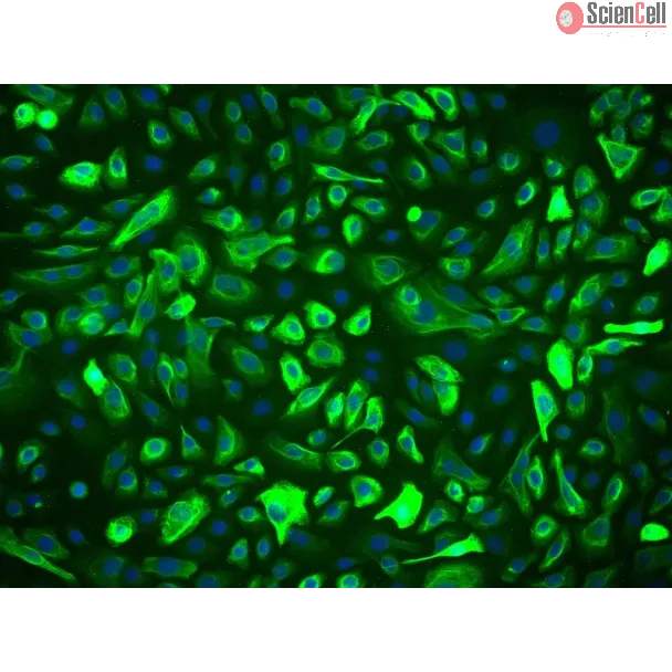
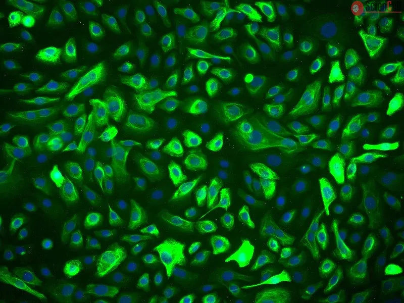
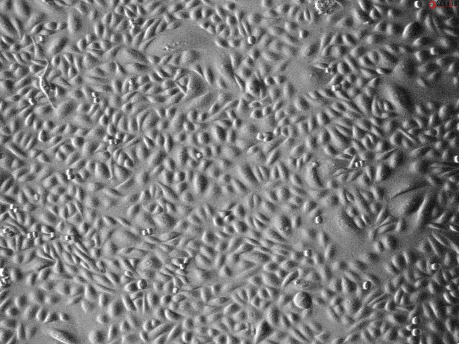
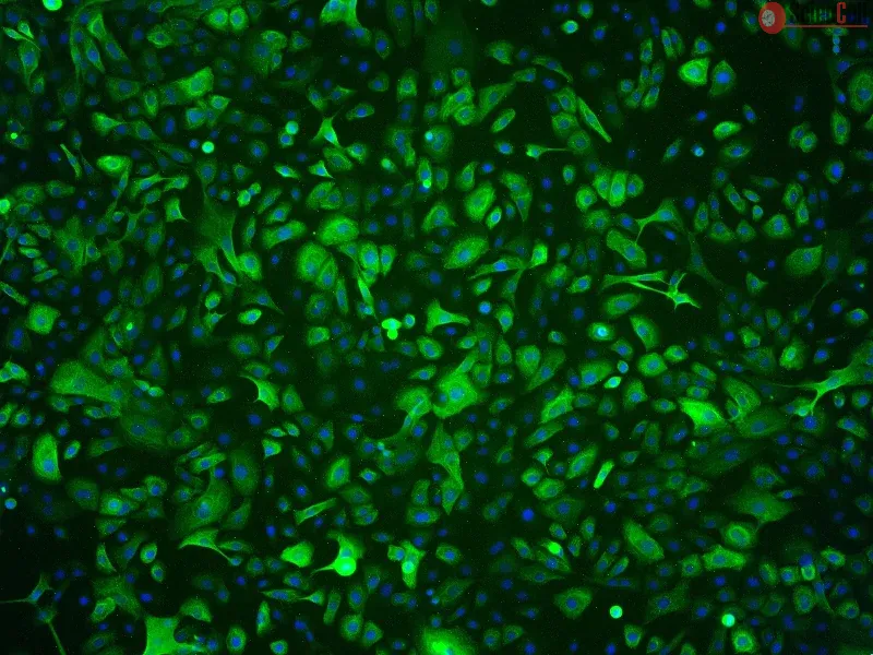
HPrEpiC from ScienCell Research Laboratories are isolated from normal human prostate tissue. HPrEpiC are cryopreserved at passage one and delivered frozen. Each vial contains >5 x 10⁵ cells in 1 ml volume. HPrEpiC are characterized by immunofluorescence with antibodies specific to cytokeratin-18 and/or cytokeratin-19. HPrEpiC are negative for HIV-1, HBV, HCV, mycoplasma, bacteria, yeast and fungi. HPrEpiC are guaranteed to further expand for 15 population doublings under the conditions provided by ScienCell Research Laboratories.
Recommeneded Medium
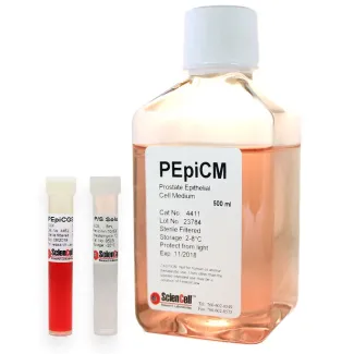
It is recommended to use Prostate Epithelial Cell Medium (PEpiCM, Cat. #4411) for culturing HPrEpiC in vitro.
Product Sheets
 4410
4410
Search for a Certificate of Analysis by Lot Number below:
Purpose
Bladder outlet obstruction (BOO) leads to detrusor overactivity (DO) and structural remodeling of the bladder. However, the molecular mechanisms underlying this ... More
Purpose
Bladder outlet obstruction (BOO) leads to detrusor overactivity (DO) and structural remodeling of the bladder. However, the molecular mechanisms underlying this process remain incompletely understood.
Materials and Methods
In a rat model of partial BOO, we assessed bladder function via cystometry and evaluated histological changes using hematoxylin and eosin staining. The expression and localization of aquaporin (AQP)-2 and caveolin (CAV)-1, -2, and -3 were analyzed by Western blotting and immunofluorescence in bladder tissues. Additionally, immunostaining was performed in cultured human bladder smooth muscle cells to assess protein co-localization.
Results
BOO rats exhibited elevated detrusor contraction pressure and shortened intercontraction intervals. Histologically, hypertrophied smooth muscle bundles and increased extracellular matrix were observed. Western blot analysis revealed significant upregulation of AQP2 and CAV1–3 in BOO bladders. Tissue immunofluorescence demonstrated increased expression and redistribution of these proteins in the detrusor muscle. Cultured cell analysis confirmed subcellular co-localization of AQP2 with CAV1–3.
Conclusions
DO induced by BOO is associated with upregulated expression of AQP2 and CAV1–3 in the urinary bladder. The coexpression observed in bladder cells suggests potential molecular interactions between AQP2 and CAVs that may contribute to the pathophysiology of BOO-related bladder dysfunction.
Keywords: Aquaporin-2; Bladder outlet obstruction; Caveolin; Urinary bladder, overactive Less
This study reports on probing the utility of in situ chromatin texture features such as nuclear DNA methylation and chromatin condensation patterns — visualized by fluo... More
This study reports on probing the utility of in situ chromatin texture features such as nuclear DNA methylation and chromatin condensation patterns — visualized by fluorescent staining and evaluated by dedicated three-dimensional (3D) quantitative and high-throughput cell-by-cell image analysis — in assessing the proliferative capacity, i.e. growth behavior of cells: to provide a more dynamic picture of a cell population with potential implications in basic science, cancer diagnostics/prognostics and therapeutic drug development. Two types of primary cells and four different cancer cell lines were propagated and subjected to cell-counting, flow cytometry, confocal imaging, and 3D image analysis at various points in culture. Additionally a subset of primary and cancer cells was accelerated into senescence by oxidative stress. DNA methylation and chromatin condensation levels decreased with declining doubling times when primary cells aged in culture with the lowest levels reached at the stage of proliferative senescence. In comparison, immortal cancer cells with constant but higher doubling times mostly displayed lower and constant levels of the two in situ-derived features. However, stress-induced senescent primary and cancer cells showed similar levels of these features compared with primary cells that had reached natural growth arrest. With regards to global DNA methylation and chromatin condensation levels, aggressively growing cancer cells seem to take an intermediate level between normally proliferating and senescent cells. Thus, normal cells apparently reach cancer-cell equivalent stages of the two parameters at some point in aging, which might challenge phenotypic distinction between these two types of cells. Companion high-resolution molecular profiling could provide information on possible underlying differences that would explain benign versus malign cell growth behaviors. Keywords: DNA methylation, chromatin condensation, cell proliferation, aging, senescence, cancer, 3D imaging, cell-by-cell analysis Less
ScienCell Research Laboratories (SRL) takes pride in being a resource for researchers all over the world. The publications listed here are not meant as an endorsement or confirmation of the reliability of the products.






,-1-mg-ml--2.jpg)