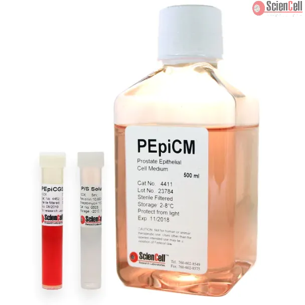
Prostate Epithelial Cell Medium (PEpiCM), when used with Prostate Epithelial Cell Growth Supplement (PEpiCGS, Cat #4452), is a complete medium designed for optimal growth of normal prostate epithelial cells in vitro. It is a sterile, liquid medium which contains essential and non-essential amino acids, vitamins, organic and inorganic compounds, hormones, growth factors and trace minerals. The medium is HEPES and bicarbonate buffered and has a pH of 7.4 when equilibrated in an incubator with an atmosphere of 5% CO2/95% air. The medium is formulated (quantitatively and qualitatively) to provide an optimally balanced nutritional environment that selectively supports the growth of normal prostate epithelial cells in vitro.
RNA activation, as a method of regulating gene expression at the transcriptional level, is far less widely used than RNA interference because of the insufficient understa... More
RNA activation, as a method of regulating gene expression at the transcriptional level, is far less widely used than RNA interference because of the insufficient understanding of the mechanism and the unstable success rate. It is necessary to analyze the failure cases of RNA activation to promote the application of RNA activation. When we validated the saRNAs designed to induce KLK1 expression, we found that saKLK1-374 can upregulate KLK1 expression in prostate tumor cell lines, but failed in normal prostate cell lines. To determine whether the RNA activation of normal cells is difficult only when the target gene is KLK1, we tested p21WAF1/CIP1 as the target gene in RNA activation experiments of normal and cancer prostate cells. Next, to determine whether the above phenomenon exists in other tissues, we used normal and cancerous bladder cells to perform RNA activation experiments with KLK1 and p21WAF1/CIP1 as targets. We have also extended the time from transfection to detection to evaluate whether a longer incubation time can make saRNA upregulate the target genes in normal cells. Fluorescently labeled dsRNA was transfected to evaluate the transfection efficiency, and the expression of Ago2 and IPO8 necessary for RNA activation was also detected. The p21WAF1/CIP1 could be significantly upregulated by saRNA in prostate cancer cells, but not in normal prostate cells. The expression of KLK1 in bladder-derived cell lines was extremely low and could not be induced by saRNA. The p21WAF1/CIP1 was upregulated by saRNA to a higher extent in bladder cancer cells but to a lower extent in normal bladder cells. Prolonging incubation time could not make saRNA induce the expression of target genes in normal cells. Compared with tumor cells used in this study, normal cells had lower transfection efficiency or lower expression of Ago2 and IPO8. Although it has been currently found that normal cell lines in the prostate and bladder might be more difficult to be successfully induced target gene expression by exogenous saRNA than tumor cells due to low transfection efficiency or Ago2 and IPO8 expression, it is not certain that this phenomenon occurs in other types of tissue. However, researchers still need to pay attention to the transfection efficiency and/or the expression levels of Ago2 and IPO8 when conducting RNA activation experiments in normal cells. Less
This study reports on probing the utility of in situ chromatin texture features such as nuclear DNA methylation and chromatin condensation patterns — visualized by fluo... More
This study reports on probing the utility of in situ chromatin texture features such as nuclear DNA methylation and chromatin condensation patterns — visualized by fluorescent staining and evaluated by dedicated three-dimensional (3D) quantitative and high-throughput cell-by-cell image analysis — in assessing the proliferative capacity, i.e. growth behavior of cells: to provide a more dynamic picture of a cell population with potential implications in basic science, cancer diagnostics/prognostics and therapeutic drug development. Two types of primary cells and four different cancer cell lines were propagated and subjected to cell-counting, flow cytometry, confocal imaging, and 3D image analysis at various points in culture. Additionally a subset of primary and cancer cells was accelerated into senescence by oxidative stress. DNA methylation and chromatin condensation levels decreased with declining doubling times when primary cells aged in culture with the lowest levels reached at the stage of proliferative senescence. In comparison, immortal cancer cells with constant but higher doubling times mostly displayed lower and constant levels of the two in situ-derived features. However, stress-induced senescent primary and cancer cells showed similar levels of these features compared with primary cells that had reached natural growth arrest. With regards to global DNA methylation and chromatin condensation levels, aggressively growing cancer cells seem to take an intermediate level between normally proliferating and senescent cells. Thus, normal cells apparently reach cancer-cell equivalent stages of the two parameters at some point in aging, which might challenge phenotypic distinction between these two types of cells. Companion high-resolution molecular profiling could provide information on possible underlying differences that would explain benign versus malign cell growth behaviors. Keywords: DNA methylation, chromatin condensation, cell proliferation, aging, senescence, cancer, 3D imaging, cell-by-cell analysis Less
ScienCell Research Laboratories (SRL) takes pride in being a resource for researchers all over the world. The publications listed here are not meant as an endorsement or confirmation of the reliability of the products.