Autophagy, a type II programmed cell death, is essential for cell survival under stress, e.g. lung injury, and bone marrow-derived mesenchymal stem cells (BM-MSCs) have g... More
Autophagy, a type II programmed cell death, is essential for cell survival under stress, e.g. lung injury, and bone marrow-derived mesenchymal stem cells (BM-MSCs) have great potential for cell therapy. However, the mechanisms underlying the BM-MSC activation of autophagy to provide a therapeutic effect in ischaemia/reperfusion-induced lung injury (IRI) remain unclear. Thus, we investigate the activation of autophagy in IRI following transplantation with BM-MSCs. Seventy mice were pre-treated with BM-MSCs before they underwent lung IRI surgery in vivo. Human pulmonary micro-vascular endothelial cells (HPMVECs) were pre-conditioned with BM-MSCs by oxygen-glucose deprivation/reoxygenation (OGD) in vitro. Expression markers for autophagy and the phosphoinositide 3-kinase/protein kinase B (PI3K/Akt) signalling pathway were analysed. In IRI-treated mice, administration of BM-MSCs significantly attenuated lung injury and inflammation, and increased the level of autophagy. In OGD-treated HPMVECs, co-culture with BM-MSCs attenuated endothelial permeability by decreasing the level of cell death and enhanced autophagic activation. Moreover, administration of BM-MSCs decreased the level of PI3K class I and p-Akt while the expression of PI3K class III was increased. Finally, BM-MSCs-induced autophagic activity was prevented using the inhibitor LY294002. Administration of BM-MSCs attenuated lung injury by improving the autophagy level via the PI3K/Akt signalling pathway. These findings provide further understanding of the mechanisms related to BM-MSCs and will help to develop new cell-based therapeutic strategies in lung injury. Less
Purpose Natural killer (NK) cell cytotoxicity correlates with the ligation of activating receptors (e.g., NKG2D) by their ligands (e.g., MHC class I–related chains [MIC... More
Purpose Natural killer (NK) cell cytotoxicity correlates with the ligation of activating receptors (e.g., NKG2D) by their ligands (e.g., MHC class I–related chains [MIC] A and B) on target cells. Histone deacetylase inhibitors (HDACi) at high concentrations inhibit tumor growth and can increase NKG2D ligand expression on tumor targets, but are widely regarded as toxic to NK cells. Methods We investigated the mechanism of entinostat, a benzamide-derivative narrow-spectrum HDACi, in augmenting the cytotoxicity of NK cells against human colon carcinoma and sarcoma by assessing gene and protein expression, histone acetylation, and cytotoxicity in in vitro and murine models. Results We observed that entinostat dose- and time-dependent increase in MIC expression in tumor targets and NKG2D in primary human NK cells, both correlating with increased acetylated histone 3 (AcH3) binding to associated promoters. Entinostat pretreatment of colon carcinoma and sarcoma cells, NK cells, or both led to enhanced overall cytotoxicity in vitro, which was reversed by NKG2D blockade, and inhibited growth of tumor xenografts. Lastly, we showed decreased expression of MICA and ULBP2 transcription in primary human osteosarcoma. Conclusions Entinostat enhances NK cell killing of cancer cells through upregulation of both NKG2D and its ligands, suggesting an attractive approach for augmenting NK cell immunotherapy of solid tumors such as colon carcinoma and sarcomas. Keywords: Natural killer cells, HDAC inhibitor, cancer, NKG2D, NKG2D ligands Less
Although inflammation and altered barrier functions of the vasculature, due predominantly to the infection of endothelial cell lining of small and medium-sized blood vess... More
Although inflammation and altered barrier functions of the vasculature, due predominantly to the infection of endothelial cell lining of small and medium-sized blood vessels, represent salient pathological features of human rickettsioses, the interactions between pathogenic rickettsiae and microvascular endothelial cells remain poorly understood. We have investigated the activation of nuclear transcription factor-kappa B (NF-κB) and p38 mitogen-activated protein (MAP) kinase, expression of heme oxygenase 1 (HO-1) and cyclooxygenase 2 (COX-2), and secretion of chemokines and prostaglandins after Rickettsia rickettsii infection of human cerebral, dermal, and pulmonary microvascular endothelial cells in comparison with pulmonary artery cells of macrovascular origin. NF-κB and p38 kinase activation and increased HO-1 mRNA expression were clearly evident in all cell types, along with relatively similar susceptibility to R. rickettsii infection in vitro but considerable variations in the intensities/kinetics of the aforementioned host responses. As expected, the overall activation profiles of macrovascular endothelial cells derived from human pulmonary artery and umbilical vein were nearly identical. Interestingly, cerebral endothelial cells displayed a marked refractoriness in chemokine production and secretion, while all other cell types secreted various levels of interleukin-8 (IL-8) and monocyte chemoattractant protein 1 (MCP-1) in response to infection. A unique feature of all microvascular endothelial cells was the lack of induced COX-2 expression and resultant inability to secrete prostaglandin E2 after R. rickettsii infection. Comparative evaluation thus yields the first experimental evidence for the activation of both common and unique cell type-specific host response mechanisms in macrovascular and microvascular endothelial cells infected with R. rickettsii, a prototypical species known to cause Rocky Mountain spotted fever in humans. Less
Although the liver is known to be the main site of factor VIII (FVIII) production, other organs are probably also important for the regulation of FVIII secretion. However... More
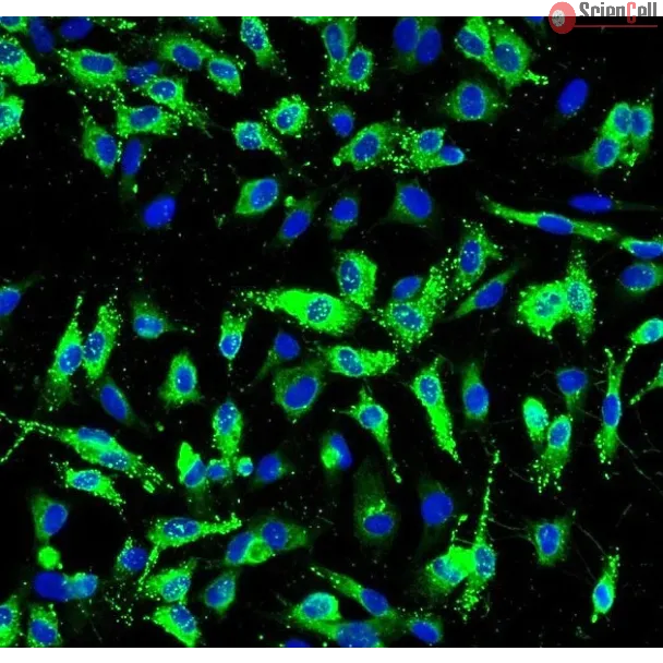
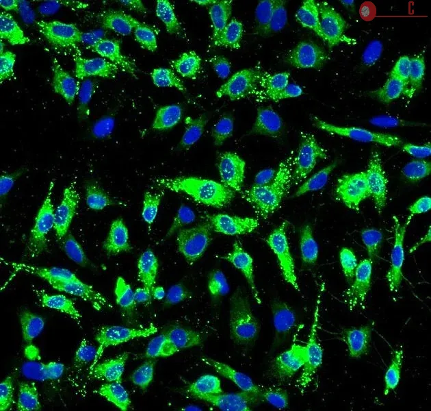
Although the liver is known to be the main site of factor VIII (FVIII) production, other organs are probably also important for the regulation of FVIII secretion. However, the study of the regulation of extrahepatic FVIII production has been hampered by the lack of definitive identification of human tissues able to secrete FVIII. Recent studies have shown that lung endothelial cells can synthesize FVIII. We therefore studied the production of FVIII by endothelial cells purified from other vascular beds. Because physiologic stress results in a rapid elevation of FVIII, we also investigated whether endothelial cells can store FVIII and secrete it after treatment with agonists. Microvascular endothelial cells from lung, heart, intestine, and skin as well as endothelial cells from pulmonary artery constitutively secreted FVIII and released it after treatment with phorbol-myristate acetate and epinephrine. By contrast, endothelial cells from the aorta, umbilical artery and umbilical vein did not constitutively secrete FVIII or release it after treatment with agonists, probably because of a lack of FVIII synthesis. Extrahepatic endothelial cells from certain vascular beds therefore appear to be an important FVIII production and storage site with the potential to regulate FVIII secretion in chronic and acute conditions. Less
Increases in endothelial cGMP prevent oxidant-mediated endothelial barrier dysfunction, but the downstream mechanisms remain unclear. To determine the role of cGMP-depend... More
Increases in endothelial cGMP prevent oxidant-mediated endothelial barrier dysfunction, but the downstream mechanisms remain unclear. To determine the role of cGMP-dependent protein kinase (PKG)(I), human pulmonary artery endothelial cells (HPAEC) lacking PKG(I) expression were infected with a recombinant adenovirus encoding PKG(Ibeta) (Ad.PKG) and compared with uninfected and control-infected (Ad.betagal) HPAEC. Transendothelial electrical resistance (TER), an index of permeability, was measured after H(2)O(2) (250 microM) exposure with or without pretreatment with 8-(4-chlorophenylthio)guanosine 3',5'-cyclic monophosphate (CPT-cGMP). HPAEC infected with Ad.PKG, but not Ad.betagal, expressed PKG(I) protein and demonstrated Ser(239) and Ser(157) phosphorylation of vasodilator-stimulated phosphoprotein after treatment with CPT-cGMP. Adenoviral infection decreased basal permeability equally in Ad.PKG- and Ad.betagal-infected HPAEC compared with uninfected cells. Treatment with CPT-cGMP (100 microM) caused a PKG(I)-independent decrease in permeability (8.2 +/- 0.6%). In all three groups, H(2)O(2) (250 microM) caused a similar approximately 35% increase in permeability associated with increased actin stress fiber formation, intercellular gaps, loss of membrane VE-cadherin, and increased intracellular Ca(2+) concentration ([Ca(2+)](i)). In uninfected and Ad.betagal-infected HPAEC, pretreatment with CPT-cGMP (100 microM) partially blocked the increased permeability induced by H(2)O(2). In Ad.PKG-infected HPAEC, CPT-cGMP (50 microM) prevented the H(2)O(2)-induced TER decrease, cytoskeletal rearrangement, and loss of junctional VE-cadherin. CPT-cGMP attenuated the peak [Ca(2+)](i) caused by H(2)O(2) similarly (23%) in Ad.betagal- and Ad.PKG-infected HPAEC, indicating a PKG(I)-independent effect. These data suggest that cGMP decreased HPAEC basal permeability by a PKG(I)-independent process, whereas the ability of cGMP to prevent H(2)O(2)-induced barrier dysfunction was predominantly mediated by PKG(I) through a Ca(2+)-independent mechanism. Less
Little is known how age alters the dynamics and function of cell adhesion molecules, especially under inflammatory and stressful conditions. One membrane constituent, int... More
Little is known how age alters the dynamics and function of cell adhesion molecules, especially under inflammatory and stressful conditions. One membrane constituent, intercellular adhesion molecule-1 (ICAM-1) is a transmembrane glycoprotein of the immunoglobulin (Ig) superfamily that regulates key outside-->in and inside-->out signals associated with cell-to-cell interactions. If conditions such as age and inflammation change usual ICAM-1 action then important downstream effects ultimately perturb endothelial cell function. In this report, ICAM-1 accumulates in late passage endothelial cells when compared to early passage endothelial cells, yet ICAM-1 protein expression is attenuated when senescent cells are challenged by TNF-alpha (10ng/ml). Importantly, age alters ICAM-1 dynamic properties from directed to random receptor motion within the membrane. Single particle tracking reveals that the average ICAM-1 mobility is 44% less in late than early passage cells after its motion is stimulated by the Protein Kinase C (PKC) activator, phorbol myristate acetate (PMA). The mechanism for altered ICAM-1 mobility partly can be explained by a reduced rate of alpha-actinin linking with ICAM-1 in late passage Human Pulmonary Artery Endothelial Cells (HPAECs). Furthermore, tyrosine phosphorylation of alpha-actinin, a requirment for ICAM-1 clustering, is markedly reduced in senescent cells. These findings support a hypothesis that senescence results in changes of ICAM-1 activation and clustering, thus resulting in an age-dependent transmembrane signaling disorder. Therefore, further understanding of age-dependent disturbances of ICAM-1 regulation during inflammation can provide important clues as to appropriate targets for therapeutic interventions and prevention of vascular disorders in elderly at the level of the endothelial surface membrane. Less
Ebola virus glycoprotein (EGP) has been implicated for the induction of cytotoxicity and injury in vascular cells. On the other hand, EGP has also been suggested to induc... More
Ebola virus glycoprotein (EGP) has been implicated for the induction of cytotoxicity and injury in vascular cells. On the other hand, EGP has also been suggested to induce massive cell rounding and detachment from the plastic surface by downregulating cell adhesion molecules without causing cytotoxicity. In this study, we have examined the cytotoxic role of EGP in primary endothelial cells by transduction with a replication-deficient recombinant adenovirus expressing EGP (Ad-EGP). Primary human cardiac microvascular endothelial cells (HCMECs) transduced with Ad-EGP displayed loss of cell adhesion from the plastic surface followed by cell death. Transfer of conditioned medium from EGP-transduced HCMEC into naive cells did not induce loss of adhesion or cell death, suggesting that EGP needs to be expressed intracellularly to exert its cytotoxic effect. Subsequent studies suggested that HCMEC death occurred through apoptosis. Results from this study shed light on the EGP-induced anoikis in primary human cardiac endothelial cells, which may have significant pathological consequences. Less
The authors determined the effect of cyclic guanosine 3′,5′-monophosphate (cGMP) on hydrogen peroxide (H2O2)-induced barrier dysfunction in bovine lung microvascular ... More
The authors determined the effect of cyclic guanosine 3′,5′-monophosphate (cGMP) on hydrogen peroxide (H2O2)-induced barrier dysfunction in bovine lung microvascular endothelial cell (BLMVEC) monolayers and compared the results to bovine pulmonary artery endothelial cells (BPAECs). In BLMVECs, H2O2 (250 μM) caused a 31.9% ± 4.8% decrease in transendothelial electrical resistance (TER) associated with increased actin stress fiber formation, intercellular gaps, and intracellular calcium concentration ([Ca2+]i). The cGMP analogue 8-(p-chlorophenylthio)-cGMP (8p-CPT-cGMP; 30 or 50 μM) prevented the H2O2-induced decrease in TER (p < .001) as well as the cytoskeletal rearrangement and intercellular gap formation. 8-pCPT-cGMP (50 μM) attenuated the peak (418.8 ± 42.1 versus 665.2 ± 38.0 nmol/L; p < .001) and eliminated the sustained increase in [Ca2+]i (193.5 ± 21.3 versus 418.8 ± 42.1 nmol/L; p < .001) caused by H2O2. 8-pCPT-cGMP also increased TER (14.2% ± 2.2%; p < .05) and decreased [Ca2+]i (201.2 ± 12.5 vs. 214.4 ± 12.1 nmol/L; p < .03) before H2O2. In BPAECs, 8p-CPT-cGMP significantly attenuated H2O2-induced increases in permeability and [C2+]i but less effectively than in BLMVECs. These results suggest that in BLMVECs, cGMP countered the adverse effects of H2O2 on barrier function by preventing actin cytoskeletal rearrangement and attenuating the increase in [Ca2+]i. Less