Fibroblasts are mesenchymal cells which perform many vital functions during development and in adulthood. They are responsible for much of the synthesis of extracellular matrix in connective tissue and play major roles in wound healing. Many diseases are associated with fibroblasts, either because fibroblasts are implicated in their etiology or because of the fibrosis that accompanies damage to other cell types. Human oral fibroblasts (HOrF), located in the oral cavity, have the ability to rapidly repair defects in the oral cavity. HOrF, in contrast to skin fibroblasts, can more quickly reorganize the extracellular matrix and migrate for wound repair. Abnormal proliferation of HOrF can lead to the development of oral squamous cell carcinoma. HOrF are a useful model for elucidating the mechanisms of fibrosis and developing treatments for oral cancers.
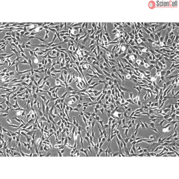
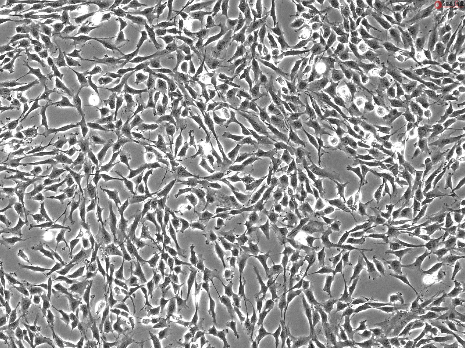
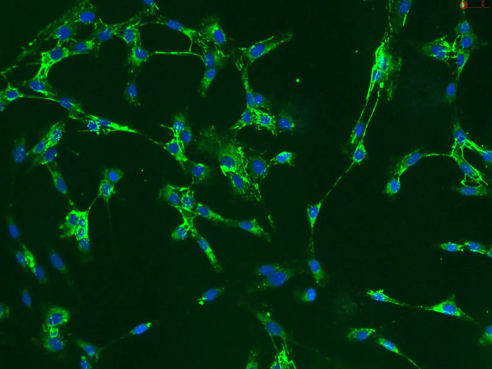
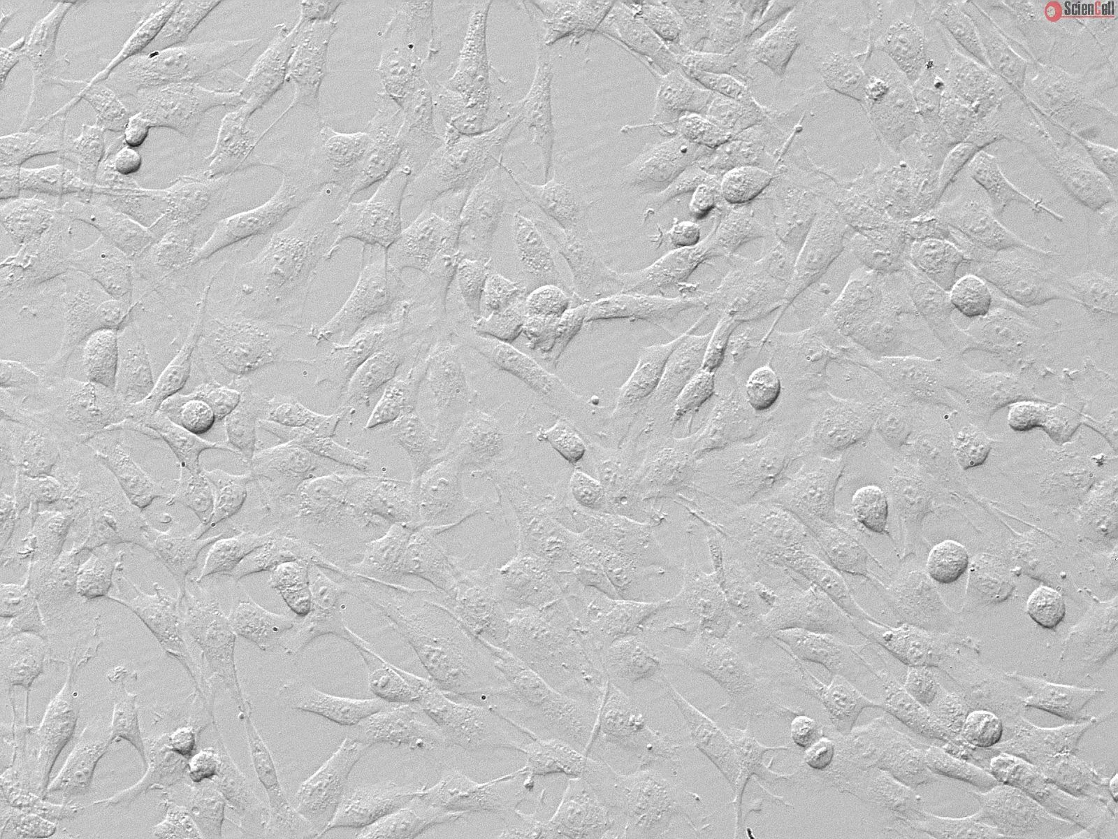
HOrF from ScienCell Research Laboratories are isolated from human oral tissue. HOrF are cryopreserved at passage one and delivered frozen. Each vial contains >5 x 10⁵ cells in 1 ml volume. HOrF are characterized by their spindle morphology and immunofluorescence with antibodies specific to fibronectin. HOrF are negative for HIV-1, HBV, HCV, mycoplasma, bacteria, yeast and fungi. HOrF are guaranteed to further expand for 15 population doublings under the conditions provided by ScienCell Research Laboratories.
Recommended Medium
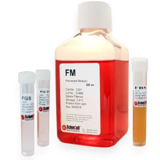
It is recommended to use Fibroblast Medium (FM, Cat. #2301) for the culturing of HOrF in vitro.
Product Sheets
 2640
2640
Search for a Certificate of Analysis by Lot Number below:
Cytoskeletal network dynamics play important roles in regulating cellular functions. Although alterations in cytoskeleton-related genes are frequently detected, limited a... More
Cytoskeletal network dynamics play important roles in regulating cellular functions. Although alterations in cytoskeleton-related genes are frequently detected, limited attention has been paid to their roles in cancer development. A novel keratin fusion variant, K6-K14/V5, was previously identified in head and neck squamous cell carcinoma (HNSCC), and its expression led to catastrophic nuclear collapse, resulting in DNA breaks and cGAS-STING activation. Such cell-killing effects can trigger autophagy induction, which, in turn, promotes cancer cell evolution/clonal selection in a dormant state. Furthermore, due to the disrupted cellular architecture and the loss of mechanosensing, these dormant cells could survive and adapt within a collagen gel. Upregulation of the partial epithelial-mesenchymal transition (pEMT) program by cytoskeleton reorganization was defined as a key step for these dormant cells to reactivate and regain their mechanical properties. Striking cell protrusions and increased MMPs were observed in the reactivated cells, facilitating the interaction with the surrounding extracellular matrix and enhancing their invasive potential. Elevated extracellular vesicles were detected in the reactivated cells, which actively stimulated tumor growth via the FGF-FGFR axis. Our study therefore offers a novel model for understanding how genetic alterations in cytoskeletal genes can directly contribute to cancer development and drive cancer evolution. Less
In head and neck cancer, intratumour lymphatic density and tumour lymphangiogenesis have been correlated with lymphatic metastasis, making lymphangiogenesis a promising t... More
In head and neck cancer, intratumour lymphatic density and tumour lymphangiogenesis have been correlated with lymphatic metastasis, making lymphangiogenesis a promising therapeutic target. However, inter-patient tumour heterogeneity makes it challenging to predict tumour progression and lymph node metastasis. Understanding the lymphangiogenic-promoting factors leading to metastasis (e.g., tumour-derived fibroblasts or TDF), would help develop strategies to improve patient outcomes. A microfluidic in vitro model of a tubular lymphatic vessel was co-cultured with primary TDF from head and neck cancer patients to evaluate the effect of TDF on lymphangiogenesis. We assessed the length and number of lymphangiogenic sprouts and vessel permeability via microscopy and image analysis. Finally, we characterised lymphatic vessel conditioning by TDF via RT-qPCR. Lymphatic vessels were conditioned by the TDF in a patient-specific manner. Specifically, the presence of TDF induced sprouting, altered vessel permeability, and increased the expression of pro-lymphangiogenic genes. Gene expression and functional responses in the fibroblast-conditioned lymphatic vessels were consistent with the patient tumour stage and lymph node status. IGF-1, upregulated among patients, was targeted to validate our personalised medicine approach. Interestingly, IGF-1 blockade was not effective across different patients. The use of lymphatic organotypic models incorporating head and neck TDF provides insight into the pathways leading to lymphangiogenesis in each patient. This model provided a platform to test anti-angiogenic therapeutics and inform of their effectiveness for individual patients. Less
ScienCell Research Laboratories (SRL) takes pride in being a resource for researchers all over the world. The publications listed here are not meant as an endorsement or confirmation of the reliability of the products.

,-1-mg-ml--2.jpg)