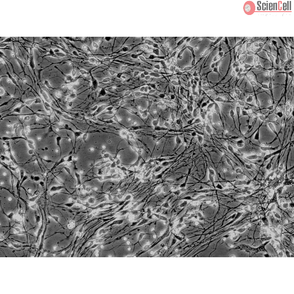
The tissue of the central nervous system is made up of two classes of cells that may be broadly categorized as neurons and glia. Neurons are anatomic, functional, and trophic units of the brain. Despite great variability in size and shape, all neurons share common morphologic features, the key elements of a highly complex communication network. Neurons are dynamically polarized cells that serve as the major signaling unit of the nervous system. The human brain contains about 1 x 10^11 neurons and each are able to contact at least 10,000 other neurons. The hippocampal neurons play a special role in learning and memory. Cultured hippocampal neurons are a useful model for studying neuronal phenomena such as differentiation, survival, process growth, and synaptogenesis.
HN-h from ScienCell Research Laboratories are isolated from human hippocampus. HN-h are cryopreserved at P0 and delivered frozen. Each vial contains >1 x 10⁶ cells in 1 ml volume. HNh are characterized by immunofluorescence with antibodies specific to β-tubulin III. HN-h are negative for HIV-1, HBV, HCV, mycoplasma, bacteria, yeast, and fungi. HN-h are guaranteed to further culture under the conditions provided by ScienCell Research Laboratories; however, HNh are not recommended for expanding or long-term cultures since the cells do not proliferate in culture.
Recommended Medium
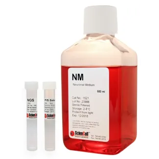
It is recommended to use Neuronal Medium (NM, Cat. #1521) for culturing HN-h in vitro.
Product Sheets
 1540
Search for a Certificate of Analysis by Lot Number below:
Sevoflurane is a common anesthetic agent used in surgical settings and previous studies have indicated that it exerts a neurotoxic effect. However, the molecular mechanis... More
Sevoflurane is a common anesthetic agent used in surgical settings and previous studies have indicated that it exerts a neurotoxic effect. However, the molecular mechanism underlying this side effect is unknown. In addition, the human microRNA‑302 (hsa‑miR‑302) family members have been reported to be involved in neuronal cell development and biology. Thus, the present study aimed to investigate the potential implication of hsa‑miR‑302e in the sevoflurane‑induced cytotoxicity on human hippocampal cells (HN‑h). HN‑h cells were transfected with hsa‑miR‑302e mimic, hsa‑miR‑302e inhibitor or negative controls and subsequently exposed to different concentrations of sevoflurane. An MTT assay was used to assess the cytotoxicity of sevoflurane on HN‑h cells. Cell apoptosis was determined by flow cytometry. The levels of lactate dehydrogenase release, reactive oxygen species, lipid peroxidation and intracellular calcium (Ca2+) were additionally detected. Reverse transcription‑quantitative polymerase chain reaction and western blotting were conducted to determine mRNA and protein expression, respectively. A luciferase assay was performed for validating the targeting of OXR1 by hsa‑miR‑302e. The results indicated that sevoflurane induced a decrease in cell viability, malondialdehyde and reactive oxygen species production, lactate dehydrogenase release, intracellular Ca2+ production, calcium/calmodulin‑dependent protein kinase II phosphorylation and apoptosis. In addition, treatment with sevoflurane induced the expression of hsa‑miR‑302e while the expression of its target, oxidation resistance gene 1 (OXR1), was significantly downregulated. Inhibition of hsa‑miR‑302e expression protected neuronal cells from sevoflurane cytotoxicity. Mechanistic studies demonstrated that OXR1 was a direct target of hsa‑miR‑302e. Furthermore, the overexpression of OXR1 abolished the effect of sevoflurane on neuronal cells. The results of the present study indicated that sevoflurane exerts its neurotoxic effect by regulating the hsa‑miR‑302e/OXR1 axis. Therefore, the manipulation of the hsa‑miR‑302e/OXR1 pathway will be useful for preventing sevoflurane‑induced neurotoxicity. Less
Protein kinase Cϵ (PKCϵ) promotes synaptic maturation and synaptogenesis via activation of synaptic growth factors such as BDNF, NGF, and IGF. However, many of the deta... More
Protein kinase Cϵ (PKCϵ) promotes synaptic maturation and synaptogenesis via activation of synaptic growth factors such as BDNF, NGF, and IGF. However, many of the detailed mechanisms by which PKCϵ induces synaptogenesis are not fully understood. Accumulation of PSD-95 to the postsynaptic density (PSD) is known to lead to synaptic maturation and strengthening of excitatory synapses. Here we investigated the relationship between PKCϵ and PSD-95. We show that the PKCϵ activators dicyclopropanated linoleic acid methyl ester and bryostatin 1 induce phosphorylation of PSD-95 at the serine 295 residue, increase the levels of PSD-95, and enhance its membrane localization. Elimination of the serine 295 residue in PSD-95 abolished PKCϵ-induced membrane accumulation. Knockdown of either PKCϵ or JNK1 prevented PKCϵ activator-mediated membrane accumulation of PSD-95. PKCϵ directly phosphorylated PSD-95 and JNK1 in vitro. Inhibiting PKCϵ, JNK, or calcium/calmodulin-dependent kinase II activity prevented the effects of PKCϵ activators on PSD-95 phosphorylation. Increase in membrane accumulation of PKCϵ and phosphorylated PSD-95 (p-PSD-95S295) coincided with an increased number of synapses and increased amplitudes of excitatory post-synaptic potentials (EPSPs) in adult rat hippocampal slices. Knockdown of PKCϵ also reduced the synthesis of PSD-95 and the presynaptic protein synaptophysin by 30 and 44%, respectively. Prolonged activation of PKCϵ increased synapse number by 2-fold, increased presynaptic vesicle density, and greatly increased PSD-95 clustering. These results indicate that PKCϵ promotes synaptogenesis by activating PSD-95 phosphorylation directly through JNK1 and calcium/calmodulin-dependent kinase II and also by inducing expression of PSD-95 and synaptophysin. Less
ScienCell Research Laboratories (SRL) takes pride in being a resource for researchers all over the world. The publications listed here are not meant as an endorsement or confirmation of the reliability of the products.