Fibroblasts are mesenchymal cells derived from the embryonic mesoderm. Mammary fibroblasts (MF) synthesize components of the stromal extracellular matrix of the mammary gland. More importantly, such stromal extracellular matrix regulates the proliferation and differentiation of mammary epithelial cells by influencing their gene expression. In addition, previous studies have shown that mammary fibroblasts play a role in recruiting macrophages for immune surveillance which, in concert with extracellular matrix turnover and angiogenesis, is associated with a supportive mammary microenvironment for tumor cell progression. These results suggest that MF play a stimulatory functional role in breast cancer development and invasion.
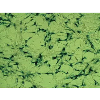
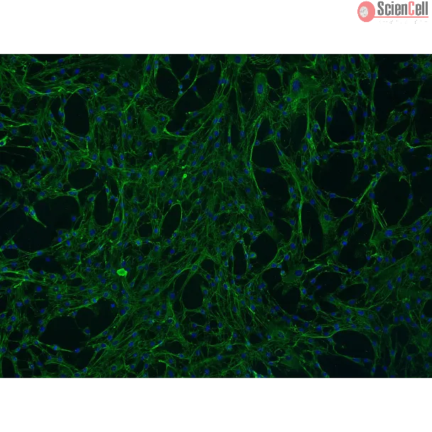
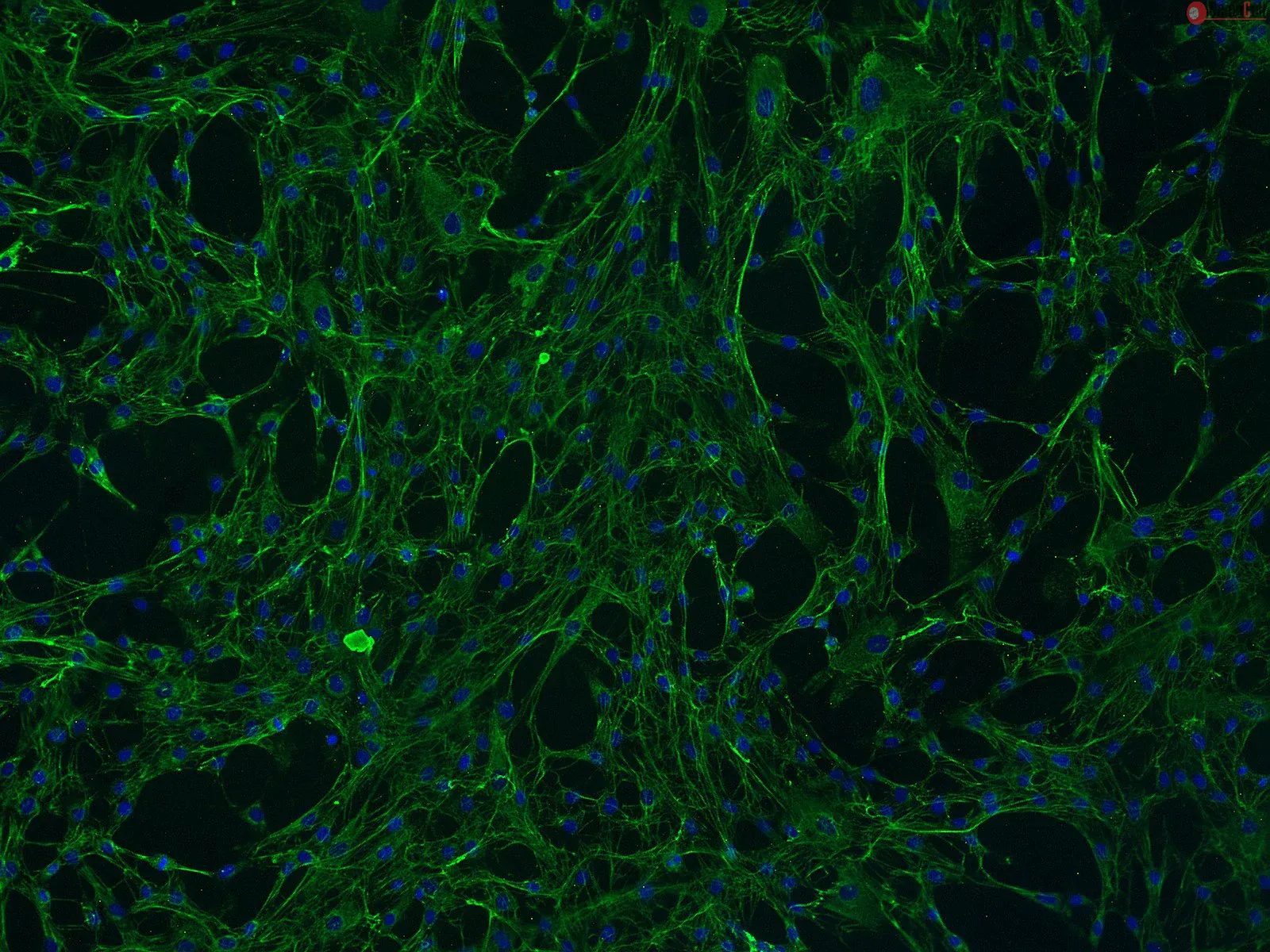
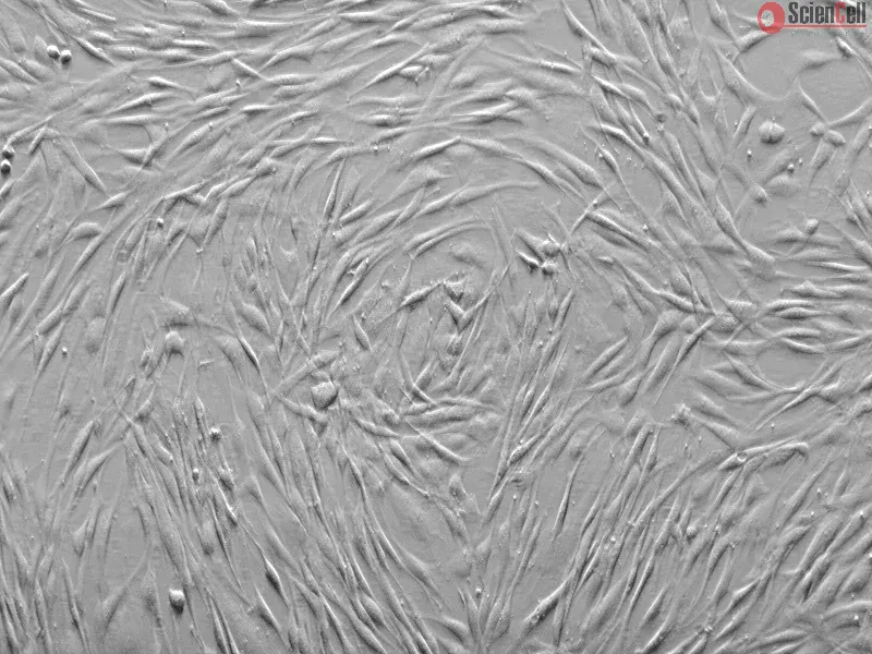
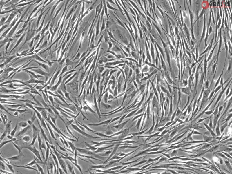
HMF from ScienCell Research Laboratories are isolated from human breast. HMF are cryopreserved at passage one and delivered frozen. Each vial contains >5 x 10⁵ cells in 1 ml volume. HMF are characterized by immunofluorescence with antibodies specific to fibronectin. HMF are negative for HIV-1, HBV, HCV, mycoplasma, bacteria, yeast, and fungi. HMF are guaranteed to further expand for 15 population doublings under the conditions provided by ScienCell Research Laboratories.
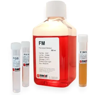
Recommended Medium
It is recommended to use Fibroblast Medium (FM, Cat. #2301) for culturing HMF in vitro.
A mounting body of evidence in cancer research suggests that the local microenvironment of tumor cells has a profound influence on cancer progression and metastasis. In v... More
A mounting body of evidence in cancer research suggests that the local microenvironment of tumor cells has a profound influence on cancer progression and metastasis. In vitro studies on the tumor microenvironment and its pharmacological modulation, however, are often hampered by the technical challenges associated with creating physiological cell culture environments that integrate cancer cells with the key components of their native niche such as neighboring cells and extracellular matrix (ECM) to mimic complex microarchitecture of cancerous tissue. Using earlystage breast cancer as a model disease, here we describe a biomimetic microengineering strategy to reconstitute three-dimensional (3D) structural organization and microenvironment of breast tumors in human cell-based in vitro models. Specifically, we developed a microsystem that enabled co-culture of breast tumor spheroids with human mammary ductal epithelial cells and mammary fibroblasts in a compartmentalized 3D microfluidic device to replicate microarchitecture of breast ductal carcinoma in situ (DCIS). We also explored the potential of this breast cancer-on-a-chip system as a drug screening platform by evaluating the efficacy and toxicity of an anticancer drug (paclitaxel). Our microengineered disease model represents the first critical step towards recapitulating pathophysiological complexity of breast cancer, and may serve as an enabling tool to systematically examine the contribution of the breast cancer microenvironment to the progression of DCIS to an invasive form of the disease. Less
Collagen-based three-dimensional (3D) in vitro models that recapitulate the structural and functional context of normal and malignant tissues provide a relevant surrogate... More
Collagen-based three-dimensional (3D) in vitro models that recapitulate the structural and functional context of normal and malignant tissues provide a relevant surrogate to animal models in the study of developmental and carcinogenic processes. Human breast epithelial MCF10A cells embedded in a collagen gel formed both acinar and tubular structures only when the gel was detached (floating) from the cell culture plate's well, and allowed to be contracted by the cells. Epithelial phenotype depended upon the time and the location within the gel, as ducts formed exclusively on the upper layer of the gel while ductal branching occurred earlier in the central area of the gel, and gradually progressed toward the periphery. The addition of fibroblasts accelerated tubulogenesis. MCF10A cells facilitated the organization of thick collagen fibers packed into large bundles at the tip of the ducts and parallel to the direction of ductal elongation. In gels that were not detached from the well's wall, MCF10A cells organized in monolayer and collagen fibers were aligned along the axis of outstretched sprouts stemming from those cellular aggregates. Partial gel release induced uniaxial tubulogenesis associated with orderly aligned collagen fibers. These results suggest that proper collagen organization is necessary for epithelial morphogenesis to occur, and that biomechanical interactions between fibers and cells mediated duct formation, elongation and branching. Less
Background: Stromal-epithelial interactions mediate breast development, and the initiation and progression of breast cancer. In the present study, we developed 3-dimensio... More
Background: Stromal-epithelial interactions mediate breast development, and the initiation and progression of breast cancer. In the present study, we developed 3-dimensional (3D) in vitro models to study breast cancer tissue organization and the role of the microenvironment in phenotypic determination. Less
Epithelial-stromal interactions play a crucial role in normal embryonic development and carcinogenesis of the human breast while the underlying mechanisms of these events... More
Epithelial-stromal interactions play a crucial role in normal embryonic development and carcinogenesis of the human breast while the underlying mechanisms of these events remain poorly understood. To address this issue, we constructed a physiologically relevant, three-dimensional (3D) culture surrogate of complex human breast tissue that included a tri-culture system made up of human mammary epithelial cells (MCF10A), human fibroblasts and adipocytes, i.e., the two dominant breast stromal cell types, in a Matrigel/collagen mixture on porous silk protein scaffolds. The presence of stromal cells inhibited MCF10A cell proliferation and induced both alveolar and ductal morphogenesis and enhanced casein expression. In contrast to the immature polarity exhibited by co-cultures with either fibroblasts or adipocytes, the alveolar structures formed by the tri-cultures exhibited proper polarity similar to that observed in breast tissue in vivo. Only alveolar structures with reverted polarity were observed in MCF10A monocultures. Consistent with their phenotypic appearance, more functional differentiation of epithelial cells was also observed in the tri-cultures, where casein alpha- and -beta mRNA expression was significantly increased. This in vitro tri-culture breast tissue system sustained on silk scaffold effectively represents a more physiologically relevant 3D microenvironment for mammary epithelial cells and stromal cells than either co-cultures or monocultures. This experimental model provides an important first step for bioengineering an informative human breast tissue system, with which to study normal breast morphogenesis and neoplastic transformation. Copyright 2010 Elsevier Ltd. All rights reserved. Less
ScienCell Research Laboratories (SRL) takes pride in being a resource for researchers all over the world. The publications listed here are not meant as an endorsement or confirmation of the reliability of the products.

,-1-mg-ml--2.jpg)