Cardiac myocytes are the most physically energetic cells in the body. They are highly specialized high-oxygen-content cells that house a large number of mitochondria. They occupy as much as 75% of the cardiac mass, but constitute only about one third of the total cell number in the heart. Differentiated cardiac myocytes have little capacity to proliferate; however, hypertrophic growth has been shown to respond to alpha1-adrenergic stimuli via the Ras/MEK pathway. All cardiac myocytes are capable of spontaneous rhythmic depolarization and repolarization of their membranes. Contraction of cardiac myocytes is myogenic, which is independent of nervous stimulation. There is a complex network of signals in cardiac myocytes regulating the rhythmic pumping of the heart. Cardiac myocyte hypertrophy and apoptosis have been implicated in the loss of contractile function during heart failure. A better understanding of the cardiac signaling network will help reveal the cellular mechanisms leading to cardiac myocyte death.
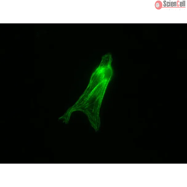
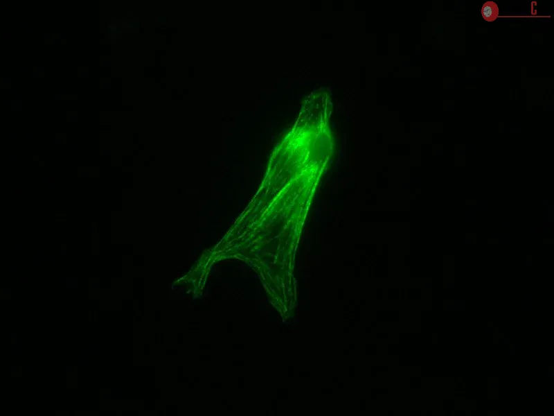
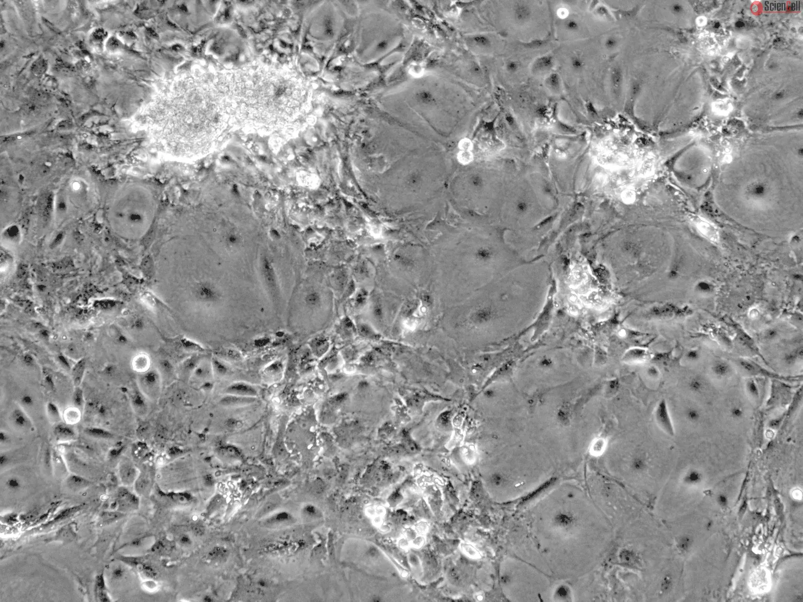
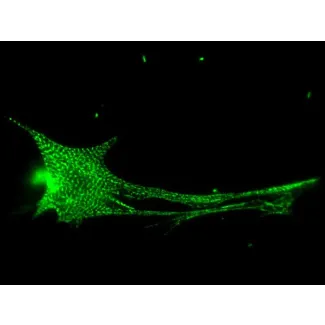
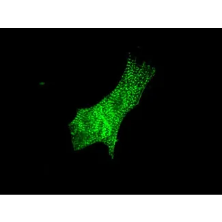
Mouse Cardiac Myocytes from CD1 (MCM) ScienCell Research Laboratories are isolated from postnatal day 2 or postnatal day 8 CD-1 mouse heart. MCM are cryopreserved at P0 and delivered frozen. Each vial contains >1 x 10⁶ cells in 1 ml volume. MCM are characterized by immunofluorescence with antibodies specific to sarcomeric alpha-actinin and/or α-smooth muscle actin. MCM are negative for mycoplasma, bacteria, yeast, and fungi. MCM are guaranteed to further culture under the conditions provided by ScienCell Research Laboratories; however, MCM are not recommended for expanding or long-term cultures since the cells do not proliferate in culture.
Recommended Medium
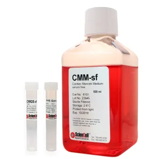
It is recommended to use Cardiac Myocyte Medium-serum free (CMM-sf, Cat. #6101) for culturing MCM in vitro.
心肌细胞是人体内能量代谢最为活跃的细胞之一。它们高度特化,具有较高的耗氧量,并含有大量线粒体。心肌细胞约占心脏组织总质量的75%,但仅约占心脏细胞总数的三分之一。分化成熟的心肌细胞增殖能力较弱;然而,研究表明,心肌细胞的肥大性生长可通过Ras/MEK信号通路响应α1肾上腺素能刺激。所有心肌细胞均具有自发性、有节律的膜去极化和复极化能力。心肌细胞的收缩具有肌源性,不依赖神经刺激。心肌细胞内存在复杂的信号调控网络,共同调节心脏的节律性搏动。心肌细胞肥大和凋亡与心力衰竭过程中收缩功能的丧失密切相关。进一步深入了解心肌细胞中的信号调控网络,有助于揭示导致心肌细胞死亡的细胞机制。
CD1小鼠心肌细胞(MCM)由ScienCell Research Laboratories提供,分离自出生后第2天(P2)或第8天(P8)的CD-1小鼠心脏。MCM在P0代进行冻存,并以冷冻状态运输。每瓶含有超过1 × 10⁶个细胞,体积为1 ml。MCM采用免疫荧光法进行鉴定,使用针对肌节α-辅肌动蛋白(sarcomeric alpha-actinin)和/或α-平滑肌肌动蛋白(α-smooth muscle actin)的特异性抗体进行检测。MCM经检测不含支原体、细菌、酵母和真菌。按照ScienCell Research Laboratories提供的培养条件,MCM可进一步进行体外培养;但由于这些细胞在培养过程中不具有增殖能力,因此不建议用于细胞扩增或长期培养。
推荐培养基
建议使用无血清心肌细胞培养基(Cardiac Myocyte Medium-serum free,CMM-sf,产品编号6101)对MCM进行体外培养。
Product Sheets
 M6200
M6200
Search for a Certificate of Analysis by Lot Number below:
The clinical controversy of rosiglitazone as a hypoglycemic agent is potentially associated with heart failure, mainly due to its potent activation of peroxisome prolifer... More
The clinical controversy of rosiglitazone as a hypoglycemic agent is potentially associated with heart failure, mainly due to its potent activation of peroxisome proliferator-activated receptor γ (PPARγ). PPARγ partial agonists showed superior pharmacological profiles to rosiglitazone. This study compared differences in cardiac morphology and function of the PPARγ partial agonist CMHX008 with rosiglitazone. High-fat diet (HFD) induced obese mice, ob/ob mice and cardiomyocytes overexpressing PPARγ2 were treated with CMHX008 or rosiglitazone. Heart function, myocardial morphology, and hypertrophy-related gene expression were examined. Clinical information from patients with type 2 diabetes mellitus (T2DM) who had taken rosiglitazone and undergone Doppler echocardiography was collected. HFD and ob/ob mice significantly developed cardiac contractile dysfunction, with upregulated PPARγ2 protein levels in heart tissues. Cardiomyocytes of HFD and ob/ob mice were disorderly arranged, the cell areas expanded, and collagen accumulated. In vitro cardiomyocytes overexpressing PPARγ2 displayed obvious structural abnormalities and high mRNA levels of ANP and BNP, critical cardiac hypertrophy-related genes. HFD-fed mice treated with rosiglitazone or CMHX008 had significantly improved cardiac function, but rosiglitazone induced higher expression of ANP and βMHC and hypertrophic cardiomyopathy, while CMHX008 did not. Patients with T2DM taking rosiglitazone exhibited increased thickness of the posterior wall and the ventricular septum, suggesting cardiac hypertrophy. Our findings show that diabetic cardiomyopathy was associated with ectopic overexpression of PPARγ2. The full agonist rosiglitazone prevents cardiac dysfunction at the expense of compensatory hypertrophy, while the partial agonist CMHX008 shared a comparable protective effect without altering the structure of cardiomyocytes. Less
Myocardial ischemia/reperfusion (I/R) injury may cause the apoptosis of cardiomyocytes as well as cardiac fibrosis, which is characterized as the transdifferentiation of ... More
Myocardial ischemia/reperfusion (I/R) injury may cause the apoptosis of cardiomyocytes as well as cardiac fibrosis, which is characterized as the transdifferentiation of fibroblasts to myofibroblasts and collagen deposition. MicroRNAs (miRNAs or miRs) have been demonstrated to be involved in myocardial I/R injury. However, the underlying molecular mechanism remains largely unclear. In the present study, mouse cardiomyocyte M6200 cells were treated with hypoxia/reoxygenation (H/R). Our data indicated that H/R treatment led to cell apoptosis, the increased expression of fibrosis‑related proteins, namely collagen I, II, III, and fibronectin, as well as the downregulation of miR-142-3p in M6200 cells. Overexpression of miR-142-3p suppressed the H/R-induced apoptosis and fibrosis of M6200 cells. Bioinformatics analysis and a Dual‑Luciferase reporter assay further identified high mobility group box 1 (HMGB1) as a direct target gene of miR-142-3p, and miR-142-3p negatively regulated the protein level of HMGB1 in M6200 cells. Furthermore, knockdown of HMGB1 enhanced cell proliferation whereas it inhibited the apoptosis and fibrosis of M6200 cells. In addition, TGF-β1/Smad3 signaling was suggested to be involved in the miR-142-3p/HMGB1-mediated apoptosis and fibrosis of M6200 cells treated with H/R. Taken together, the findings of the present study demonstrate that miR-142-3p inhibits H/R-induced apoptosis and fibrosis of cardiomyocytes, partly at least, by the direct inhibition of HMGB1 expression. Therefore, these findings have increased our understanding of the pathogenesis of H/R-induced myocardial injury. Less
ScienCell Research Laboratories (SRL) takes pride in being a resource for researchers all over the world. The publications listed here are not meant as an endorsement or confirmation of the reliability of the products.