Cardiac myocytes are the most physically energetic cells in the body. They are highly specialized high-oxygen-content cells that house a large number of mitochondria. They occupy as much as 75% of the cardiac mass, but constitute only about one third of the total cell number in the heart. Differentiated cardiac myocytes have little capacity to proliferate; however, hypertrophic growth has been shown to respond to alpha1-adrenergic stimuli via the Ras/MEK pathway. All cardiac myocytes are capable of spontaneous rhythmic depolarization and repolarization of their membranes. Contraction of cardiac myocytes is myogenic, which is independent of nervous stimulation. There is a complex network of signals in cardiac myocytes regulating the rhythmic pumping of the heart. Cardiac myocyte hypertrophy and apoptosis have been implicated in the loss of contractile function during heart failure. A better understanding of the cardiac signaling network will help reveal the cellular mechanisms leading to cardiac myocyte death.
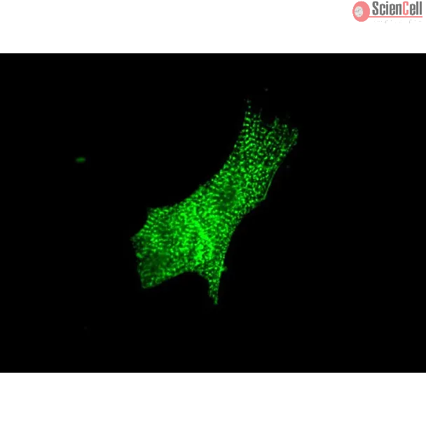
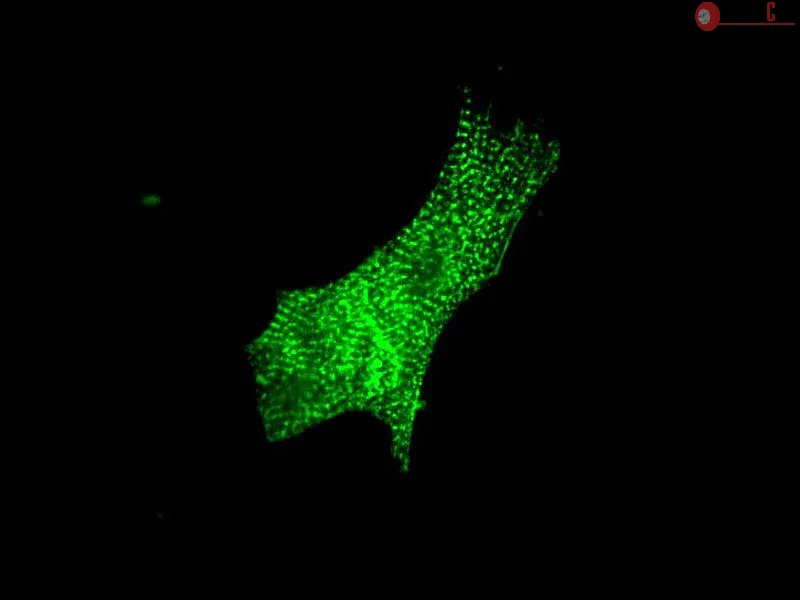
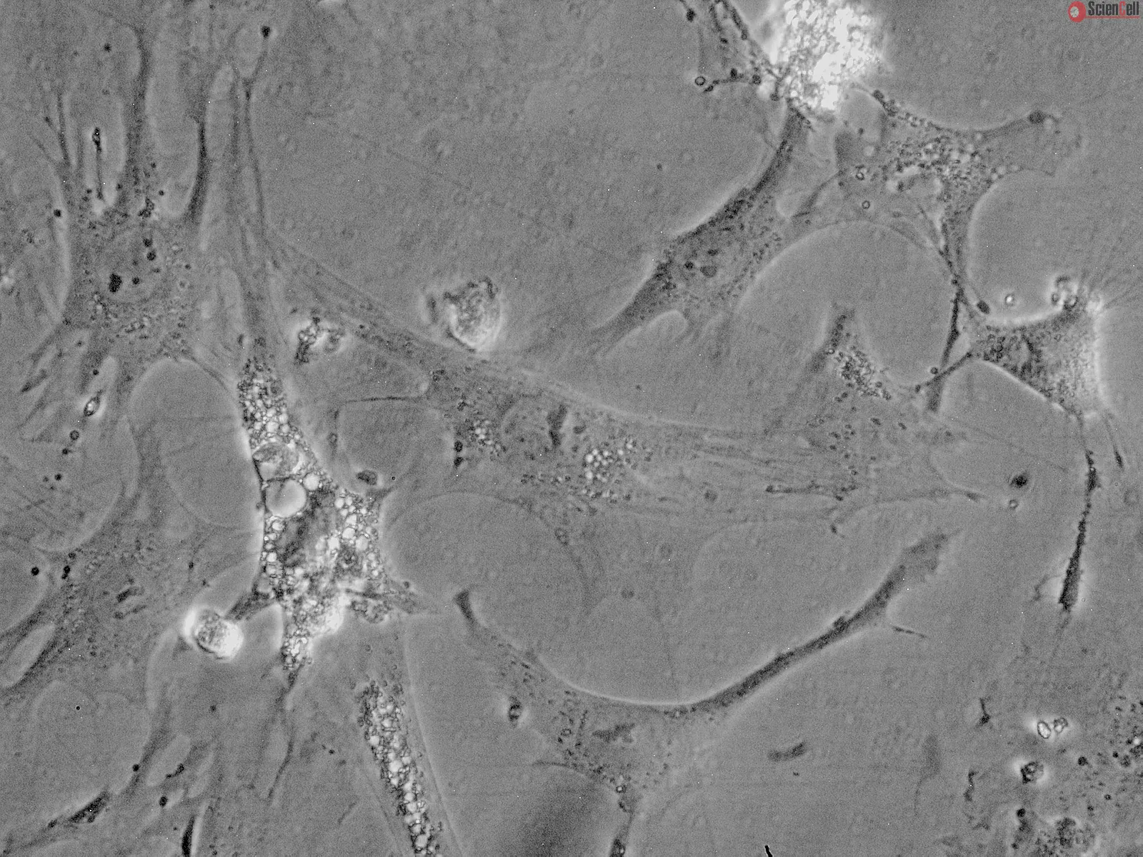
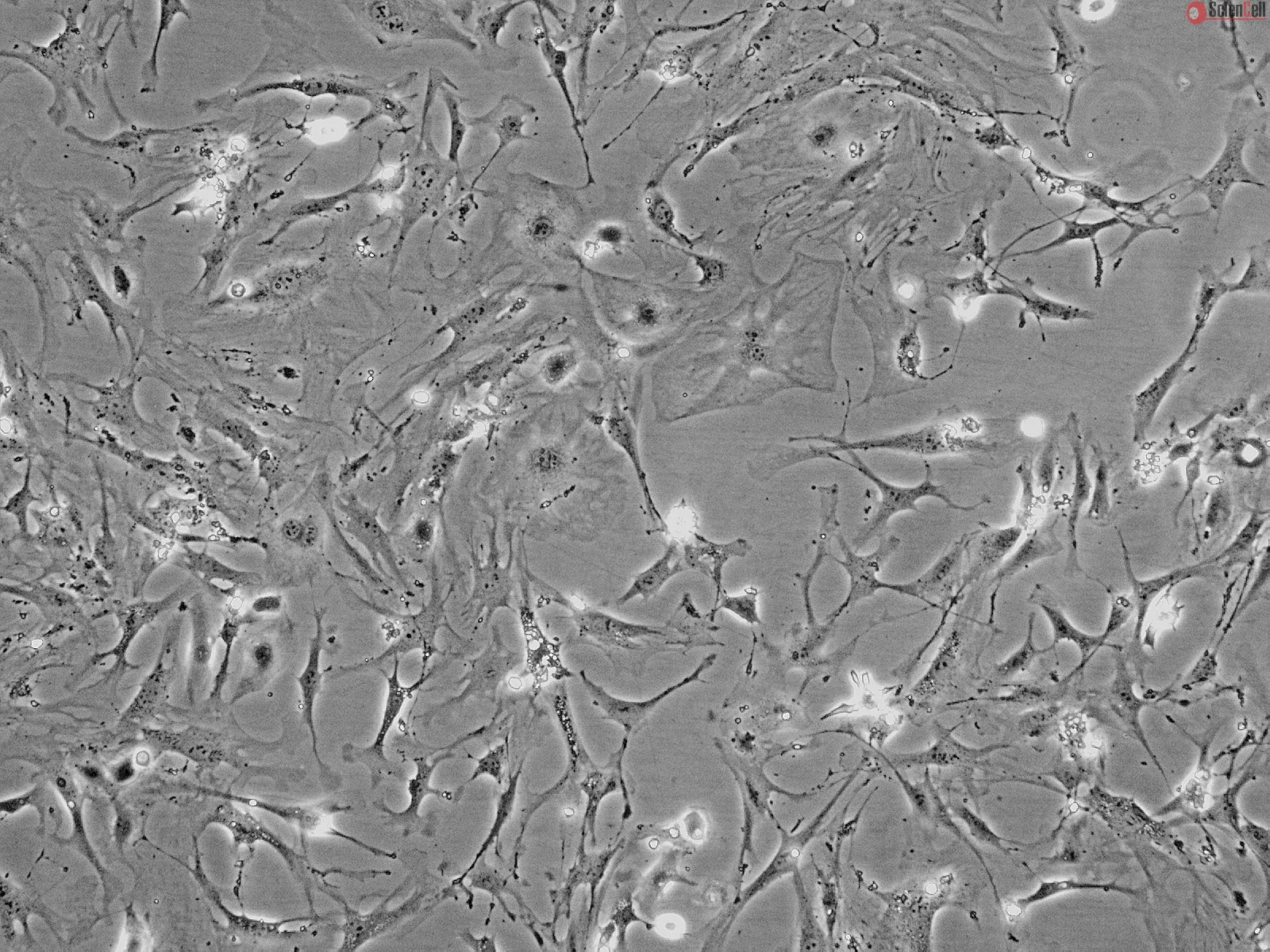
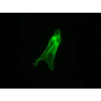
MCM from ScienCell Research Laboratories are isolated from postnatal day 2 or postnatal day 8 C57BL/6 mouse heart. MCM are cryopreserved at P0 and delivered frozen. Each vial contains >1 x 10⁶ cells in 1 ml volume. MCM are characterized by immunofluorescence with antibodies specific to smooth muscle actin and/or sacromeric alpha-actinin. MCM are negative for mycoplasma, bacteria, yeast, and fungi. MCM are guaranteed to further culture under the conditions provided by ScienCell Research Laboratories; however, MCM are not recommended for expanding or long-term cultures since the cells do not proliferate in culture.
Recommended Medium
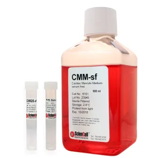
It is recommended to use Cardiac Myocyte Medium-serum free (CMM-sf, Cat. #6101) for culturing MCM in vitro.
心肌细胞是人体内能量代谢最为活跃的细胞之一。它们高度特化,具有较高的耗氧量,并含有大量线粒体。心肌细胞约占心脏组织总质量的75%,但仅约占心脏细胞总数的三分之一。分化成熟的心肌细胞增殖能力较弱;然而,研究表明,心肌细胞的肥大性生长可通过Ras/MEK信号通路响应α1肾上腺素能刺激。所有心肌细胞均具有自发性、有节律的膜去极化和复极化能力。心肌细胞的收缩具有肌源性,不依赖神经刺激。心肌细胞内存在复杂的信号调控网络,共同调节心脏的节律性搏动。心肌细胞肥大和凋亡与心力衰竭过程中收缩功能的丧失密切相关。进一步深入了解心肌细胞中的信号调控网络,有助于揭示导致心肌细胞死亡的细胞机制。
C57BL/6小鼠心肌细胞(MCM)由ScienCell Research Laboratories提供,分离自出生后第2天(P2)或第8天(P8)的C57BL/6小鼠心脏。MCM在P0代进行冻存,并以冷冻状态运输。每瓶含有超过1 × 106个细胞,体积为1 ml。MCM采用免疫荧光法进行鉴定,使用针对平滑肌肌动蛋白(smooth muscle actin)和/或肌节α-辅肌动蛋白(sarcomeric alpha-actinin)的特异性抗体进行检测。MCM经检测不含支原体、细菌、酵母和真菌。按照ScienCell Research Laboratories提供的培养条件,MCM可进一步进行体外培养;但由于这些细胞在培养过程中不具有增殖能力,因此不建议用于细胞扩增或长期培养。
推荐培养基
建议使用无血清心肌细胞培养基(Cardiac Myocyte Medium-serum free,CMM-sf,产品编号6101)对MCM进行体外培养。
Product Sheets
 M6200-57
Search for a Certificate of Analysis by Lot Number below:
Background Doxorubicin (DOX) cardiotoxicity is a major complication of cancer therapy and involves macrophage-driven inflammation and myocardial remodeling. The macrophag... More
Background Doxorubicin (DOX) cardiotoxicity is a major complication of cancer therapy and involves macrophage-driven inflammation and myocardial remodeling. The macrophage surface protein Mac-2 (galectin-3) is upregulated in cardiac injury, but its role in regulating macrophage function and downstream injury pathways remains undefined. Methods We used CRISPR/Cas9-engineered Mac-2-null macrophages to evaluate chemotaxis, cytokine gene expression, and lysosomal stress signaling in vitro. To examine paracrine injury mechanisms, we performed co-culture assays with cardiomyocytes and fibroblasts. In vivo, we studied homozygous Mac-2-mutant mice and used CD45.1/CD45.2 bone marrow transplantation with lineage tracking to define hematopoietic versus stromal contributions to DOX-induced inflammation, apoptosis, fibrosis, and systolic dysfunction. Results Doxorubicin induced Mac-2 and inflammatory transcripts (Il6, Tnf, Ccl2) in wild-type macrophages, whereas Mac-2 knockout reduced DOX uptake, chemotaxis, and cytokine induction. In co-culture, DOX-treated WT macrophages increased caspase-3/7 activity in cardiomyocytes and phospho-TFEB in fibroblasts, both attenuated with Mac-2 deletion. In vivo, Mac-2-null mice exhibited less cardiac inflammation, apoptosis, and fibrosis with preserved systolic function and reduced mortality. Bone marrow transplantation demonstrated that hematopoietic Mac-2 suppressed cardiac Tfeb and upregulated Sqstm1 and Tgfb1, enhancing inflammatory and apoptotic responses, whereas Mac-2-deficient marrow restored Tfeb, limited Sqstm1/Tgfb1, and protected cardiac function. Conclusions Mac-2 promotes DOX-induced cardiac injury by facilitating inflammatory activation in macrophages, driving fibroblast lysosomal stress via TFEB and SQSTM1, and augmenting caspase-3–associated apoptosis in cardiomyocytes. Loss of Mac-2 in hematopoietic cells reduces inflammation, fibrosis, and systolic dysfunction in vivo. Less
Aims: Pathological cardiac fibrosis and hypertrophy are common features of left ventricular remodeling that often progress to heart failure (HF). Endothelial cells (ECs) ... More
Aims: Pathological cardiac fibrosis and hypertrophy are common features of left ventricular remodeling that often progress to heart failure (HF). Endothelial cells (ECs) are the most abundant non-myocyte cells in adult mouse heart. Simvastatin, a strong inducer of Krüppel-like Factor 2 (Klf2) in ECs, ameliorates pressure overload induced maladaptive cardiac remodeling and dysfunction. This study aims to explore the detailed molecular mechanisms of the anti-remodeling effects of simvastatin. Methods and Results: RGD-magnetic-nanoparticles were used to endothelial specific delivery of siRNA and we found absence of simvastatin's protective effect on pressure overload induced maladaptive cardiac remodeling and dysfunction after in vivo inhibition of EC-Klf2. Mechanism studies showed that EC-Klf2 inhibition reversed the simvastatin-mediated reduction of fibroblast proliferation and myofibroblast formation, as well as cardiomyocyte size and cardiac hypertrophic genes, which suggested that EC-Klf2 might mediate the anti-fibrotic and anti-hypertrophy effects of simvastatin. Similar effects were observed after Klf2 inhibition in cultured ECs. Moreover, Klf2 regulated its direct target gene TGFβ1 in ECs and mediated the protective effects of simvastatin, and inhibition of EC-Klf2 increased the expression of EC-TGFβ1 leading to simvastatin losing its protective effects. Also, EC-Klf2 was found to regulate EC-Foxp1 and loss of EC-Foxp1 attenuated the protective effects of simvastatin similar to EC-Klf2 inhibition. Conclusions: We conclude that cardiac microvasculature ECs are important in the modulation of pressure overload induced maladaptive cardiac remodeling and dysfunction, and the endothelial Klf2-TGFβ1 or Klf2-Foxp1-TGFβ1 pathway mediates the preventive effects of simvastatin. This study demonstrates a novel mechanism of the non-cholesterol lowering effects of simvastatin for HF prevention. Keywords: Heart failure (HF), Maladaptive cardiac remodeling (cardiac fibrosis and hypertrophy), HMG-CoA reductase inhibitors, Simvastatin, Vascular endothelial cells (ECs), Krüppel-like Factor 2 (Klf2), Transforming growth factor-beta 1 (TGFβ1), Forkhead Box P1 (Foxp1) Less
The hypoxic conditions induced by reduced blood flow decreases oxygen availability in target tissues. Cellular hypoxia leads to mitochondrial dysfunction, decreased energ... More
The hypoxic conditions induced by reduced blood flow decreases oxygen availability in target tissues. Cellular hypoxia leads to mitochondrial dysfunction, decreased energy production, and increased production of reactive oxygen species. To determine the alteration in expression of mitochondrial genes after hypoxia in cardiomyocytes, we developed a rodent mitochondrial gene chip (RoMitoChip). The chip had 1088 probe sets including 46 probe sets representing 37 mouse mitochondrial DNA transcripts and the remaining probe sets representing mouse nuclear genes contributing to the mitochondrial structure and function. Mouse cardiomyocytes isolated from neonatal C57BL/6 mice that were subjected to hypoxia (1% oxygen) for different time intervals demonstrated a dichotomy in the expression profile of tRNA and mRNA transcripts. We report a total of 483 signature genes that were altered by hypoxia in the cardiac myocytes and related to mitochondrial structure and function. This includes 23 transcripts on mitochondrial DNA. Pathway analysis demonstrated predominant changes in the expression of genes involved in oxidative phosphorylation, glucose and fatty acid metabolism, and apoptosis. The most upregulated genes after 24 h of hypoxia included hypoxia-inducible factor 1, alpha subunit, inducible genes Bnip3, Pdk1, and Aldoc. Whereas Bnip3 is important in the cardiomyocyte death pathway, Pdk1 enzyme is critical in conserving mitochondrial function by diverting metabolic intermediates to glycolysis. This study identifies the participation of two important pathways, cell death and glycolytic, and two key proteins, Bnip3 and Pdk1, playing critical roles in these pathways in cardiomyocytes after severe hypoxia. Less
ScienCell Research Laboratories (SRL) takes pride in being a resource for researchers all over the world. The publications listed here are not meant as an endorsement or confirmation of the reliability of the products.