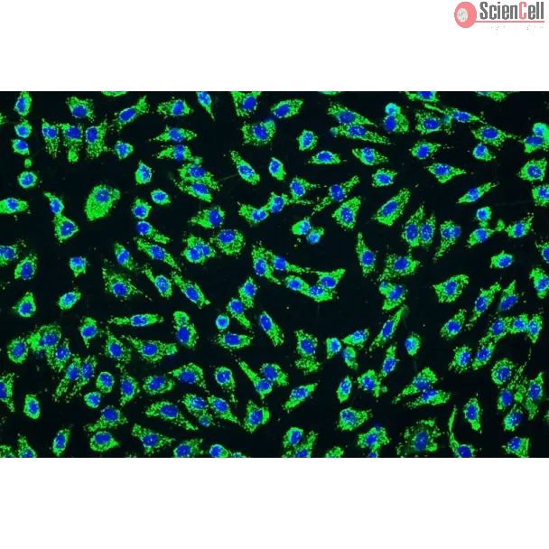
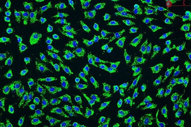
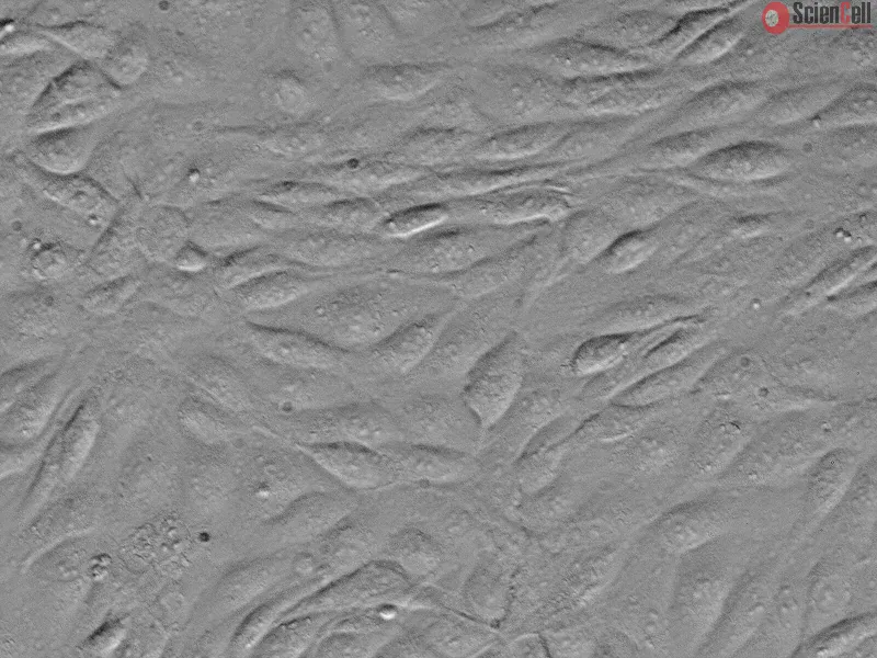
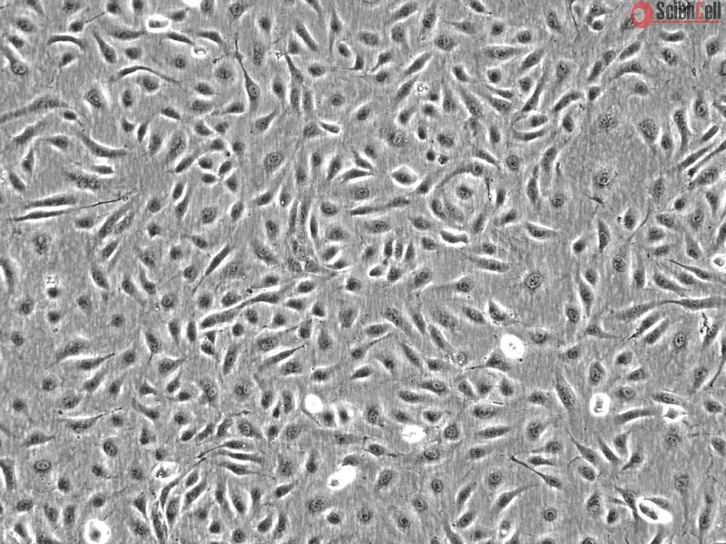
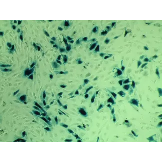
Vascular endothelial cells contribute to the maintenance of vascular homeostasis. Vascular endothelial cells produce and secrete activators and inhibitors of the coagulation and fibrinolysis system. In addition, they mediate the adhesion and aggregation of blood platelets. Endothelial cells also release molecules that regulate cell proliferation and control vessel wall tone. Human umbilical vein endothelial cells (HUVEC) and human umbilical artery endothelial cells (HUAEC) are the most commonly used cell type for the study of endothelial cell processes in vitro. HUAEC have a “cobblestone” morphology, show positive staining for vWF/Factor VIII and CD-31, and the ability to take up acetylated low-density lipoprotein. Cells pretreated with IL-1 or TNF-alpha also selectively express E-selectin and VCAM.
HUAEC from ScienCell Research Laboratories are isolated from human umbilical arteries. HUAEC are cryopreserved at passage one and delivered frozen. Each vial contains >5 x 10⁵ cells in 1 ml volume. HUAEC are characterized by immunofluorescence with antibodies specific to vWF/Factor VIII and/or CD31 (PECAM1). HUAEC are negative for HIV-1, HBV, HCV, mycoplasma, bacteria, yeast and fungi. HUAEC are guaranteed to further expand for 15 population doublings under the conditions provided by ScienCell Research Laboratories.
Recommended Medium
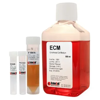
It is recommended to use Endothelial Cell Medium (ECM, Cat. #1001) for the culturing of HUAEC in vitro.
血管内皮细胞参与维持血管稳态。血管内皮细胞可产生并分泌凝血和纤溶系统的激活因子及抑制因子。此外,内皮细胞还参与介导血小板的黏附和聚集,并释放调节细胞增殖及控制血管壁张力的分子。人脐静脉内皮细胞(HUVEC)和人脐动脉内皮细胞(HUAEC)是体外研究内皮细胞生物学过程最常用的细胞类型。HUAEC 呈典型的“铺路石”样形态,对 vWF/凝血因子 VIII 和 CD-31 呈阳性染色,并具有摄取乙酰化低密度脂蛋白(acetylated low-density lipoprotein)的能力。经 IL-1 或 TNF-α 预处理后,HUAEC 还可选择性表达 E-选择素(E-selectin)和血管细胞黏附分子(VCAM)。
ScienCell Research Laboratories 的人脐动脉内皮细胞(HUAEC)分离自人脐动脉。HUAEC 以第 1 代(P1)冻存,并以冷冻状态运输。每瓶含有 >5 × 10⁵ 个细胞,体积为 1 ml。HUAEC 采用针对血管性血友病因子(vWF)/凝血因子 VIII 和/或 CD31(PECAM1)的特异性抗体进行免疫荧光鉴定。HUAEC 经检测对 HIV-1、HBV、HCV、支原体、细菌、酵母菌和真菌均呈阴性。在 ScienCell Research Laboratories 提供的培养条件下,HUAEC 可进一步扩增 15 个群体倍增(population doublings)。
推荐培养基
推荐使用内皮细胞培养基(Endothelial Cell Medium,ECM,产品编号:1001)在体外培养 HUAEC。
Product Sheets
 8010
8010
Search for a Certificate of Analysis by Lot Number below:
Increasing evidence supports the hypothesis that inflammatory reactions serves an important function in the formation, progression and plaque rupture of atherosclerosis. ... More
Increasing evidence supports the hypothesis that inflammatory reactions serves an important function in the formation, progression and plaque rupture of atherosclerosis. Interleukin (IL)‑1 primarily induces inflammation and is closely associated with the inflammatory environment and the formation of atherosclerosis. The present study aimed to establish an in vitro model for the evaluation of drug efficacy in the intervention of atherosclerosis from the inflammatory perspective, and to observe the anti‑inflammatory effects of tanshinone IIA and andrographolide on atherosclerosis. The IL‑1β‑induced inflammation‑activated endothelial cell (EC)‑smooth muscle cell (SMC)‑mononuclear cell (MC) co‑culture model was established, based on the changes in a series of atherosclerosis‑associated inflammatory markers secreted by ECs and SMCs. The expression of connexin in ECs, adhesion of MCs and changes in inflammatory signalling molecules were selected as evaluation indices for the inflammatory microenvironment of atherosclerosis. The use of this model revealed that tanshinone IIA exhibited significant efficacy against atherosclerosis and its inflammatory reactions. Inflammatory reactions were regarded as the primary mechanism underlying atherosclerosis. The established model simulated a series of relevant changes in the arterial wall under the inflammatory cytokines with oxidized low‑density lipoprotein during the atherosclerotic process. The present study presented a reliable method for the identification of drugs with potential anti‑inflammatory activity in atherosclerosis, for investigating the mechanisms of action, considering the improvement of the inflammatory state and the increase in plaque stability observed. Less
In the phase III clinical trial, RELAX-AHF, serelaxin caused rapid and long-lasting haemodynamic changes. However, the cellular mechanisms involved are unclear in humans.... More
In the phase III clinical trial, RELAX-AHF, serelaxin caused rapid and long-lasting haemodynamic changes. However, the cellular mechanisms involved are unclear in humans. This study examined the effects of serelaxin in co-cultures of human primary endothelial cells (ECs) and smooth muscle cells (SMCs) on cAMP and cGMP signalling. Stimulation of HUVECs or human coronary artery endothelial cells (HCAECs) with serelaxin, concentration-dependently increased cGMP accumulation in co-cultured SMCs to a greater extent than in monocultures of either cell type. This was not observed in human umbilical artery endothelial cells (HUAECs) that do not express the relaxin receptor, RXFP1. Treatment of ECs with l-NG-nitro arginine (NOARG; 30 μM, 30 min) inhibited serelaxin-mediated (30 nM) cGMP accumulation in HUVECs, HCAECs and co-cultured SMCs. In HCAECs, but not HUVECs, pre-incubation with indomethacin (30 μM, 30 min) also inhibited cGMP accumulation in SMCs. Pre-incubation of SMCs with the guanylate cyclase inhibitor ODQ (1 μM, 30 min) had no effect on serelaxin-mediated (30 nM) cGMP accumulation in HUVECs and HCAECs but inhibited cGMP accumulation in SMCs. Serelaxin stimulation of HCAECs, but not HUVECs, increased cAMP accumulation concentration-dependently in SMCs. Pre-incubation of HCAECs with indomethacin, but not l-NOARG, abolished cAMP accumulation in co-cultured SMCs, suggesting involvement of prostanoids. In co-cultures, treatment of ECs with serelaxin caused marked cGMP accumulation in SMCs and with HCAEC also cAMP accumulation. Responses involved EC-derived NO and with HCAEC prostanoid production. Thus, serelaxin differentially modulates vascular tone in different vascular beds. Less
Although the liver is known to be the main site of factor VIII (FVIII) production, other organs are probably also important for the regulation of FVIII secretion. However... More
Although the liver is known to be the main site of factor VIII (FVIII) production, other organs are probably also important for the regulation of FVIII secretion. However, the study of the regulation of extrahepatic FVIII production has been hampered by the lack of definitive identification of human tissues able to secrete FVIII. Recent studies have shown that lung endothelial cells can synthesize FVIII. We therefore studied the production of FVIII by endothelial cells purified from other vascular beds. Because physiologic stress results in a rapid elevation of FVIII, we also investigated whether endothelial cells can store FVIII and secrete it after treatment with agonists. Microvascular endothelial cells from lung, heart, intestine, and skin as well as endothelial cells from pulmonary artery constitutively secreted FVIII and released it after treatment with phorbol-myristate acetate and epinephrine. By contrast, endothelial cells from the aorta, umbilical artery and umbilical vein did not constitutively secrete FVIII or release it after treatment with agonists, probably because of a lack of FVIII synthesis. Extrahepatic endothelial cells from certain vascular beds therefore appear to be an important FVIII production and storage site with the potential to regulate FVIII secretion in chronic and acute conditions. Less
ScienCell Research Laboratories (SRL) takes pride in being a resource for researchers all over the world. The publications listed here are not meant as an endorsement or confirmation of the reliability of the products.