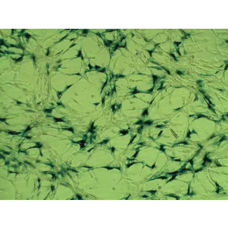
Fibroblasts are mesenchymal cells derived from the embryonic mesoderm. They synthesize components of the extracellular matrix, maintain the structural integrity of connective tissues, and actively participate in wound healing. Accumulating evidence indicates that ovarian fibroblasts can be induced and activated by ovarian cancer cells to secrete a variety of growth factors, chemokines, collagens, and matrix-modifying enzymes to establish a pro-tumorigenic, anti-immunogenic niche for cancer progression and metastasis. These results suggest that fibroblasts play a stimulatory functional role in cancer development and invasive behavior.
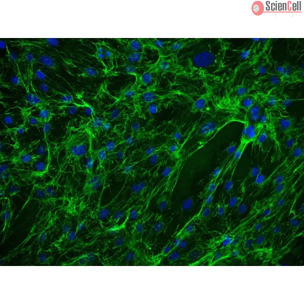
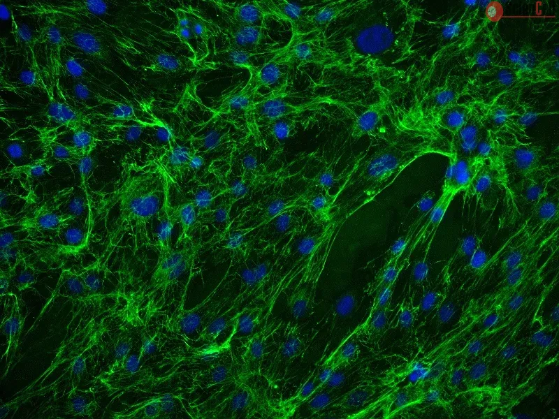
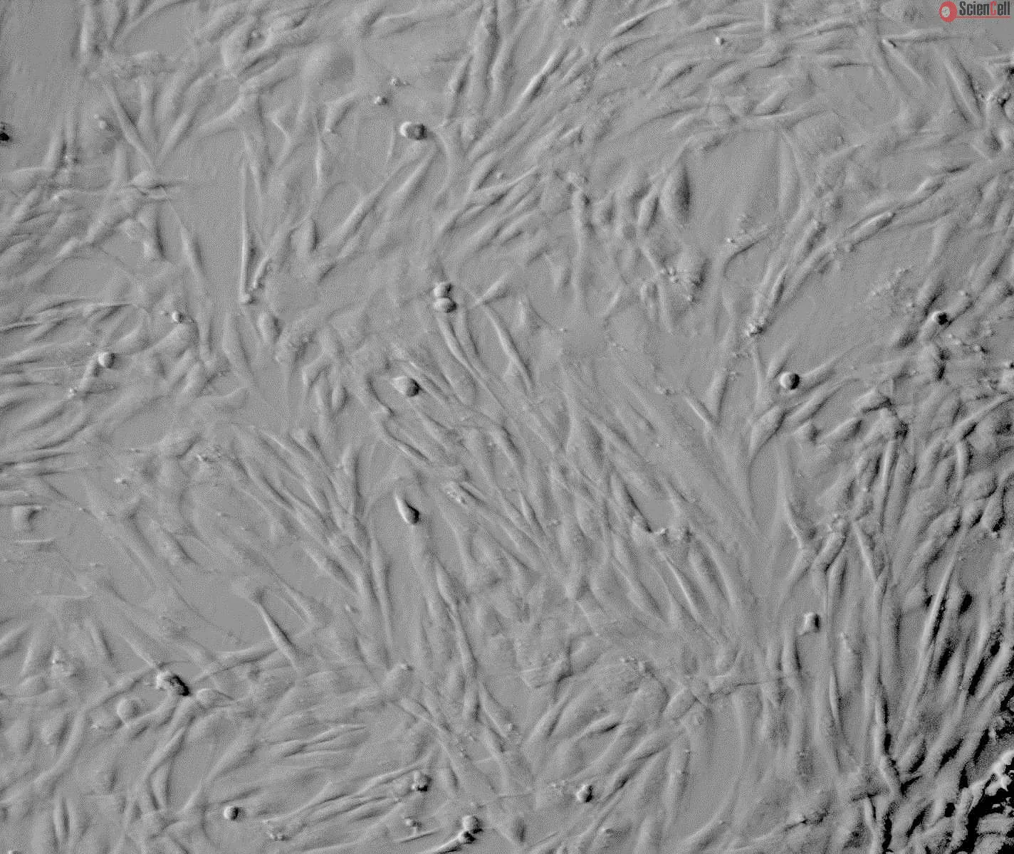
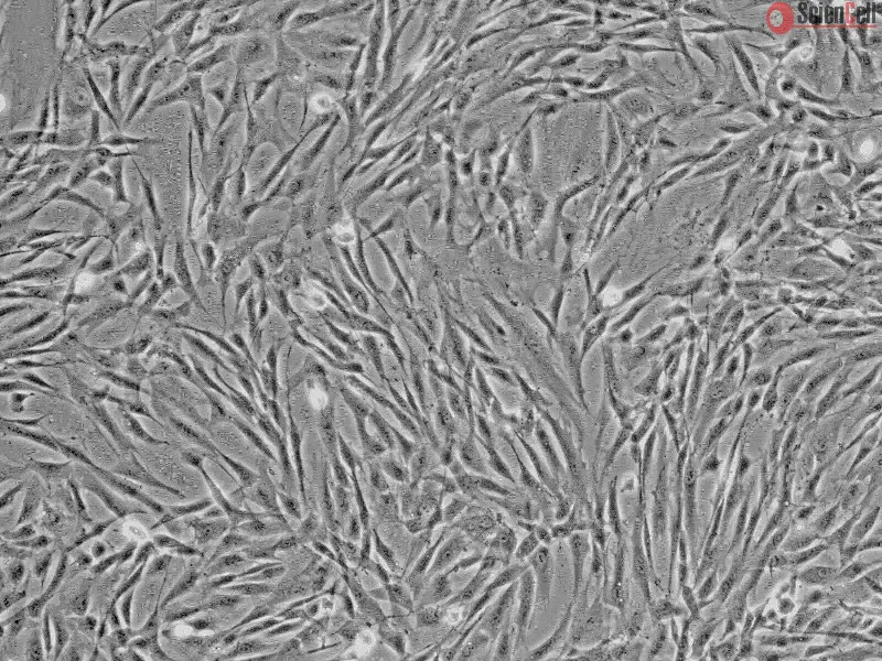
HOF from ScienCell Research Laboratories are isolated from human ovary. HOF are cryopreserved at passage one and delivered frozen. Each vial contains >5 x 10⁵ cells in 1 ml volume. HOF are characterized by their spindle morphology and immunofluorescence with antibody specific to fibronectin. HOF are negative for HIV-1, HBV, HCV, mycoplasma, bacteria, yeast, and fungi. HOF are guaranteed to further expand for 15 population doublings under the conditions provided by ScienCell Research Laboratories.
Recommended Medium
It is recommended to use Fibroblast Medium (FM, Cat. #2301) for culturing HOF in vitro.
成纤维细胞是一类来源于胚胎中胚层的间充质细胞。它们可合成细胞外基质成分,维持结缔组织的结构完整性,并在伤口愈合过程中发挥重要作用。越来越多的研究表明,卵巢成纤维细胞可受到卵巢癌细胞的诱导和激活,分泌多种生长因子、趋化因子、胶原蛋白以及基质调控酶,从而建立有利于肿瘤发生、免疫抑制的微环境,促进癌症进展和转移。这些研究结果表明,成纤维细胞在癌症发生发展及侵袭过程中具有重要的促进作用。
ScienCell Research Laboratories 提供的人卵巢成纤维细胞(HOF)分离自人卵巢组织。HOF 在第一代(P1)阶段进行冻存,并以冷冻形式运输。每瓶含有超过 5 × 10⁵ 个细胞,总体积为 1 ml。HOF 通过其典型的梭形细胞形态以及使用纤维连接蛋白(fibronectin)特异性抗体进行免疫荧光染色鉴定。HOF 经检测不含 HIV-1、HBV、HCV、支原体、细菌、酵母菌及真菌。在 ScienCell Research Laboratories 提供的培养条件下,HOF 可保证进一步扩增至少 15 次群体倍增。
推荐培养基
建议使用成纤维细胞培养基(Fibroblast Medium,FM,产品编号 #2301)对 HOF 进行体外培养。
Product Sheets
 7330
7330
Search for a Certificate of Analysis by Lot Number below:
Pancreatic ductal adenocarcinoma (PDAC) is one of the most lethal cancers worldwide, with a five year survival rate below 10%. The high mortality rate of PDAC is primaril... More
Pancreatic ductal adenocarcinoma (PDAC) is one of the most lethal cancers worldwide, with a five year survival rate below 10%. The high mortality rate of PDAC is primarily due to its aggressive metastatic nature, late diagnosis, and resistance to available therapies. A strategic research direction that demands significantly more attention is the identification of novel tumor markers and the development of tools for rapid, noninvasive diagnosis of this type of cancer. Circulating tumor cells (CTCs) are a promising target for early cancer detection via liquid biopsy, but their clinical application in PDAC is limited due to their rarity and technical challenges in reliable isolation and analysis. CTCs are typically isolated based on physical properties (size, density, deformability, charge, shape, hydrodynamics) or antigenic markers using specific antibodies. Although numerous cancer-specific antibodies have been identified, a marker uniquely distinguishing PDAC has yet to be found. In this work, we present the latest results concerning the development of a microfluidic platform that enables effective immunomagnetic isolation of PDAC cells from a complex biological sample using two potential PDAC markers: Plectin-1 and EpCAM. Here, we developed a low-cost, easy-to-manufacture, and user-friendly 3D-printed microfluidic platform that integrates a micromixer and a separation chamber for rapid cell isolation. Optimization tests showed that Plectin-1 at a concentration of 0.485 μg/mL captured 102 cells/ml with a recovery of 80% ± 13.3%, EpCAM at a concentration of 0.625 μg/mL captured 103 cells/ml with a recovery of 47% ± 13.3%, and the mixture of EpCAM and Plectin-1 at concentrations of 0.3125 μg/mL and 0.242 μg/mL, respectively, achieved a recovery of 91.3% ± 4.4%. To demonstrate the applicability of the method in biological samples, blood with different anticoagulants was tested. The highest detection sensitivity was achieved in the blood sample with citrate: Plectin-1 at 0.485 μg/mL enabled detection at a concentration of 102 cells/ml (47 ± 9% recovery), and EpCAM at 1.25 μg/mL achieved a 92 ± 8% recovery. The entire isolation process from biological samples was performed in less than 1 min. Using this system combined with antibody-functionalized magnetic nanoparticles targeting EpCAM and Plectin-1, we demonstrated the efficient capture of PDAC cells from biological samples, highlighting Plectin-1 as a promising PDAC marker and the potential of immunomagnetic microfluidic separation for improved diagnostics. Less
The invasive and metastatic spread of serous ovarian cancer (SOC) results from the cooperative interactions between cancer and stroma, which include extracellular matrix ... More
The invasive and metastatic spread of serous ovarian cancer (SOC) results from the cooperative interactions between cancer and stroma, which include extracellular matrix (ECM) and cellular components, including cancer-associated fibroblasts (CAFs). Soluble factors secreted by cancer and stromal cells contribute to stroma remodeling through the secretion of ECM proteins, providing a favorable environment for cancer cell dissemination. The peptide endothelin-1 (ET-1), through two G protein-coupled receptors (GPCR), endothelin receptor type A (ETAR) and B (ETBR), acts on both cancer and stromal cells, engaging the protein β-arrestin1 (β-arr1), to bolster SOC progression. However, its role in the regulation of the ECM proteins by ovarian fibroblasts is not understood. This study delves into the role of ET-1 as a regulator of type I collagen (Col1) and fibronectin (FN). Less
Three-dimensional (3D) cellular models of cancer tissue are necessary tools to analyze new anticancer drugs under in vitro conditions. Diagnostics and treatment of ovaria... More
Three-dimensional (3D) cellular models of cancer tissue are necessary tools to analyze new anticancer drugs under in vitro conditions. Diagnostics and treatment of ovarian cancer are major challenges for current medicine. In our report we propose a new three-dimensional (3D) cellular model of ovarian cancer which can mimic a fragment of heterogeneous cancer tissue. We used Lab-on-a-chip technology to create a microfluidic system that allows cellular multilayer to be cultured. Cellular multilayer mimics the structure of two important elements of cancer tissue: flesh and stroma. For this reason, it has an advantage over other in vitro cellular models. We used human ovarian fibroblasts (HOF) and human ovarian cancer cells in our research (A2780). In the first stage of the study, we proved that the presence of non-malignant fibroblasts in co-culture with ovarian cancer cells stimulates the proliferation of cancer cells, which is important in the progression of ovarian cancer. In the next stage of the research, we tested the usefulness of the newly-developed cellular model in the analysis of anticancer drugs and therapies under in vitro conditions. We tested two photosensitizers (PS): free and nanoencapsulated meso-tetrafenylporphyrin, and we evaluated the potential of these drugs in anticancer photodynamic therapy (PDT) of ovarian cancer. We also studied the mechanism of PDT based on the analysis of the level of reactive oxygen species (ROS) in cell cultures. Our research confirmed that the use of new-generation PS can significantly increase the efficacy of PDT in the treatment of ovarian cancer. We also proved that the newly-developed 3D cellular model is suitable for rapid screening of anticancer drugs and has the potential to be used clinically in the future, e.g. in the selection of treatment methods for anticancer personalized medicine. Less
ScienCell Research Laboratories (SRL) takes pride in being a resource for researchers all over the world. The publications listed here are not meant as an endorsement or confirmation of the reliability of the products.