Human Myometrial Microvascular Endothelial Cells
Isolated from human uterus. HMMEC are cryopreserved at passage one and delivered frozen. Each vial contains >5 x 10⁵ cells in 1 ml volume.
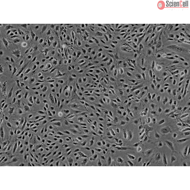
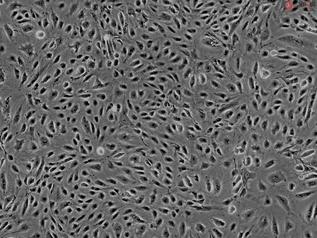
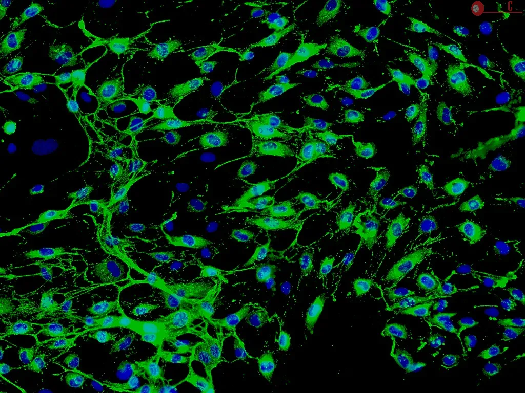
Myometrial microvascular endothelial cells (MMEC) are located in the myometrium between the endometrium and the serosa of the uterus and exhibit typical cobblestone morphology. During pregnancy, angiogenesis occurs through MMEC proliferation in order to support the hypertrophy of the myometrial smooth muscle cell layer. Significant evidence also indicates that endothelial cell dysfunction in the myometrium may contribute to the development of preeclampsia. In addition, MMEC proliferation and angiogenesis may play a role in the formation of leiomyomata. Studies have shown that MMEC express the oxytocin receptor, which may promote vasodilation in the microvasculature of the myometrium. Human MMEC are a useful in vitro model to elucidate the mechanisms of angiogenesis and to develop treatments for female reproductive tract disorders.
HMMEC from ScienCell Research Laboratories are isolated from human uterus. HMMEC are cryopreserved at passage one and delivered frozen. Each vial contains >5 x 10⁵ cells in 1 ml volume. HMMEC are characterized by immunofluorescence with antibodies specific to vWF/Factor VIII and/or CD31 (PECAM1). HMMEC are negative for HIV-1, HBV, HCV, mycoplasma, bacteria, yeast, and fungi. HMMEC are guaranteed to further expand for 10 population doublings under the conditions provided by ScienCell Research Laboratories.
Recommended Medium
It is recommended to use Endothelial Cell Medium (ECM, Cat. #1001) for culturing HMMEC in vitro.
| 目录编号 | 7000 |
|---|---|
| 制造国家 | 美国 |
| 产品编码 | HMMEC |
| 规格/数量 | 5 x 10^5 cells/vial |
| 产品用途 | 本产品仅供研究使用。未经批准,不得用于人体、动物或体外诊断程序。 |
| 储存 | 收到后,立即将细胞从干冰转移到液氮中,并在液氮中保存,直到需要进行实验。 |
| 运输 | 干冰。 |