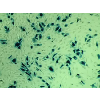
Vascular endothelial cells lining the blood vessels actively participate in many vital biological processes. Studies have shown that mammary vascular endothelial cells (MVEC) secrete a mammaryderived growth inhibitor to modulate endothelial cell proliferation and differentiation of the mammary gland. MVEC also regulate trafficking of lymphocytes and the inflammatory response, and are thus implicated in mastitis. Further studies demonstrate a critical role for MVEC in breast cancer development through angiogenesis. Human MVEC (HMVEC) cultures are a great model for identifying targets for therapeutic development in breast cancer.
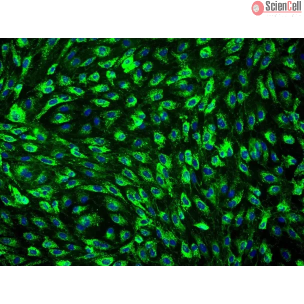
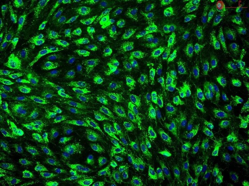
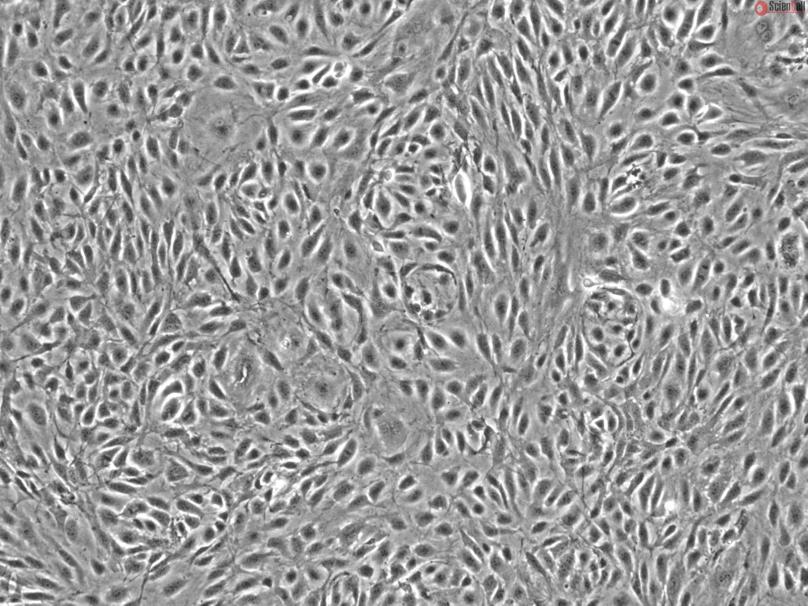
HMVEC from ScienCell Research Laboratories are isolated from human breast. HMVEC are cryopreserved at passage one and delivered frozen. Each vial contains >5 x 10⁵ cells in 1 ml volume. HMVEC are characterized by immunofluorescence with antibodies specific to vWF/Factor VIII and/or CD31 (PECAM1). HMVEC are negative for HIV-1, HBV, HCV, mycoplasma, bacteria, yeast, and fungi. HMVEC are guaranteed to further expand for 10 population doublings under the conditions provided by ScienCell Research Laboratories.
Recommended Medium
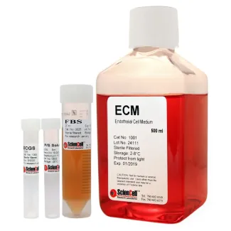
It is recommended to use Endothelial Cell Medium (ECM, Cat. #1001) for culturing HMVEC in vitro.
血管内皮细胞排列于血管内壁,参与多种重要的生物学过程。研究表明,乳腺血管内皮细胞(MVEC)可分泌乳腺源性生长抑制因子,从而调节内皮细胞的增殖以及乳腺组织的分化过程。MVEC 还参与调控淋巴细胞迁移和炎症反应,因此与乳腺炎的发生密切相关。进一步研究显示,MVEC 可通过促进血管生成,在乳腺癌的发生发展过程中发挥关键作用。人乳腺血管内皮细胞(HMVEC)培养体系可作为研究模型,用于发现乳腺癌治疗开发中的潜在靶点。
ScienCell Research Laboratories 提供的人乳腺血管内皮细胞(HMVEC)分离自人乳腺组织。HMVEC 在第一代(P1)阶段进行冻存,并以冷冻形式运输。每瓶含有超过 5 × 10⁵ 个细胞,总体积为 1 ml。HMVEC 通过使用针对 vWF/Ⅷ因子(Factor VIII)和/或 CD31(PECAM1)的特异性抗体进行免疫荧光染色鉴定。HMVEC 经检测不含 HIV-1、HBV、HCV、支原体、细菌、酵母菌及真菌。在 ScienCell Research Laboratories 提供的培养条件下,HMVEC 可保证进一步扩增至少 10 次群体倍增。
推荐培养基
建议使用内皮细胞培养基(Endothelial Cell Medium,ECM,产品编号 #1001)对 HMVEC 进行体外培养。
Product Sheets
 7600
7600
Search for a Certificate of Analysis by Lot Number below:
Kaposi's sarcoma (KS), a highly angiogenic and invasive tumor often involving different organ sites, including the oral cavity, is caused by infection with Kaposi's sarco... More
Kaposi's sarcoma (KS), a highly angiogenic and invasive tumor often involving different organ sites, including the oral cavity, is caused by infection with Kaposi's sarcoma-associated herpesvirus (KSHV). Diverse cell markers have been identified on KS tumor cells, but their origin remains an enigma. We previously showed that KSHV could efficiently infect, transform, and reprogram rat primary mesenchymal stem cells (MSCs) into KS-like tumor cells. In this study, we showed that human primary MSCs derived from diverse organs, including bone marrow (MSCbm), adipose tissue (MSCa), dental pulp, gingiva tissue (GMSC), and exfoliated deciduous teeth, were permissive to KSHV infection. We successfully established long-term cultures of KSHV-infected MSCa, MSCbm, and GMSC (LTC-KMSCs). While LTC-KMSCs had lower proliferation rates than the uninfected cells, they expressed mixtures of KS markers and displayed differential angiogenic, invasive, and transforming phenotypes. Genetic analysis identified KSHV-derived microRNAs that mediated KSHV-induced angiogenic activity by activating the AKT pathway. These results indicated that human MSCs could be the KSHV target cells in vivo and established valid models for delineating the mechanism of KSHV infection, replication, and malignant transformation in biologically relevant cell types. Less
Most synthetic polymeric materials currently used for bone tissue engineering lack specific signals through which cells can identify and interact with the surface, result... More
Most synthetic polymeric materials currently used for bone tissue engineering lack specific signals through which cells can identify and interact with the surface, resulting in incompatibility and compromised osteogenic activity. Soluble inductive factors also have issues including a short half-live in vivo. Bone forming peptide-1 is a truncated peptide from the immature form of bone morphogenetic protein-7 (BMP-7) that displays higher osteogenic activity than full-length, mature BMP-7. In this study, we used a mussel-inspired immobilization strategy mediated by polymerization of dopamine to introduce recently discovered stimulators of bone forming peptide-1 (BFP-1) onto the surface of poly-lactic-co-glycolic acid (PLGA) substrate to form a biomaterial that overcomes these challenges. Human adipose-derived stem cells (hASCs), being abundant and easy accessible, were used to test the osteogenic activity of BFP-1 and the novel biomaterial. Under osteoinductive conditions, cells treated with both BFP-1 alone and BFP-1-coated biomaterials displayed elevated expression of the osteogenic markers alkaline phosphatase (ALP), osteocalcin (OC), and RUNX2. Furthermore, hASCs associated with poly-dopamine-assisted BFP-1-immobilized PLGA (pDA-BFP-1-PLGA) scaffolds promoted in vivo bone formation in nude mice. Our novel materials may hold great promise for future bone tissue engineering applications. Less
ScienCell Research Laboratories (SRL) takes pride in being a resource for researchers all over the world. The publications listed here are not meant as an endorsement or confirmation of the reliability of the products.