成纤维细胞是来源于胚胎中胚层的间充质细胞。由于其在培养中较易生长,成纤维细胞被广泛用于各种细胞和分子研究。其较强的耐受性也使其适用于多种实验操作,例如基因转染或显微注射等。一般而言,成纤维细胞分泌一种富含 I 型和/或 III 型胶原蛋白的非刚性细胞外基质。有研究表明,不同器官的成纤维细胞在本质上存在差异。在伤口愈合过程中,真皮成纤维细胞会从增殖和迁移阶段转换为收缩及基质重塑阶段。此外,它们在炎症刺激下会大量分泌透明质酸。
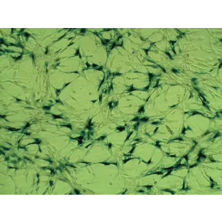
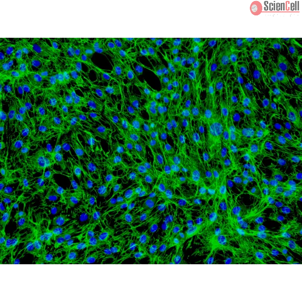
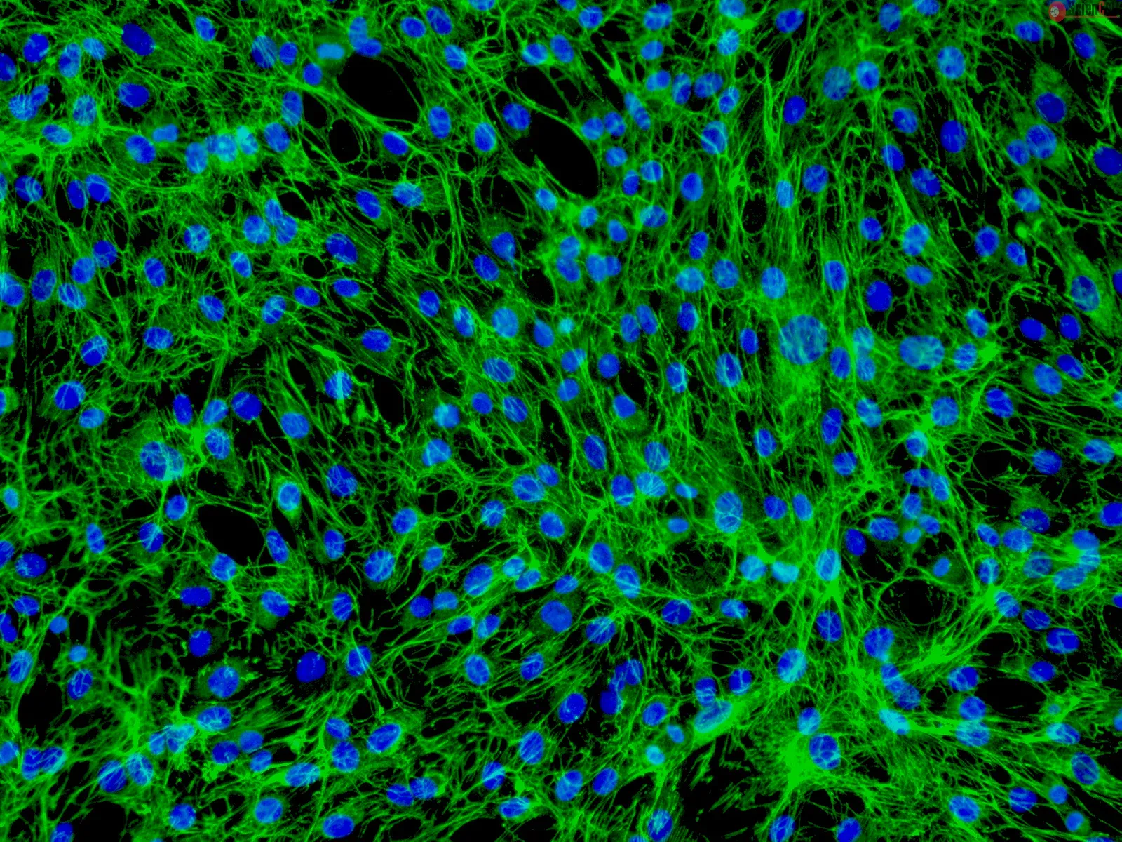
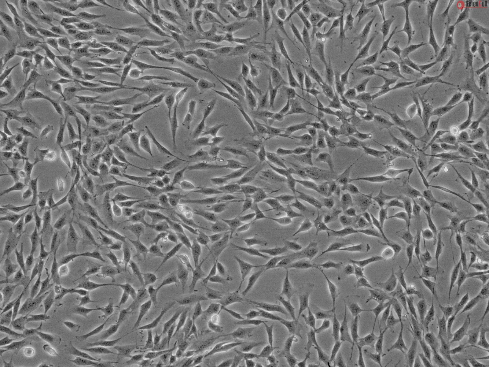
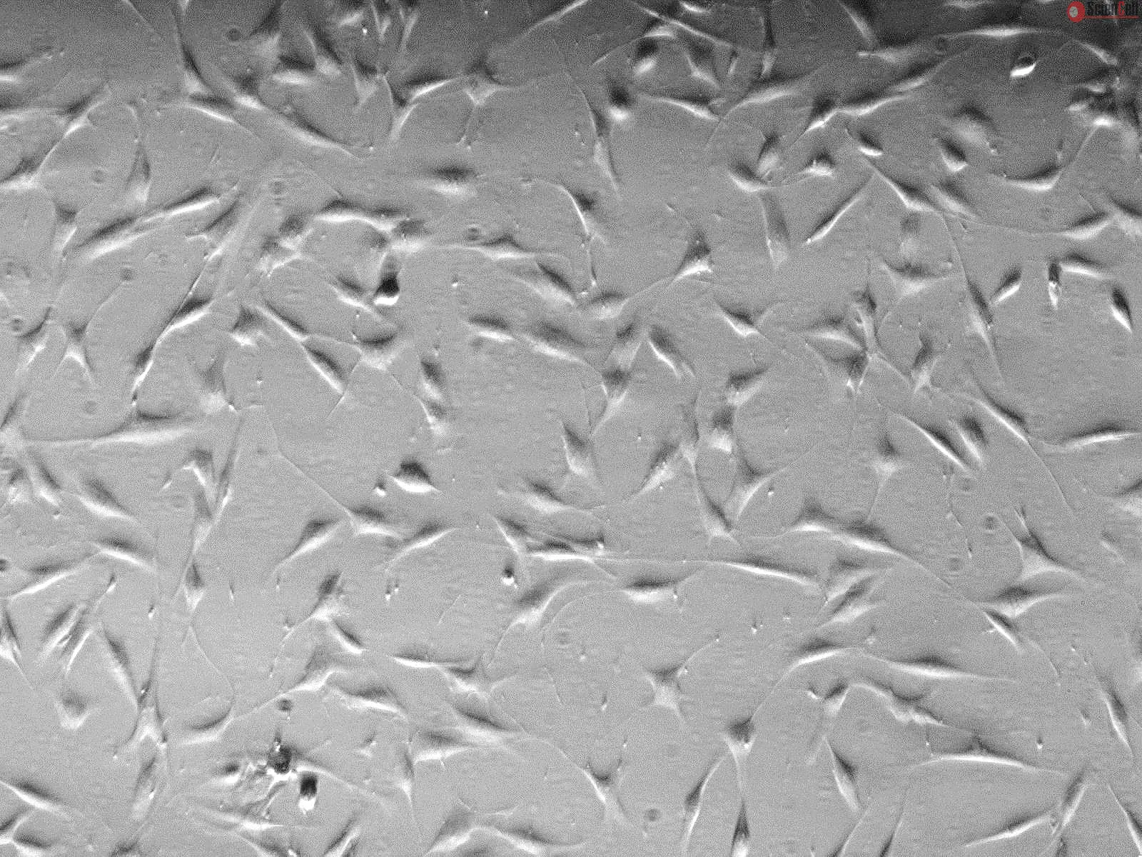
HDF-n(新生儿人真皮成纤维细胞)由 ScienCell Research Laboratories 提供,来源于新生儿人皮肤。在第一代时即冷冻保存并以冷冻形式运输。每瓶包含超过 5 x 10⁵ 个细胞,体积为 1 毫升。HDF-n 的特征是具有梭形形态,并可通过针对纤维连接蛋白(fibronectin)的抗体进行免疫荧光鉴定。HDF-n 对 HIV-1、HBV、HCV、支原体、细菌、酵母菌和真菌检测均为阴性。根据 ScienCell 提供的条件,HDF-n 可保证扩增 15 次群体倍增。
推荐培养基
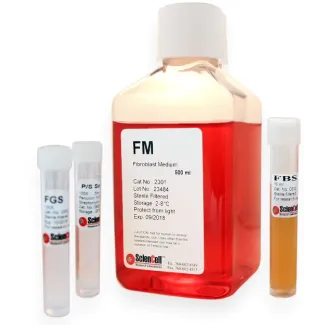
建议使用成纤维细胞培养基(Fibroblast Medium, FM,货号 #2301)进行 HDF-n 的 体外 培养。
Product Sheets
 2310
2310
Search for a Certificate of Analysis by Lot Number below:
Background: Calcium electroporation describes the use of high voltage electric pulses to introduce supraphysiological calcium concentrations into cells. This promising me... More
Background: Calcium electroporation describes the use of high voltage electric pulses to introduce supraphysiological calcium concentrations into cells. This promising method is currently in clinical trial as an anti-cancer treatment. One very important issue is the relation between tumor cell kill efficacy–and normal cell sensitivity. Methods: Using a 3D spheroid cell culture model we have tested the effect of calcium electroporation and electrochemotherapy using bleomycin on three different human cancer cell lines: a colorectal adenocarcinoma (HT29), a bladder transitional cell carcinoma (SW780), and a breast adenocarcinoma (MDA-MB231), as well as on primary normal human dermal fibroblasts (HDF-n). Results: The results showed a clear reduction in spheroid size in all three cancer cell spheroids three days after treatment with respectively calcium electroporation (p<0.0001) or electrochemotherapy using bleomycin (p<0.0001). Strikingly, the size of normal fibroblast spheroids was neither affected after calcium electroporation nor electrochemotherapy using bleomycin, indicating that calcium electroporation, like electrochemotherapy, will have limited adverse effects on the surrounding normal tissue when treating with calcium electroporation. The intracellular ATP level, which has previously been shown to be depleted after calcium electroporation, was measured in the spheroids after treatment. The results showed a dramatic decrease in the intracellular ATP level (p<0.01) in all four spheroid types—malignant as well as normal. Less
Cultured human skin fibroblasts were irradiated twice successively with the 1.5 J/cm(2) of 532-nm and 1,064-nm lasers, respectively. The mRNA of procollagen, matrix metal... More
Cultured human skin fibroblasts were irradiated twice successively with the 1.5 J/cm(2) of 532-nm and 1,064-nm lasers, respectively. The mRNA of procollagen, matrix metalloproteinases (MMPs), tissue inhibitors of metalloproteinases (TIMPs), heat-shock protein 70 (Hsp70), interleukin-6 (IL-6) and transforming growth factor beta (TGF-beta) were analyzed at 24 and 48 h post-irradiation by using RT-PCR. Both lasers significantly increased the expression of type I and III procollagen, TIMP1, and TIMP2, but decreased MMP1 and MMP2 expression. The 1,064-nm laser initiated TGF-beta expression while the 532-nm laser elicited the increase of Hsp70 and IL-6. The increase/decrease rates of procollagen, TIMPs and MMPs for the 1,064-nm laser were higher than that of the 532-nm laser. Thus, both lasers effectively accelerated collagen synthesis and inhibited collagen degradation. Collagen synthesis induced by the 1,064-nm laser might be partly due to the upregulation of TGF-beta expression, while the increase of Hsp70 and IL-6 might be partly responsible for collagen synthesis stimulated by the 532-nm laser. With the parameters used in this study, the 1,064-nm infrared laser is more effective in promoting the beneficial molecular activities than the 532-nm visible laser. Less
ScienCell Research Laboratories (SRL) takes pride in being a resource for researchers all over the world. The publications listed here are not meant as an endorsement or confirmation of the reliability of the products.

-1.jpg)