Conjunctival fibroblasts, like other fibroblasts, are responsible for fibrosis, matrix production, and tissue remodeling. They are also actively involved as an effector in ocular allergies. In chronic allergic conjunctivitis, conjunctival fibroblasts responded to histamine to increase proliferation and induce the production of proinflammatory cytokines, such as IL-1, IL-6, and IL-8, to perpetuate allergic inflammation. Conjunctival fibroblast cultures can be used as an in vitro model for rapid screening of drugs and may provide a basis for the development of new treatments for ocular proliferative disorders.
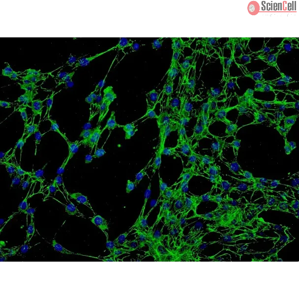
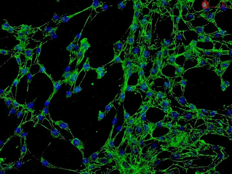
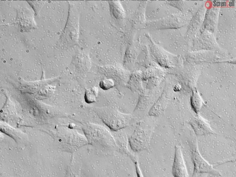
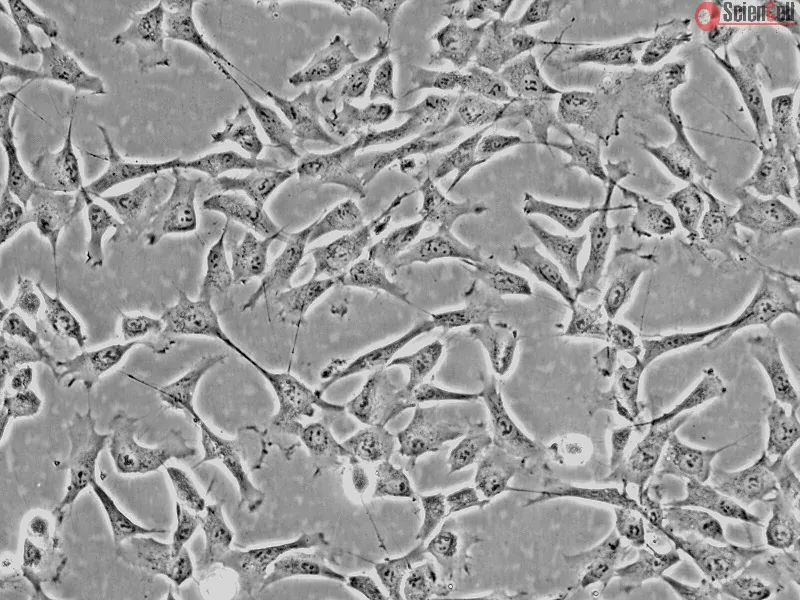
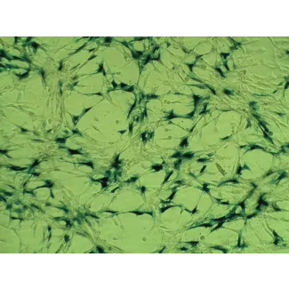
HConF from ScienCell Research Laboratories are isolated from human conjunctiva. HConF are cryopreserved at passage one and delivered frozen. Each vial contains >5 x 10⁵ cells in 1 ml volume. HConF are characterized by their spindle morphology and immunofluorescence with antibody specific to fibronectin. HConF are negative for HIV-1, HBV, HCV, mycoplasma, bacteria, yeast, and fungi. HConF are guaranteed to further expand for 15 population doublings under the conditions provided by ScienCell Research Laboratories.
Recommended Medium
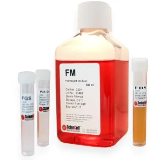
It is recommended to use Fibroblast Medium (FM, Cat. #2301) for culturing HConF in vitro.
结膜成纤维细胞与其他成纤维细胞一样,参与纤维化、细胞外基质生成和组织重塑等过程,同时也是眼部过敏反应的重要效应细胞。在慢性过敏性结膜炎中,结膜成纤维细胞可对组胺产生反应,促进细胞增殖,并诱导白细胞介素-1(IL-1)、白细胞介素-6(IL-6)和白细胞介素-8(IL-8)等促炎性细胞因子的产生,从而持续加剧过敏性炎症。结膜成纤维细胞培养物可作为一种体外模型,用于快速筛选药物,并为开发治疗眼部增殖性疾病的新方法提供基础。
ScienCell Research Laboratories 的 HConF 分离自人结膜,于第一代(P1)进行冻存,并以冷冻状态提供。每瓶含有 >5 × 10⁵ 个细胞,体积为 1 mL。HConF 通过其梭形细胞形态以及采用针对纤连蛋白(fibronectin)的特异性抗体进行免疫荧光染色来表征。HConF 经检测为 HIV-1、HBV、HCV、支原体、细菌、酵母菌和真菌阴性。在 ScienCell Research Laboratories 提供的培养条件下,HConF 可进一步扩增至少 15 个群体倍增。
推荐培养基
建议使用成纤维细胞培养基(FM,产品编号 2301)进行 HConF 的体外培养。
Product Sheets
 6570
6570
Search for a Certificate of Analysis by Lot Number below:
Purpose: Vernal keratoconjunctivitis (VKC) is a severe, recurrent allergic conjunctivitis. Previously, we found high concentrations of oncostatin M (OSM) in the tears of ... More
Purpose: Vernal keratoconjunctivitis (VKC) is a severe, recurrent allergic conjunctivitis. Previously, we found high concentrations of oncostatin M (OSM) in the tears of patients with VKC. Here, we investigated the role of OSM in VKC by focusing on epithelial barrier function and IL-33 production. Less
Purpose: To determine the ocular anti-allergic effects of mapracorat, a novel selective glucocorticoid receptor agonist (SEGRA) in primary human conjunctival fibroblasts ... More
Purpose: To determine the ocular anti-allergic effects of mapracorat, a novel selective glucocorticoid receptor agonist (SEGRA) in primary human conjunctival fibroblasts and epithelial cells. Less
Purpose: BOL-303242-X is a novel selective glucocorticoid receptor agonist under clinical evaluation for the treatment of inflammatory skin and eye diseases. Data from in... More
Purpose: BOL-303242-X is a novel selective glucocorticoid receptor agonist under clinical evaluation for the treatment of inflammatory skin and eye diseases. Data from in vitro and in vivo studies suggest an improved side-effect profile of this compound compared to classical glucocorticoids. The aim of this study was to determine the anti-inflammatory effect of BOL-303242-X in ocular cells. Methods: Four primary human ocular cell cultures, including human conjunctival fibroblasts (HConFs), human corneal epithelial cells (HCEpiCs), human optic nerve astrocytes (HONAs), and human retinal endothelial cells (HRECs), as well as a human monocytic cell line, THP-1, were challenged with either lipopolysacharide (LPS) or interleukin-1ß (IL-1ß). Luminex technology was used to determine the effect of BOL-303242-X on LPS- or IL-1ß-induced cytokine release and intercellular adhesion molecule-1 (ICAM-1) levels. Effects of BOL-303242-X on nuclear factor kappa B (NFκB) and mitogen-activated protein kinase (MAPK) in HCEpiCs were also assessed by measuring inhibitory kappa B protein-α (IκB-α), phosphorylated p65 NFκB, and MAPK levels by western blotting. Dexamethasone (DEX) or triamcinolone acetonide (TA) was used as the control. Results: LPS or IL-1ß induced multiple cytokine release in all cell types studied. BOL-303242-X significantly reduced LPS- or IL-1ß-induced inflammatory cytokine release in a dose-dependent manner, including granulocyte colonystimulating factor (G-CSF), IL-1ß, IL-6, IL-8, IL-12p40, monocyte chemotactic protein-1 (MCP-1), and tumor necrosis factor-α (TNF-α). BOL-303242-X showed activity and potency comparable to that observed for DEX or TA. A statistically significant inhibitory effect of BOL-303242-X was observed at doses ranging from 1 to 100 nM in HConFs, HCEpiCs, HONAs, and THP-1. The IC50 values for these effects were in the low nM range. BOL-303242-X also significantly reduced LPS-induced IL-1ß release and ICAM-1 levels in HRECs. Furthermore, BOL-303242-X inhibited IL-1ß-induced decreases in IκB-α levels, as well as IL-1ß-induced phosphorylation of NFκB, p38, and c-Jun-N-terminal kinase (JNK) MAPKs in HCEpiCs. Conclusions: BOL-303242-X acts as a potent anti-inflammatory agent in various primary human ocular cells with similar activity and potency compared to classical steroids. Results also suggest that MAPK (p38 and JNK) and NFκB signaling pathways are involved in the anti-inflammatory properties of BOL-303242-X in HCEpiCs. An improved side effect profile of this novel SEGRA compound has been reported recently. Thus, BOL-303242-X may provide a new option for the treatment of ophthalmic conditions with an inflammatory component. Less
Sphingosine-1-phosphate (S1P) is a pleiotropic lysolipid that has recently been implicated in the regulation of tissue fibrosis. However, the fibrogenic potential of S1P ... More
Sphingosine-1-phosphate (S1P) is a pleiotropic lysolipid that has recently been implicated in the regulation of tissue fibrosis. However, the fibrogenic potential of S1P in the eye has not previously been investigated. In the current study, we evaluated cells from the anterior and posterior segments of the eye for the presence of S1P and their potential ability to produce and respond to S1P. In addition, we investigated the regulatory role of S1P as a mediator of proliferation, cellular transformation and pro-fibrotic protein expression in human retinal pigmented epithelial cells. Expression of S1P receptors and sphingosine kinases (the enzymes that produce S1P) was examined using RT-PCR, and intracellular localization of S1P was examined using immunoblotting, immunohistochemistry and ELISA in primary human retinal pigmented epithelial (RPE) cells, primary human conjunctival fibroblasts (ConF), and primary human corneal fibroblasts (CF). RPE cell proliferation was determined using an MTT-based cell proliferation assay, and RPE myofibroblast transformation, collagen type I production and profibrotic protein expression were assessed using immunofluorescence, ELISA and immunoblot. S1P(1-3, 5) receptors and sphingosine kinases 1 and 2 were expressed and intracellular pools of S1P were detected in RPE cells, ConF and CF. S1P stimulated RPE cell proliferation in a dose- and time-dependent manner. S1P induced myofibroblast transformation of RPE cells, as indicated by increased alpha-smooth muscle actin (alpha-SMA) expression and its incorporation into prominent stress fibers, and promoted collagen type I production. S1P stimulated the expression of plasminogen activator inhibitor-1 (PAI-1) and heat shock protein 47 (HSP47), two proteins that are linked to increased tissue fibrosis. Combined, these data demonstrate that RPE cells, ConF and CF from the human eye not only have the molecular ability to produce and respond to S1P, but also contain S1P. Furthermore, S1P promotes proliferation, myofibroblast transformation, collagen production and pro-fibrotic protein expression by human RPE cells. These data suggest that S1P is a previously unrecognized mediator of profibrotic cellular function and signaling in the eye. Less
ScienCell Research Laboratories (SRL) takes pride in being a resource for researchers all over the world. The publications listed here are not meant as an endorsement or confirmation of the reliability of the products.