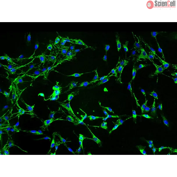
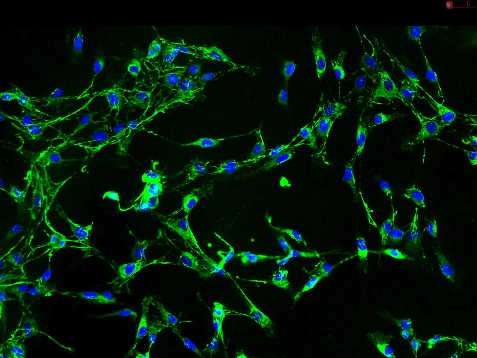
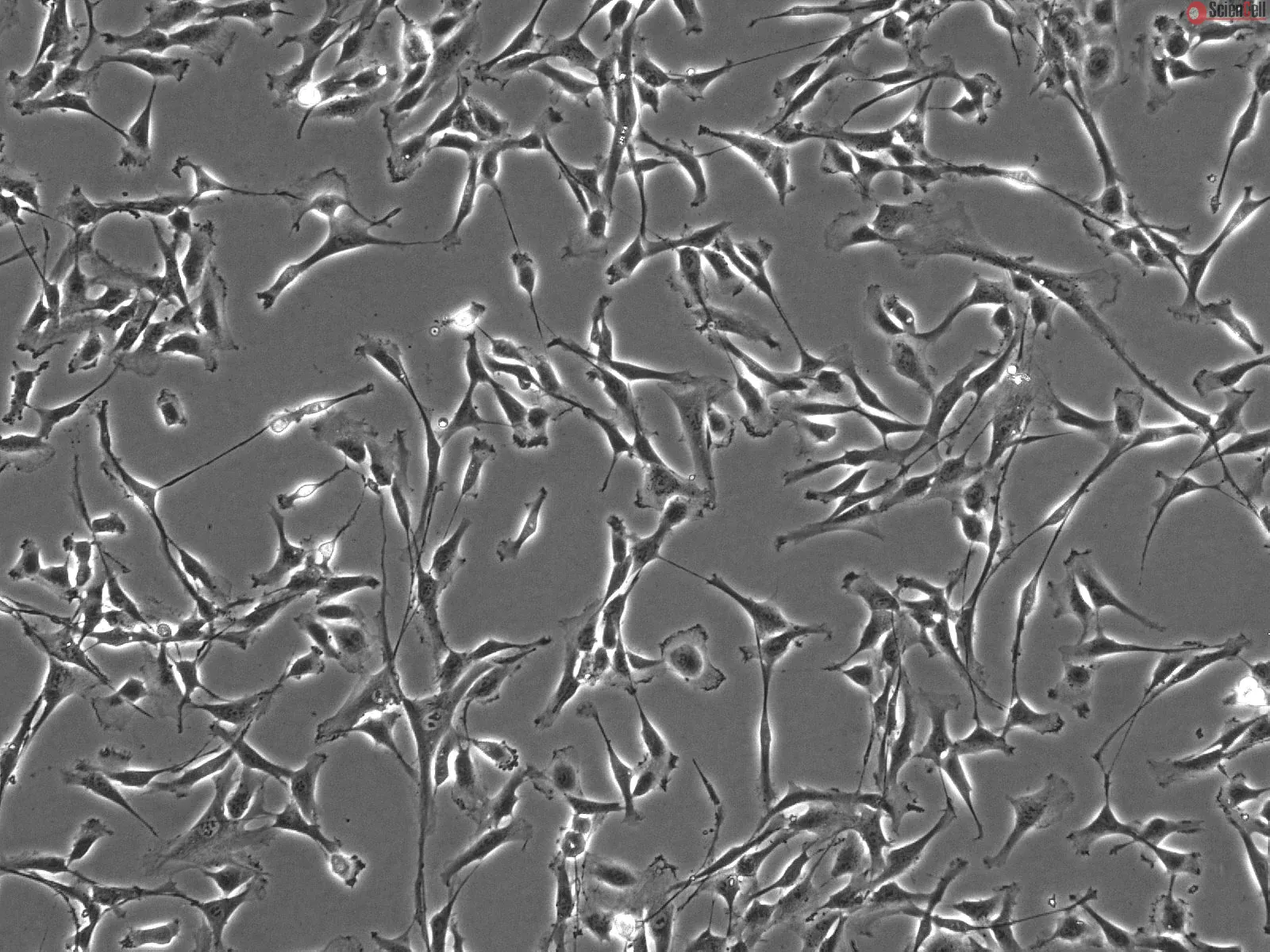
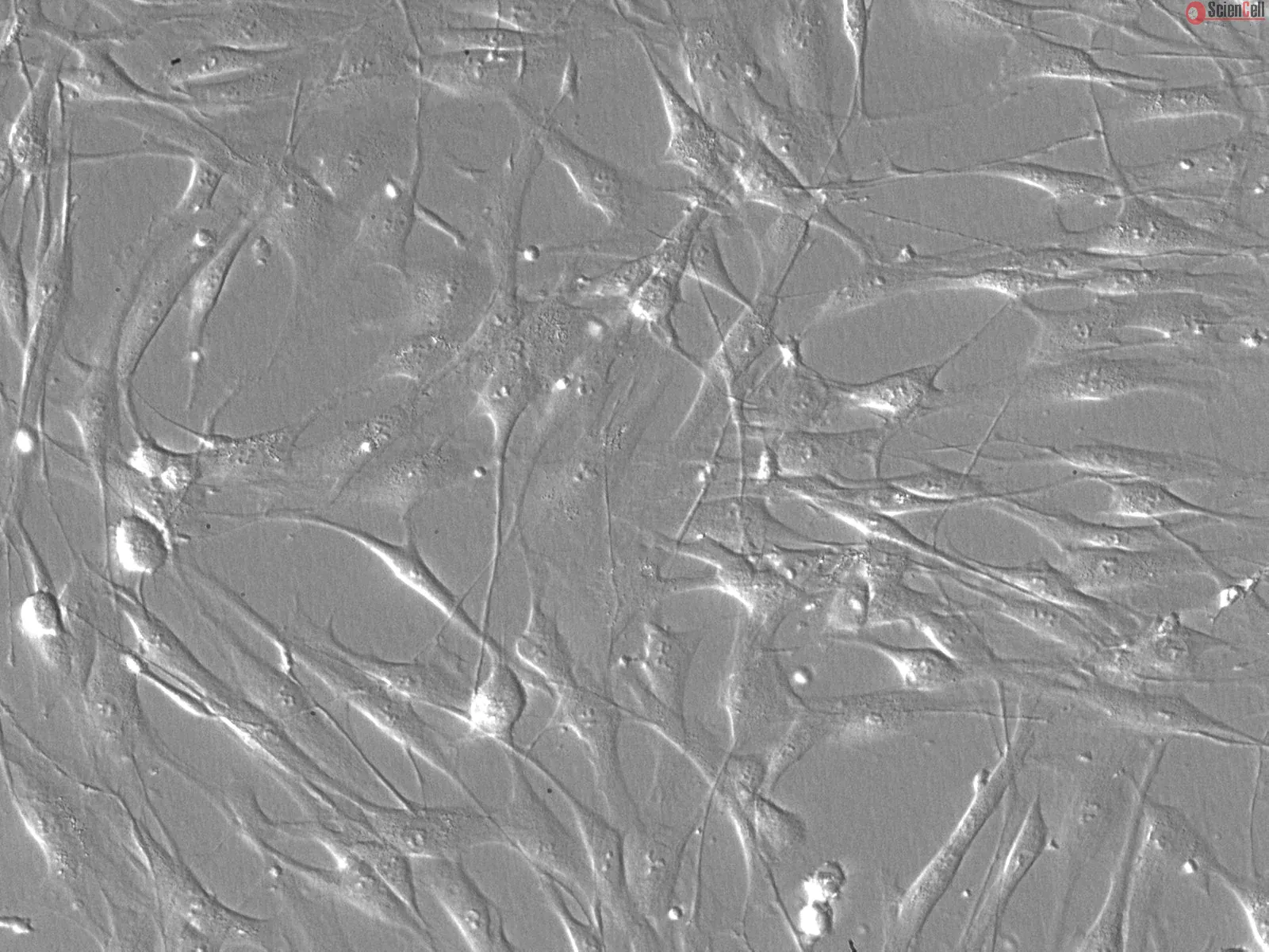
在血管外膜中,作为覆盖血管最外层的结缔组织,外膜成纤维细胞(AF)通过产生胶原蛋白为血管提供结构支撑,并将血管固定于邻近组织。AF 是血管壁中最早对高血压和血管损伤作出反应的细胞,通过激活和增殖参与应答。在病理条件下,AF 会产生细胞因子和趋化因子,诱导免疫细胞大量浸润至血管壁的外膜层。免疫细胞浸润外膜可导致外膜炎症,并可能引发心血管疾病。AF 的这些重要特性使其培养物成为研究心血管疾病发病机制及开发新型治疗方法的理想工具。
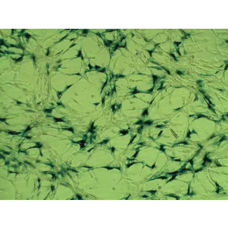
ScienCell Research Laboratories 提供的 HAAF 分离自人主动脉。HAAF 在第一代传代时冷冻保存,并以冷冻状态提供。每瓶含有 >5 x 10⁵ 个细胞,体积为 1 ml。HAAF 具有纺锤形形态,并通过针对纤维连接蛋白的特异性抗体进行免疫荧光鉴定。HAAF 对 HIV-1、HBV、HCV、支原体、细菌、酵母和真菌检测均为阴性。在 ScienCell Research Laboratories 提供的培养条件下,保证 HAAF 可进一步扩增至 15 次群体倍增。
推荐培养基
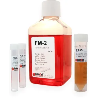
建议在体外培养 HAAF 时使用 Fibroblast Medium-2(FM-2,货号:2331)。
Product Sheets
 6120
6120
Search for a Certificate of Analysis by Lot Number below:
Introduction Heightened inflammatory and thrombotic processes are common hallmarks of vascular diseases. The interaction between these two processes remains unclear and a... More
Introduction Heightened inflammatory and thrombotic processes are common hallmarks of vascular diseases. The interaction between these two processes remains unclear and a better understanding of these links can allow for the design of more effective treatment options. Activation of complement component 1 (C1) leads to the initiation of the classical arm of the complement cascade, availability of plasma C1q, and the potential association of C1q and receptors for C1q. The association of C1q and gC1qR, the receptor for the globular head of C1q, is notable and has been associated with a wide range of disturbed physiological processes. We have recently shown that when this interaction occurs on vascular wall cells, including adventitial fibroblasts and vascular smooth muscle cells, there is a significant up-regulation of tissue factor (TF) expression. However, whether or not this TF is biologically active and can facilitate extrinsic coagulation activation remains unknown. We hypothesized that TF expressed via gC1qR-C1q association would support the progression of extrinsic coagulation. Methods We quantified the association of Factor VII/VIIa (FVII/FVIIa) with adventitial fibroblast and vascular smooth muscle cell TF, using colorimetric assays. Further, we observed the formation of Factor Xa and Factor IIa (thrombin), as well as the concentration of intracellular Akt (protein kinase B) and phosphorylated Akt. Results/Conclusions Our results indicate that TF expression in response to C1q exposure accelerates zymogen formation within the extrinsic coagulation cascade and alters Akt/p-Akt expression. Overall, these findings highlight a significant connection between altered innate inflammation and heightened thrombin generation. Summary Research reported in this publication was supported by the National Institute of Allergy and Infectious Disease of the National Institutes of Health under award number R21AI146535. The authors (BG) receive royalties from the sale of monoclonal antibodies against gC1qR clone 60.11. The authors (BG) hold a patent for the development of these antibodies for therapy against cancer and angioedema, respectively (US patent 8,883,153-B2, “Methods for Prevention and Treatment of Angioedema”). The data that support the findings of this study are available from the corresponding author upon reasonable request. Less
The aim of this study was to investigate and characterize the efficacy and mechanism of action of asiaticoside in combination with rapamycin in the inhibition of in-stent... More
The aim of this study was to investigate and characterize the efficacy and mechanism of action of asiaticoside in combination with rapamycin in the inhibition of in-stent restenosis (ISR). The effects of asiaticoside combined with rapamycin on cell proliferation in vitro were evaluated by MTT assay. The mRNA expression was analyzed by quantitative polymerase chain reaction (qPCR). Enzyme-linked immunosorbent assay (ELISA) was used to confirm protein synthesis. The cell growth inhibition rate in the combination group was significantly higher compared with those in the asiaticoside and rapamycin groups for human aortic fibroblasts (HAFs; 63.50±3.83, 53.06±8.10 and 60.34±4.9%, respectively) and human aortic smooth muscle cells (HASMCs; 33.12±1.35, 26.21±7.59 and 28.27±4.92, respectively; P<0.05). However, for human coronary artery endothelial cells (HCAECs), the cell growth inhibition rates in the combination, asiaticoside and rapamycin groups were 11.09±1.17, 26.22±4.24 and 34.80±2.80%, respectively (P<0.05), as detected by MTT assay. The qPCR assay showed that in the combination group the level of von Willebrand factor (vWF) mRNA was downregulated, while platelet endothelial cell adhesion molecule (PECAM-1) and endothelial nitric oxide synthase (eNOS) mRNAs were upregulated in HCAECs compared with the rapamycin group (P<0.05). Transforming growth factor (TGF)-β1 and TIMP1 mRNAs were downregulated while Smad7 and matrix metalloproteinase 1 (MMP1) mRNAs were upregulated in HAFs compared with the rapamycin and AT groups (P<0.05). The ELISA showed that the type I collagen level was significantly reduced in HASMCs and HAFs (P<0.05). The data suggest that asiaticoside combined with rapamycin may be effective in the reduction of ISR. Keywords: in-stent restenosis, asiaticoside, rapamycin, Smad7 Less
ScienCell Research Laboratories (SRL) takes pride in being a resource for researchers all over the world. The publications listed here are not meant as an endorsement or confirmation of the reliability of the products.