Cardiac myocytes are the most physically energetic cells in the body. They are highly specialized high-oxygen-content cells that house a large number of mitochondria [1]. They occupy as much as 75% of the cardiac mass, but constitute only about one third of the total cell number in the heart. Differentiated cardiac myocytes have little capacity to proliferate; however, hypertrophic growth has been shown to respond to alpha1-adrenergic stimuli via the Ras/MEK pathway [2]. All cardiac myocytes are capable of spontaneous rhythmic depolarization and repolarization of their membranes. Contraction of cardiac myocytes is myogenic, which is independent of nervous stimulation. There is a complex network of signals in cardiac myocytes regulating the rhythmic pumping of the heart [3]. Cardiac myocyte hypertrophy and apoptosis have been implicated in the loss of contractile function during heart failure. A better understanding of the cardiac signaling network will help reveal the cellular mechanisms leading to cardiac myocyte death.
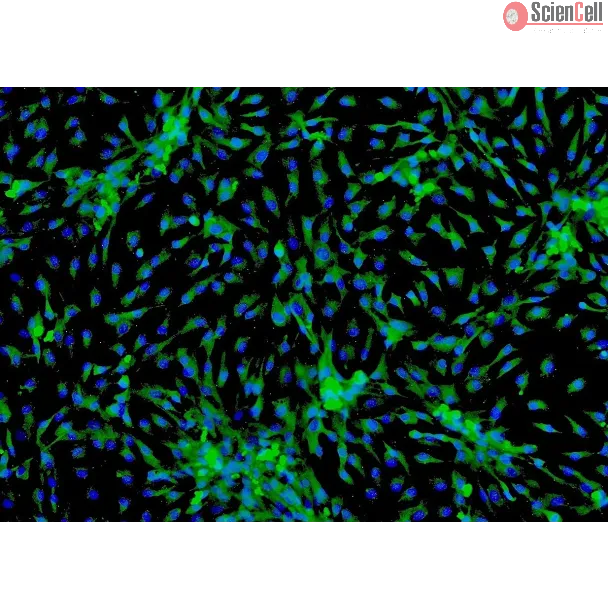
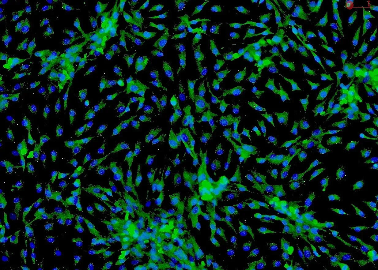
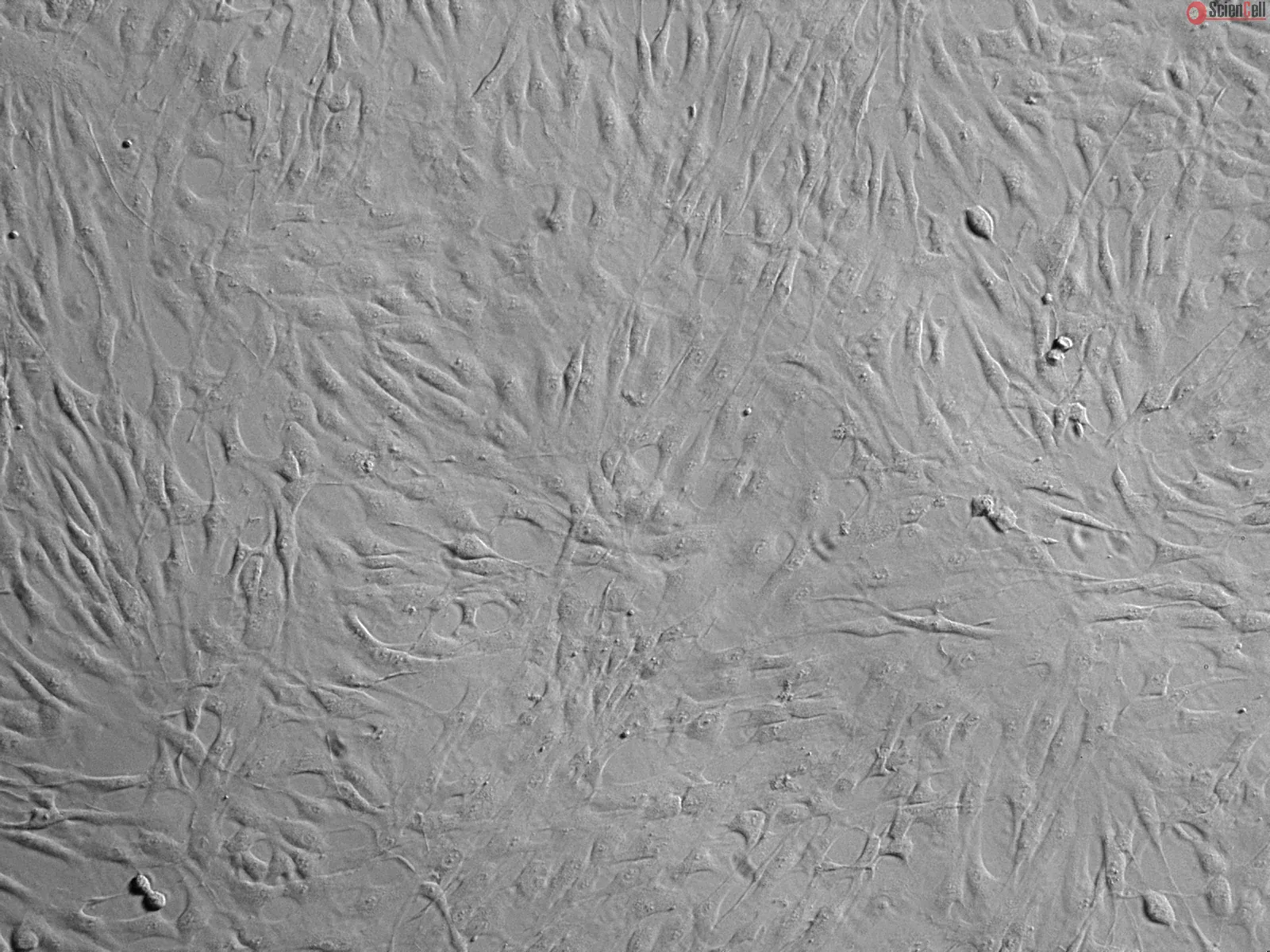
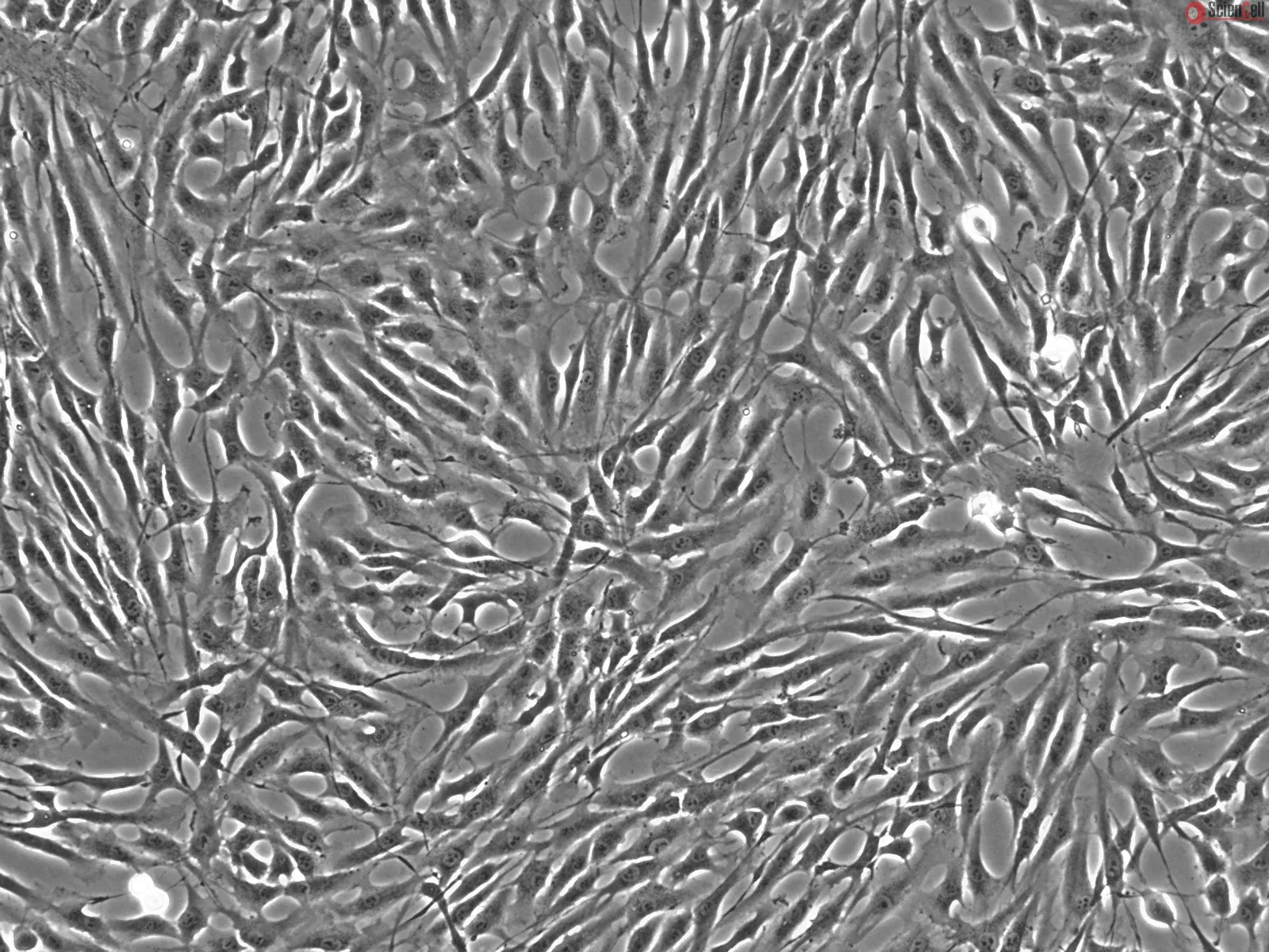
HCM-a from ScienCell Research Laboratories are isolated from human adult heart. HCM-a are cryopreserved after purification and delivered frozen. Each vial contains >1 x 10^6 cells in 1 ml volume. HCM-a are characterized by immunofluorescence with antibody specific to sarcomeric α-actinin. HCM-a are negative for HIV-1, HBV, HCV, mycoplasma, bacteria, yeast, and fungi. HCM-a are guaranteed to further culture under the conditions provided by ScienCell Research Laboratories; however, HCM-a are not recommended for expanding or long-term cultures since the cells do not proliferate in culture.
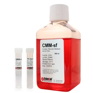
Recommended Medium
It is recommended to use Cardiac Myocyte Medium-serum free (CMM-sf, Cat. #6101) for culturing HCM-a in vitro.
Molecular mechanisms protecting cardiomyocytes from stress-induced death, including tension stress, are essential for cardiac physiology and defects in these protective m... More
Molecular mechanisms protecting cardiomyocytes from stress-induced death, including tension stress, are essential for cardiac physiology and defects in these protective mechanisms can result in pathological alterations. Bcl2-associated athanogene 3(BAG3) is expressed in cardiomyocytes and is a component of the chaperone-assisted autophagy pathway, essential for homeostasis of mechanically altered cells. BAG3 ablation in mice results in a lethal cardiomyopathy soon after birth and mutations of this gene have been associated with different cardiomyopathies including stress-induced Takotsubo cardiomyopathy (TTC). The pathogenic mechanism leading to TTC has not been defined, but it has been suggested that the heart can be damaged by excessive epinephrine (epi) spillover in the absence of a protective mechanism. The aim of this study was to provide more evidence for a role of BAG3 in the pathogenesis of TTC. Therefore, we sequenced BAG3 gene in 70 TTC patients and in 81 healthy donors with the absence of evaluable cardiovascular disease. Mutations and polymorphisms detected in the BAG3 gene included a frequent nucleotide change g2252c in the BAG3 3′-untranslated region (3′-UTR) of Takotsubo patients (Po0.05), resulting in loss of binding of microRNA-371a-5p (miR-371a-5p) as evidenced by dual-luciferase reporter assays and argonaute RNA-induced silencing complex catalytic component 2/pull-down assays. Moreover, we describe a novel signaling pathway in cardiomyocytes that leads to BAG3 upregulation on exposure to epi through an ERK-dependent upregulation of miR-371a-5p. In conclusion, the presence of a g2252c polymorphism in the BAG3 3′-UTR determines loss of miR-371a-5p binding and results in an altered response to epi, potentially representing a new molecular mechanism that contributes to TTC pathogenesis. Cell Death and Disease (2015) 6, e1948; doi:10.1038/cddis.2015.280; published online 29 October 2015 Less
Although it has been observed that aggregate size affects cardiac development, an incomplete understanding of the cellular mechanisms underlying human pluripotent stem ce... More
Although it has been observed that aggregate size affects cardiac development, an incomplete understanding of the cellular mechanisms underlying human pluripotent stem cell-derived cardiomyogenesis has limited the development of robust defined-condition cardiac cell generation protocols. Our objective was thus to elucidate cellular and molecular mechanisms underlying the endogenous control of human embryonic stem cell (hESC) cardiac tissue development, and to test the hypothesis that hESC aggregate size influences extraembryonic endoderm (ExE) commitment and cardiac inductive properties. hESC aggregates were generated with 100, 1000, or 4000 cells per aggregate using microwells. The frequency of endoderm marker (FoxA2 and GATA6)-expressing cells decreased with increasing aggregate size during early differentiation. Cardiogenesis was maximized in aggregates initiated from 1000 cells, with frequencies of 0.49±0.06 cells exhibiting a cardiac progenitor phenotype (KDR(low)/C-KIT(neg)) on day 5 and 0.24±0.06 expressing cardiac Troponin T on day 16. A direct relationship between ExE and cardiac differentiation efficiency was established by forming aggregates with varying ratios of SOX7 (a transcription factor required for ExE development) overexpressing or knockdown hESCs to unmanipulated hESCs. We demonstrate, in a defined, serum-free cardiac induction system, that robust and efficient cardiac differentiation is a function of endogenous ExE cell concentration, a parameter that can be directly modulated by controlling hESC aggregate size. Less
ScienCell Research Laboratories (SRL) takes pride in being a resource for researchers all over the world. The publications listed here are not meant as an endorsement or confirmation of the reliability of the products.


,-1-mg-ml--2.jpg)