Fibroblasts are mesenchymal cells derived from the embryonic mesoderm. They have been extensively used for a wide range of cellular and molecular studies as they are one of easiest types of cells to grow in culture. Their durability also makes them amenable to a variety of manipulations ranging from studies employing gene transfection to microinjection. In general, fibroblasts secrete a non-rigid extracellular matrix which is rich in type I and/or type III collagen. There is evidence showing that fibroblasts in various organs are intrinsically different. Dermal fibroblasts switch from a proliferative, migratory phase to a contractile, matrix-remodeling phase during wound healing. In addition, they secrete large quantities of hyaluronan in response to inflammatory stimuli.
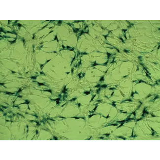
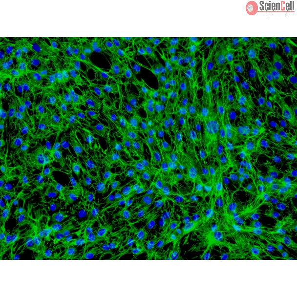
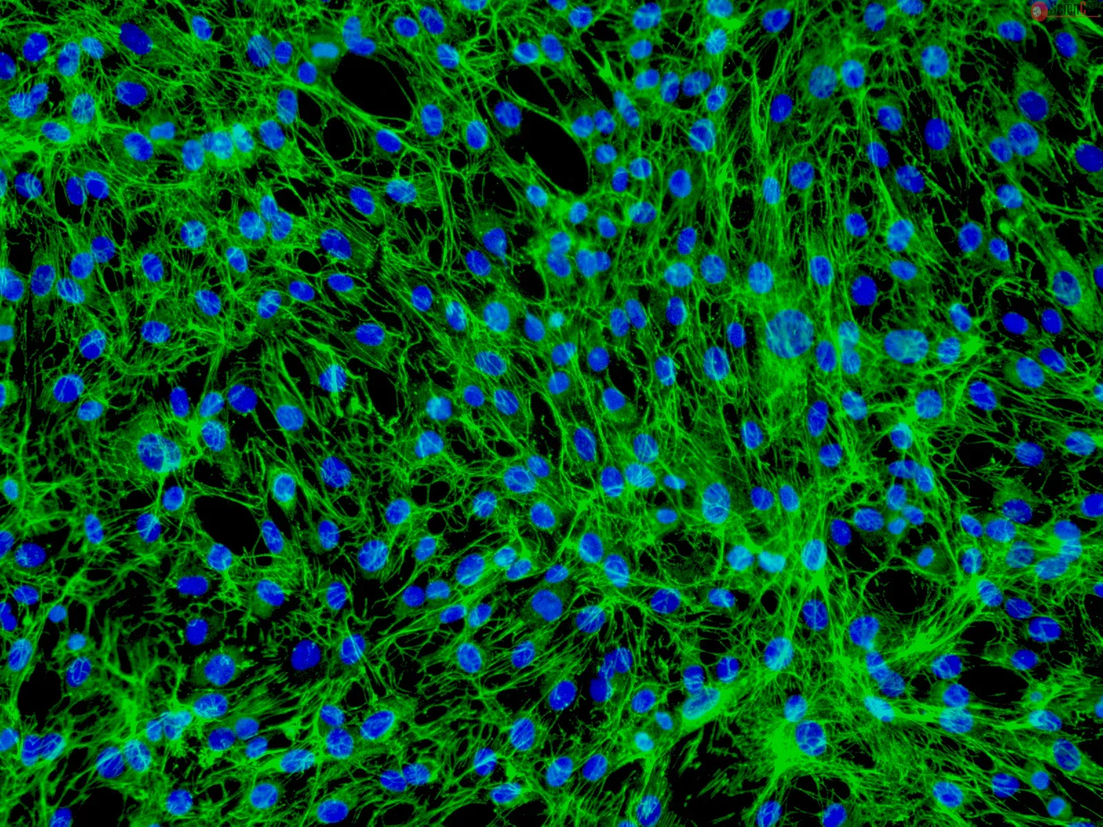
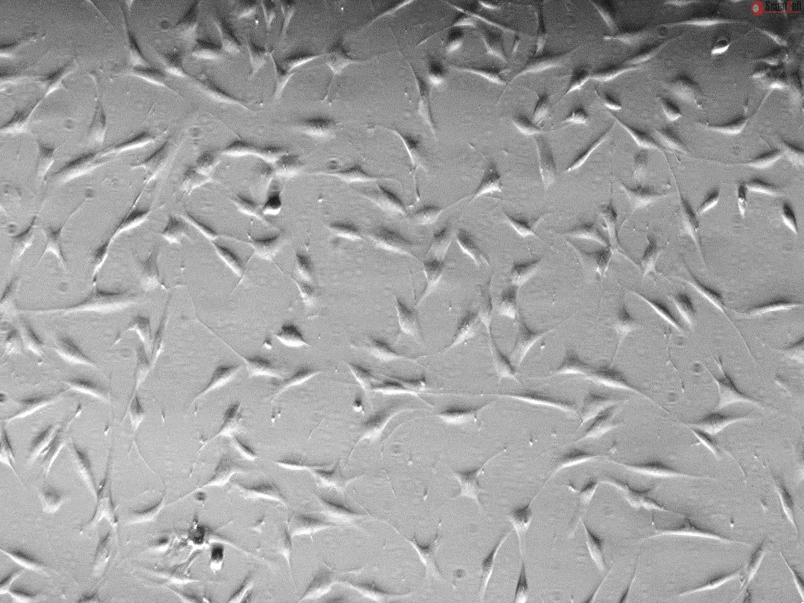
HDF-n from ScienCell Research Laboratories are isolated from neonatal human skin. HDF-n are cryopreserved at passage one and delivered frozen. Each vial contains > 5 x 10⁵ cells in 1 ml volume. HDF-n are characterized by their spindle morphology and immunofluorescence with antibody specific to fibronectin. HDF-n are negative for HIV-1, HBV, HCV, mycoplasma, bacteria, yeast, and fungi. HDF-n are guaranteed to further expand for 15 population doublings under the conditions provided by ScienCell Research Laboratories.
Recommended Medium
It is recommended to use Fibroblast Medium (FM, Cat. #2301) for culturing HDF-n in vitro.
Background: Calcium electroporation describes the use of high voltage electric pulses to introduce supraphysiological calcium concentrations into cells. This promising me... More
Background: Calcium electroporation describes the use of high voltage electric pulses to introduce supraphysiological calcium concentrations into cells. This promising method is currently in clinical trial as an anti-cancer treatment. One very important issue is the relation between tumor cell kill efficacy–and normal cell sensitivity. Methods: Using a 3D spheroid cell culture model we have tested the effect of calcium electroporation and electrochemotherapy using bleomycin on three different human cancer cell lines: a colorectal adenocarcinoma (HT29), a bladder transitional cell carcinoma (SW780), and a breast adenocarcinoma (MDA-MB231), as well as on primary normal human dermal fibroblasts (HDF-n). Results: The results showed a clear reduction in spheroid size in all three cancer cell spheroids three days after treatment with respectively calcium electroporation (p<0.0001) or electrochemotherapy using bleomycin (p<0.0001). Strikingly, the size of normal fibroblast spheroids was neither affected after calcium electroporation nor electrochemotherapy using bleomycin, indicating that calcium electroporation, like electrochemotherapy, will have limited adverse effects on the surrounding normal tissue when treating with calcium electroporation. The intracellular ATP level, which has previously been shown to be depleted after calcium electroporation, was measured in the spheroids after treatment. The results showed a dramatic decrease in the intracellular ATP level (p<0.01) in all four spheroid types—malignant as well as normal. Less
Cultured human skin fibroblasts were irradiated twice successively with the 1.5 J/cm(2) of 532-nm and 1,064-nm lasers, respectively. The mRNA of procollagen, matrix metal... More
Cultured human skin fibroblasts were irradiated twice successively with the 1.5 J/cm(2) of 532-nm and 1,064-nm lasers, respectively. The mRNA of procollagen, matrix metalloproteinases (MMPs), tissue inhibitors of metalloproteinases (TIMPs), heat-shock protein 70 (Hsp70), interleukin-6 (IL-6) and transforming growth factor beta (TGF-beta) were analyzed at 24 and 48 h post-irradiation by using RT-PCR. Both lasers significantly increased the expression of type I and III procollagen, TIMP1, and TIMP2, but decreased MMP1 and MMP2 expression. The 1,064-nm laser initiated TGF-beta expression while the 532-nm laser elicited the increase of Hsp70 and IL-6. The increase/decrease rates of procollagen, TIMPs and MMPs for the 1,064-nm laser were higher than that of the 532-nm laser. Thus, both lasers effectively accelerated collagen synthesis and inhibited collagen degradation. Collagen synthesis induced by the 1,064-nm laser might be partly due to the upregulation of TGF-beta expression, while the increase of Hsp70 and IL-6 might be partly responsible for collagen synthesis stimulated by the 532-nm laser. With the parameters used in this study, the 1,064-nm infrared laser is more effective in promoting the beneficial molecular activities than the 532-nm visible laser. Less
ScienCell Research Laboratories (SRL) takes pride in being a resource for researchers all over the world. The publications listed here are not meant as an endorsement or confirmation of the reliability of the products.


,-1-mg-ml--2.jpg)

-1.jpg)