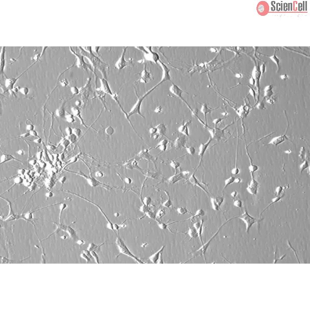
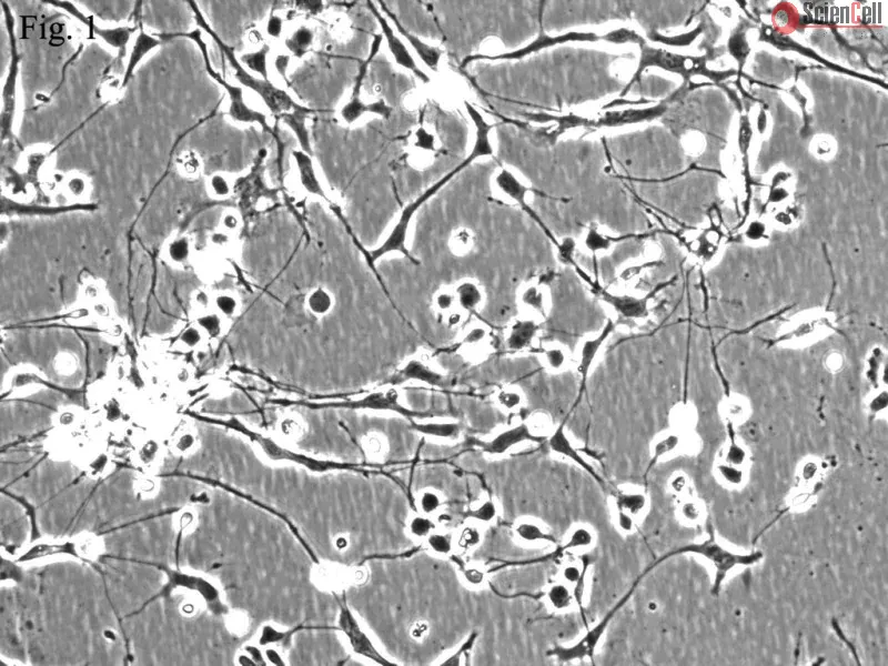
The development of the cerebellum involves a set of coordinated cell movements and two separate proliferation zones: the ventricular zone and the external granule cell layer (EGL), a rhombic-lip-derived progenitor pool. The EGL appears segregated during early cerebellum formation and produces only granule cells. Cerebellar granule cells (CGC) are the most abundant neurons in the brain, about 1 x 10¹¹ in humans. Their axons run as parallel fibres along the coronal axis, and the one-dimensional spread of excitation that results from this arrangement is a key assumption in theories of cerebellar function. CGC receive inhibitory synaptic input from Golgi cells, which are mediated by gamma-aminobutyric acid (GABA). During both in vivo and in vitro development, CGC depend on the activity of the NMDA glutamate receptor subtype for survival and full differentiation. Cultured CGC are widely used as a model system for studying neuronal apoptosis.
HCGC from ScienCell Research Laboratories are isolated from human cerebellum. HCGC are cryopreserved at P0 and delivered frozen. Each vial contains >1 x 10⁶ cells in 1 ml volume. HCGC are characterized by immunofluorescence with antibodies specific to β-tubulin III. HCGC are negative for HIV-1, HBV, HCV, mycoplasma, bacteria, yeast, and fungi. HCGC are guaranteed to further culture under the conditions provided by ScienCell Research Laboratories; however, HCGC are not recommended for expanding or long-term cultures since the cells do not proliferate in culture.
Recommended Medium
It is recommended to use Neuronal Medium (NM, Cat. #1521) for culturing HCGC in vitro.
Purpose: To determine whether human amniotic membranes (AMs) can induce human and rat iris pigment epithelial (IPE) cells grown on them to develop characteristics of RPE ... More
Purpose: To determine whether human amniotic membranes (AMs) can induce human and rat iris pigment epithelial (IPE) cells grown on them to develop characteristics of RPE cells in situ better than IPE cells grown on plastic plates, and to determine whether subretinal transplantation of IPE cell sheets grown on AMs can protect photoreceptor cells in dystrophic Royal College of Surgeons (RCS) rats. Methods: IPE cells from humans and Long-Evans rats were cultured on the basement membrane side of dispase-treated AMs. Two weeks after seeding, ultrastructural changes were evaluated by transmission electron microscopy, and the level of expression of several genes present in differentiated retinal pigment epithelial (RPE) cells was determined by real time PCR and western blotting. IPE cell sheets cultured on AMs were transplanted into the subretinal space of 4-week-old RCS rats, and eyes were analyzed histologically 12 weeks after grafting. Results: IPE cells cultured on AMs showed ultrastructural features like intercellular junctions, similar to RPE cells in situ. IPE cells grown on AMs had a greater upregulation in the expression of genes important for the function of differentiated RPE cells (e.g., pigment epithelium-derived factor [PEDF], RPE65, bestrophin, VEGF, and BDNF) than IPE cells grown on plastic plates. The number of photoreceptors present in RCS rats after subretinal transplantation of IPE cell sheets grown on AMs was significantly higher than that of sham injected rats and rats receiving transplantation of AMs without IPE cells. Conclusions: The more advanced degree of differentiation of IPE cells grown on AMs indicates that AMs are a better substrate to culture IPE cells than plastic plates. This was supported by the greater protection of photoreceptors of RCS rats when IPE cell sheets cultured on AMs were transplanted in the subretinal space. Less
The HIV-1 gene products Tat and gp120 are toxic to neurons and can activate cells of myeloid origin, properties that are thought to contribute to the clinical manifestati... More
The HIV-1 gene products Tat and gp120 are toxic to neurons and can activate cells of myeloid origin, properties that are thought to contribute to the clinical manifestations of HIV-1-associated dementia (HAD). To investigate the intracellular signaling mechanisms involved in these events, the effect of Tat and gp120 on mixed lineage kinase (MLK) 3 activation was examined. Tat and gp120 were shown to induce autophosphorylation of MLK3 in primary rat neurons; this was abolished by the addition of an inhibitor of MLK3 (CEP1347). CEP1347 also enhanced survival of both rat and human neurons and inhibited the activation of human monocytes after exposure to Tat and gp120. Furthermore, overexpression of wild-type MLK3 led to the induction of neuronal death, whereas expression of a dominant negative MLK3 mutant protected neurons from the toxic effects of Tat. MLK3-dependent downstream signaling events were implicated in the neuroprotective and monocyte-deactivating pathways triggered by CEP1347. Thus, the inhibition of p38 MAPK and JNK protected neurons from Tat-induced apoptosis, whereas the inhibition of p38 MAPK, but not of JNK, was sufficient to prevent Tat- and gp120-mediated activation of monocytes. These results suggest that the normal function of MLK3 is compromised by HIV-1 neurotoxins (Tat, gp120), resulting in the activation of downstream signaling events that result in neuronal death and monocyte activation (with release of inflammatory cytokines). In aggregate, our data define MLK3 as a promising therapeutic target for intervention in HAD. Less
ScienCell Research Laboratories (SRL) takes pride in being a resource for researchers all over the world. The publications listed here are not meant as an endorsement or confirmation of the reliability of the products.


,-1-mg-ml--2.jpg)