Glioblastoma (GB) is the most malignant form of human brain tumor, characterized by heterogeneous cell populations, including undifferentiated cells defined as GB stem ce... More
Glioblastoma (GB) is the most malignant form of human brain tumor, characterized by heterogeneous cell populations, including undifferentiated cells defined as GB stem cells (GSCs), responsible for initiating the neoplastic process as well as recurrence. Previous studies demonstrated that the activation of M2 muscarinic acetylcholine receptor (M2 mAChR) by the orthosteric agonist arecaidine propargyl ester (APE) and the dualsteric agonist Iper-N-naphthalimide (N-8-Iper) caused a significant decrease in cell proliferation and survival in both GSCs and GB cell lines. Interestingly, N-8-Iper activates M2 mAChR with higher efficacy and at a lower concentration than APE. The work aimed to better investigate the mechanisms downstream of M2 mAChR activation by both agonists responsible for cytotoxic and pro-apoptotic effects in both U251 cell line and G166 cells (GSCs). To this end, we assessed mitochondrial function by using cell-based assays. Our results demonstrate the ability of N-8-Iper, both at the high (100 μM) and low (25 μM) dose, to induce alteration of mitochondrial morphology and activity, affecting both cellular respiration and ROS production in U251 and G166 cells. Instead, APE causes the same alterations but only in the U251 cell line. Given the relevance of lipid metabolism analysis in the study of cancer, lipid droplets (LDs) were evaluated in the presence or absence of the two M2 agonists. LDs accumulation within the cells was detected after N-8-Iper treatment in both cell lines, whereas APE produced similar effects only in the U251 cell line. No cytotoxic effects and mitochondrial alteration were detected on normal human astrocytes. These results clearly suggest that N-8-Iper has a more potent action on GSCs than APE, thus making this dual-acting agonist a promising muscarinic ligand able to better characterize the inhibitory effects of the M2 muscarinic receptor in glioblastoma cells as well as in other tumor types. Less
In vascularized microphysiological systems, co-culture of fibroblasts and endothelial cells is commonly employed to induce capillary network formation through angiogenesi... More
In vascularized microphysiological systems, co-culture of fibroblasts and endothelial cells is commonly employed to induce capillary network formation through angiogenesis. However, when recapitulating organs such as brain or pancreas, which contain few or no fibroblasts, conventional fibroblast-dependent co-culture poses a limitation to physiological relevance. Here, we developed a modular microfluidic platform that allows reversible connection and disconnection of fibroblast and endothelial cell modules. When the modules were connected, fibroblast-induced angiogenesis of endothelial cells was promoted, and upon disconnection, a vascularized module composed solely of endothelial cells for fibroblast-free capillary network was prepared and able to be maintained for a certain period. We analyzed vascular morphogenesis under different conditions of fibroblast concentration and co-culture duration. We also confirmed that the preformed capillary network remained stable for up to 2 days after module disconnection. Furthermore, upon module reconnection after 2-day of disconnection, angiogenic activity was reactivated through the reestablished co-culture. This approach overcomes the limitations of conventional co-culture methods, enabling the application to organ-specific fibroblast-free vascularization conditions, and provides a foundation for investigating the interaction between preformed capillary networks and other tissues or organs. Less
Radiation-induced neurocognitive dysfunction after brain radiotherapy is a growing concern among the increasing numbers of long-term cancer survivors, particularly in chi... More
Radiation-induced neurocognitive dysfunction after brain radiotherapy is a growing concern among the increasing numbers of long-term cancer survivors, particularly in children. This dysfunction significantly impacts memory, learning, and overall quality of life. Neural stem and progenitor cells (NSPCs) play a vital role in maintaining neurogenesis and plasticity, processes essential for memory formation and cognitive resilience. Currently, no effective treatments exist, highlighting the urgent need for strategies to mitigate these effects. One potential contributing factor to this dysfunction is the depletion or dysregulation of NSPCs following radiation. Here, we developed an in vitro microfluidic neurogenic niche setup to investigate how non-irradiated NSPCs respond to the inflammatory secretome produced by irradiated human fetal astrocytes (HFA) and human brain microvascular endothelial cells (HBMEC). NSPCs viability was dose-dependently affected when exposed to conditioned media from irradiated cells. Notably, NSPCs exposed to conditioned media from cells irradiated at 2 Gy and 8 Gy exhibited increased expression of SOX9 and S100B, respectively, suggesting a shift toward a gliogenic fate. Our findings suggest that this microfluidic model is valuable for exploring radiation-induced neurocognitive dysfunction and identifying potential therapeutic targets. Less
Accumulation of amyloid beta 1–42 (Aβ42) peptide in the extracellular space in the brain is a major observation in Alzheimer’s Disease (AD)-related pathology. Astroc... More
Accumulation of amyloid beta 1–42 (Aβ42) peptide in the extracellular space in the brain is a major observation in Alzheimer’s Disease (AD)-related pathology. Astrocytes are known to play pivotal role in clearing the extracellular aβ peptide from the brain, and the underlying mechanism of Aβ42 peptide clearance remains underappreciated. Like other cell types in the brain, astrocytes have primary cilia, a nonmotile microtubule-based organelle. Aβ42 peptide is reported to affect cilia length or structure in multiple cell types including neurons and inhibit ciliary p75 neurotrophin receptor (p75NTR). To date, the relationship between the extracellular Aβ42 and the astrocytic cilia has not been established. In this work, using primary human hippocampal astrocytes and post-mortem brain specimens obtained from AD patients, we performed molecular, flow cytometry and imaging approaches to investigate the relationship of astrocytic cilia and extracellular Aβ42 peptide. Our data demonstrate that the exogenous Aβ42 peptide treatment in vitro, induces expression of p75NTR in astrocyte cilia in a dose-dependent fashion. We also observed the enrichment of exogenous Aβ42 peptide in the astrocyte cilia and the plasma membrane of astrocytes. In exogenous Aβ42 peptide-treated groups, we observed aberrant proliferation and cell cycle, increased oxidative stress and apoptosis. Interestingly, we observed an enrichment of astrocytic p75NTR expression in the human post-mortem AD-brain. Silencing RNA (siRNA)-mediated knockdown of p75NTR gene significantly minimized the enrichment of exogenous Aβ peptide and the oxidative stress in primary hippocampal astrocytes in vitro. These studies unravel a molecular signaling mechanism that involves Aβ42 peptide-induced p75NTR-mediated oxidative stress that affects overall astrocyte health in AD-associated pathology. Less
The blood vessels of the central nervous (CNS) system form a tight, protective blood-brain barrier (BBB). This barrier is essential for healthy CNS function but also pose... More
The blood vessels of the central nervous (CNS) system form a tight, protective blood-brain barrier (BBB). This barrier is essential for healthy CNS function but also poses a hurdle in the treatment of increasingly common neurological disorders. Additionally, BBB dysfunction is a hallmark of many neurological diseases, further emphasizing a need for a better understanding of BBB function in health and disease. We present a human self-assembling 3D model of the BBB in a microfluidic cell culture platform that allows culture of 48 models in parallel on one tissue culture plate. Human brain microvascular endothelial cells, pericytes, and astrocytes form highly reproducible BBB vascular networks under unidirectional perfusion and remain viable for a minimum of 14 days. Immunostaining reveals close cell-cell interactions with pericytes and astrocyte endfeet in direct contact with the brain microvasculature. Compared to endothelial monocultures, co-cultures with astrocytes or pericytes result in improved barrier function, lower vessel diameters, increased branching, and alignment of the vessels in the direction of fluid flow. These results were most pronounced in tri-cultures containing all three cell types. Unlike similar models previously reported, this brain microvasculature model allows for unidirectional perfusion without the need for pumps and syringes. Combined with its high-throughput nature, this feature renders the model suitable for studies of BBB function in health and disease, and assessment of potential BBB restorative therapies. Less
Mass spectrometry imaging enables spatially resolved, label-free detection of metabolites in tissue and culture systems, providing insight into their metabolic landscape ... More
Mass spectrometry imaging enables spatially resolved, label-free detection of metabolites in tissue and culture systems, providing insight into their metabolic landscape and spatial distribution. However, conventional approaches often lack the spatial resolution and specificity needed to investigate metabolic heterogeneity at the single-cell level, particularly in physiologically relevant models. Here, we present a single-cell ambient mass spectrometry imaging platform, enabling direct chemical mapping of metabolites at 10 μm resolution. This method integrates cell labelling, high resolution microscopy and AP-MALDI Orbitrap mass spectrometry imaging to achieve cell-type-specific metabolite profiling. To demonstrate its application, we applied this approach to glioblastoma (GBM), an aggressive adult brain tumour characterised by cellular heterogeneity, metabolic adaptation, and infiltrative growth within the tumour microenvironment. A co-culture model combining patient-derived glioblastoma invasive-margin cells with human cortical astrocytes was used to recapitulate the invasive niche. Distinct metabolic signatures emerged upon glioblastoma–astrocyte interaction, involving pathways related to nucleotide metabolism, phospholipid and sphingolipid turnover, and tryptophan and tyrosine metabolism. These findings suggest cell-type-specific metabolic activity and potential intercellular metabolic interplay. Overall, this workflow offers a broadly accessible and robust approach for investigating metabolic heterogeneity at cellular resolution, enabling insights into metabolic interactions of heterogenous cell types in both disease and non-disease settings. Less
Microfluidic systems offer controlled microenvironments for cell-to-cell and cell-to-stroma interactions, which have precise physiological, biochemical, and mechanical fe... More
Microfluidic systems offer controlled microenvironments for cell-to-cell and cell-to-stroma interactions, which have precise physiological, biochemical, and mechanical features. The optimization of their conditions to best resemble tumor microenvironments constitutes an experimental modeling challenge, particularly regarding carcinogenesis in the central nervous system (CNS), given the specific features of the blood–brain barrier (BBB). Gel-free 3D microfluidic cell culture systems (gel-free 3D-mFCCSs), including features such as self-production of extracellular matrices, provide significant benefits, including promoting cell–cell communication, interaction, and cell polarity. The proposed microfluidic system consisted of a gel-free culture device inoculated with human brain microvascular endothelial cells (HBEC5i), glioblastoma multiforme cells (U87MG), and astrocytes (ScienCell 1800). The gel-free 3D-mFCCS showed a diffusion coefficient of 4.06 × 10⁻⁹ m2·s−1, and it reconstructed several features and functional properties that occur at the BBB, such as the vasculogenic ability of HBEC5i and the high duplication rate of U87MG. The optimized conditions of the gel-free 3D-mFCCS allowed for the determination of cellular proliferation, invasion, and migration, with evidence of both physical and biochemical cellular interactions, as well as the production of pro-inflammatory cytokines. In conclusion, the proposed gel-free 3D-mFCCSs represent a versatile and suitable alternative to microfluidic systems, replicating several features that occur within tumor microenvironments in the CNS. This research contributes to the characterization of microfluidic approaches and could lead to a better understanding of tumor biology and the eventual development of personalized therapies. Less
In previous work we discovered that T lymphocytes play a prominent role in the rise of brain metastases of ER-negative breast cancers. In the present study we explored ho... More
In previous work we discovered that T lymphocytes play a prominent role in the rise of brain metastases of ER-negative breast cancers. In the present study we explored how T lymphocytes promote breast cancer cell penetration through the blood brain barrier (BBB). An in vitro BBB model was employed to study the effects of T lymphocytes on BBB trespassing capacity of three different breast carcinoma cell lines. Differential protein expression was explored by comparing the proteomes of the breast cancer cells before and after co-culture with activated T lymphocytes using liquid chromatography-mass spectrometry (LC-MS). siRNA was used to silence protein expression in the breast cancer cells to study contribution to in vitro BBB passage. Furthermore, protein expression in primary breast cancer tissues was explored and related to brain-metastatic potential. Co-culturing with activated T lymphocytes or their conditioned medium (CM) resulted in increased passage through the in vitro BBB. The effects were less for cell line MDA-MB-231-B2M2 (brain affinity) as compared to MDA-MB-231 and SK-BR-7. Mass spectrometry-based proteomics revealed significant alterations in the expression of 35 proteins by the breast cancer cell lines upon T cell contact. Among the proteins is coronin-1 A, a protein related to cell motility. Knockdown of CORO1A in the breast cancer cells reduced their ability to cross the artificial BBB to 60%. The effects were significantly less for the cell line derived from breast cancer with affinity for brain. The expression of coronin-1A was confirmed by immunohistochemistry and RT-PCR of 52 breast cancer samples of patients with metastasized breast cancers, with and without brain locations. Lastly, CORO1A upregulation was validated in a publicly available mRNA expression database from 204 primary breast cancers with known metastatic sites. We conclude that T lymphocytes trigger cancer cells to express proteins including coronin-1A that enable the cancer cells to cross an in vitro BBB. In addition, a prominent role of coronin-1A in the formation of cerebral metastases in breast cancer patients is strongly suggestive by its upregulation in tissue samples of breast cancer patients with brain metastases. Less
We have previously developed a cocktail of nine small molecules to convert human fetal astrocytes into neurons, but a nine-molecule recipe is difficult for clinical appli... More
We have previously developed a cocktail of nine small molecules to convert human fetal astrocytes into neurons, but a nine-molecule recipe is difficult for clinical applications. Here, we identify a chemical formula with only three to four small molecules for astrocyte-to-neuron conversion. We demonstrate that modulation of three to four signaling pathways among Notch, glycogen synthase kinase 3, transforming growth factor β, and bone morphogenetic protein pathways is sufficient to change an astrocyte into a neuron. The chemically converted human neurons can survive >7 months in culture, fire repetitive action potentials, and display robust synaptic burst activities. Interestingly, cortical astrocyte-converted neurons are mostly glutamatergic, while midbrain astrocyte-converted neurons can yield some GABAergic neurons in addition to glutamatergic neurons. When administered in vivo through intracranial or intraperitoneal injection, the four-drug combination can significantly increase adult hippocampal neurogenesis. Together, human fetal astrocytes can be chemically converted into functional neurons using three to four small molecules, bringing us one step forward for developing future drug therapy.
Keywords: astrocyte-to-neuron conversion, small molecule, chemical conversion, reprogramming, adult neurogenesis, glutamatergic neuron Less
γ-Synuclein (γ-syn) is expressed by astrocytes in the human nervous system, and increased extracellularly in the brain and cerebrospinal fluid of individuals diagnosed ... More
γ-Synuclein (γ-syn) is expressed by astrocytes in the human nervous system, and increased extracellularly in the brain and cerebrospinal fluid of individuals diagnosed with Alzheimer's disease. Upregulation of γ-syn also coincides with proliferation of glioblastomas and other cancers. In order to better understand regulation and function of extracellular γ-syn, primary human cortical astrocytes were treated with γ-syn conditioned media at various physiological concentrations (50, 100, 150 nM) after cell synchronization. Additionally, extracellular brain-derived neurotrophic factor (BDNF), a neuroprotective growth factor released by astrocytes that has been shown to be decreased extracellularly in neurodegenerative disease, was observed in response to γ-syn treatment. Analysis of 5-bromodeoxyuridine (BrdU) and propidium iodide through flow cytometry 24 h after release from synchronization revealed an increase in G2/M phase of the cell cycle with 100 nM γ-syn during initial cell division, an effect that was reversed at 48 h. However, increased extracellular BDNF was observed at 48 h with 100 nM and 150 nM γ-syn treatment with no difference between controls at 24 h. Further analysis of cell cycle markers with immunocytochemistry of BrdU and Ki67 after treatment with 100 nM γ-syn confirmed increased initial cell proliferation and decreased non-proliferating cells. Western blot analysis demonstrated increased γ-syn levels after 100 nM treatment at 24 and 48 h, and increased pro-BDNF, mature BDNF and cell viability at 48 h. The results demonstrate that γ-syn internalization by human cortical astrocytes causes upregulation of the cell cycle, followed by subsequent BDNF expression and release. Less
Occludin is a component of tight junctions, which are essential structural components of the blood-brain barrier. However, occludin is expressed in cells without tight ju... More
Occludin is a component of tight junctions, which are essential structural components of the blood-brain barrier. However, occludin is expressed in cells without tight junctions, implying additional functions. We determined the expression and localisation of occludin in astrocytes in cell culture and in human brain tissue, and sought novel binding partners using a proteomic approach. Expression was investigated by immunocytochemistry and immunoblotting in the 1321N1 astrocytoma cell line and ScienCell human primary astrocytes, and by immunohistochemistry in human autopsy brain tissue. Recombinant N- and C-terminal occludin was used to pull-down proteins from 1321N1 cell lysates and protein-binding partners identified by mass spectrometry analysis. Occludin was expressed in both the cytoplasm and nucleus of astrocytes in vitro and in vivo. Mass spectrometry identified binding to nuclear and cytoplasmic proteins, particularly those related to RNA metabolism and nuclear function. Occludin is expressed in several subcellular compartments of brain cell-types that do not form tight junctions and the expression patterns in cell culture reflect those in human brain tissue, indicating they are suitable model systems. Proteomic analysis suggests that occludin has novel functions in neuroepithelial cells that are unrelated to tight junction formation. Further research will establish the roles of these functions in both cellular physiology and in disease states. Less
Neurovascular inflammation is a major contributor to many neurological disorders, but modeling these processes in vitro has proven to be difficult. Here, we microengineer... More
Neurovascular inflammation is a major contributor to many neurological disorders, but modeling these processes in vitro has proven to be difficult. Here, we microengineered a three-dimensional (3D) model of the human blood-brain barrier (BBB) within a microfluidic chip by creating a cylindrical collagen gel containing a central hollow lumen inside a microchannel, culturing primary human brain microvascular endothelial cells on the gel’s inner surface, and flowing medium through the lumen. Studies were carried out with the engineered microvessel containing endothelium in the presence or absence of either primary human brain pericytes beneath the endothelium or primary human brain astrocytes within the surrounding collagen gel to explore the ability of this simplified model to identify distinct contributions of these supporting cells to the neuroinflammatory response. This human 3D BBB-on-a-chip exhibited barrier permeability similar to that observed in other in vitro BBB models created with non-human cells, and when stimulated with the inflammatory trigger, tumor necrosis factor-alpha (TNF-α), different secretion profiles for granulocyte colony-stimulating factor (G-CSF) and interleukin-6 (IL-6) were observed depending on the presence of astrocytes or pericytes. Importantly, the levels of these responses detected in the 3D BBB chip were significantly greater than when the same cells were co-cultured in static Transwell plates. Thus, as G-CSF and IL-6 have been reported to play important roles in neuroprotection and neuroactivation in vivo, this 3D BBB chip potentially offers a new method to study human neurovascular function and inflammation in vitro, and to identify physiological contributions of individual cell types. Less
Chemical investigation of the cultures of marine Streptomyces sp. 182SMLY led to the discovery of two new polycyclic anthraquinones, which were elucidated as N-acetyl-N-d... More
Chemical investigation of the cultures of marine Streptomyces sp. 182SMLY led to the discovery of two new polycyclic anthraquinones, which were elucidated as N-acetyl-N-demethylmayamycin (1) and streptoanthraquinone A (2) based on the extensive spectroscopic analysis including 2D NMR, HRESIMS, and an electronic circular dichroism (ECD) calculation. Both anthraquinones remarkably suppressed the proliferation of four different glioma cell lines with IC50 values in a range from 0.5 to 7.3 μM and induced apoptosis in the glioma cells. The ratios of IC50 for normal human astrocytes to IC50 for glioma cells were 6.4-53 for 1 and >14-31 for 2. N-acetyl-N-demethylmayamycin (1) also inhibited the growth of methicillin-resistant Staphylococcus aureus with MIC 20.0 μM. Keywords: N-acetyl-N-demethylmayamycin; bioactivities against glioma cells and bacteria; marine bacterium Streptomyces sp. 182SMLY; streptoanthraquinone A. Less
Background: Human papilloma viruses (HPVs) may act early in breast oncogenesis ( “hit-and-run” phenomena). Methods: The authors used immunohistochemistry for the iden... More
Background: Human papilloma viruses (HPVs) may act early in breast oncogenesis ( “hit-and-run” phenomena). Methods: The authors used immunohistochemistry for the identification of HPV E7 oncogenic protein expression in 32 sets of benign and subsequent breast cancer specimens from the same Australian patients. Results: HPV E7 oncoprotein was clearly expressed in the nuclei of 23 (72%) of the 32 benign specimens and 20 (62.5%) of the subsequent 32 breast cancer specimens in the same patients. There was no HPV E7 protein expression in seven (30%) of the 23 breast cancer specimens that had prior HPV E7 protein-positive benign breast biopsies in the same patients. Conclusions: This observation suggests that HPV oncogenic influences occur early in some breast cancers. This finding confirms the previous observations. This early influence of HPVs may be the reason why there is no increase in the prevalence of HPV-associated breast cancer in immunocompromised patients as compared to HPV-associated cervical cancer. Less
Background Human brain glioma is the most common endocranial tumor; its mortality and morbidity are very high. The objective of this study was to determine whether miR-33... More
Background Human brain glioma is the most common endocranial tumor; its mortality and morbidity are very high. The objective of this study was to determine whether miR-338-3p can regulate malignant biological behaviors of glioma cells by targeted silencing of MACC1. Material/Methods The expression of miR-338-3p was detected by quantitative real-time PCR in brain glioma tissues and cell lines. Bioinformatics software was used to predict some potential target genes of miR-338-3p. Luciferase activities assay was used to verify the combination between target genes and miR-338-3p. And MACC1 protein expression was detected by Western blot. The apoptosis and proliferation ability were analyzed by MTT and flow cytometry assay. Results Compared with normal brain tissues and cells, miR-338-3p in glioma tissues and cell lines was confirmed to be expressed at low levels, and down-regulation of miR-338-3p tended to be correlated with worse histological grade. Up-regulation of miR-338-3p promoted apoptosis and sharply inhibited cell proliferation ability of U251 and U87 cells. The luciferase activities assay, biotin-avidin pull-down assay, and western blot analysis verified that MACC1 was a specific target gene of miR-338-3p. Subsequent experiments found that up-regulation of MACC1 significantly inhibited the apoptosis and increased the cell proliferation ability of U251 and U87 cells. The regulation effects of miR-338-3p on malignant biological behaviors of glioma cells can be partly reversed by up-regulation of MACC1. Conclusions Down-regulation of miR-338-3p was an independent prognostic biomarker associated with poor prognosis in glioma patients; miR-338-3p acted as a tumor-suppressing gene whose silencing can inhibit malignant biological behaviors of glioma cells. MACC1 was a specific target gene of miR-338-3p, which regulates malignant biological behaviors of glioma cells partly through directly silencing MACC1 expression. MeSH Keywords: Apoptosis, Cell Proliferation, Glioma, MicroRNAs Less
Spinal cord injury (SCI) induces the disruption of the blood-spinal cord barrier (BSCB) which leads to infiltration of blood cells, an inflammatory response, and neuronal... More
Spinal cord injury (SCI) induces the disruption of the blood-spinal cord barrier (BSCB) which leads to infiltration of blood cells, an inflammatory response, and neuronal cell death, resulting spinal cord secondary damage. Retinoic acid (RA) has a neuroprotective effect in both ischemic brain injury and SCI, however the relationship between BSCB disruption and RA in SCI is still unclear. In this study, we demonstrated that autophagy and ER stress are involved in the protective effect of RA on the BSCB. RA attenuated BSCB permeability and decreased the loss of tight junction (TJ) molecules such as P120, β-catenin, Occludin and Claudin5 after injury in vivo as well as in Brain Microvascular Endothelial Cells (BMECs). Moreover, RA administration improved functional recovery in the rat model of SCI. RA inhibited the expression of CHOP and caspase-12 by induction of autophagic flux. However, RA had no significant effect on protein expression of GRP78 and PDI. Furthermore, combining RA with the autophagy inhibitor chloroquine (CQ) partially abolished its protective effect on the BSCB via exacerbated ER stress and subsequent loss of tight junctions. Taken together, the neuroprotective role of RA in recovery from SCI is related to prevention of of BSCB disruption via the activation of autophagic flux and the inhibition of ER stress-induced cell apoptosis. These findings lay the groundwork for future translational studies of RA for CNS diseases, especially those related to BSCB disruption. Less
Background Glioblastoma multiforme is the most aggressive malignant primary brain tumor, characterized by rapid growth and extensive infiltration to neighboring normal br... More
Background Glioblastoma multiforme is the most aggressive malignant primary brain tumor, characterized by rapid growth and extensive infiltration to neighboring normal brain parenchyma. Both PI3K/Akt and JNK pathways are essential to glioblastoma cell survival, migration and invasion. Due to their hyperactivation in glioblastoma cells, PI3K and JNK are promising targets for glioblastoma treatment. Methods To investigate the combination effects of class IA PI3K catalytic isoforms (p110α, p110β and p110δ) and JNK inhibition on tumor cell growth and motility, glioblastoma cells and xenografts in nude mice were treated with isoform-selective PI3K inhibitors in combination with JNK inhibitor. Results We showed that combined inhibition of these PI3K isoforms and JNK exerted divergent effects on the proliferation, migration and invasion of glioblastoma cells in vitro. Pharmacological inhibition of p110β or p110δ, but not p110α, displayed synergistic inhibitory effect with JNK inhibition on glioblastoma cell proliferation and migration through decreasing phosphorylation of Akt, FAK and zyxin, leading to blockade of lamellipodia and membrane ruffles formation. No synergistic effect on invasion was observed in all the combination treatment. In vivo, combination of p110β and JNK inhibitors significantly reduced xenograft tumor growth compared with single inhibitor alone. Conclusion Concurrent inhibition of p110β and JNK exhibited synergistic effects on suppressing glioblastoma cell proliferation and migration in vitro and xenograft tumor growth in vivo. Our data suggest that combined inhibition of PI3K p110β isoform and JNK may serve as a potent and promising therapeutic approach for glioblastoma multiforme. Electronic supplementary material The online version of this article (doi:10.1186/s13046-016-0356-5) contains supplementary material, which is available to authorized users. Keywords: Glioblastoma, PI3K, p110β, JNK, Synergism, Proliferation, Migration Less
Background The cholinergic anti-inflammatory pathway (CAP) primarily functions through acetylcholine (ACh)-alpha7 nicotinic acetylcholine receptor (α7nAChR) interaction ... More
Background The cholinergic anti-inflammatory pathway (CAP) primarily functions through acetylcholine (ACh)-alpha7 nicotinic acetylcholine receptor (α7nAChR) interaction on macrophages to control peripheral inflammation. Interestingly, ACh can also bind α7nAChRs on microglia resulting in neuroprotective effects. However, ACh effects on astrocytes remain elusive. Here, we investigated the effects of nicotine, an ACh receptor agonist, on the cytokine and cholinesterase production of immunocompetent human astrocytes stimulated with interleukin 1β (IL-1β) in vitro. In addition, the potential involvement of prostaglandins as mediators of nicotine was studied using cyclooxygenase 2 (COX-2) inhibition. Methods Cultured human fetal astrocytes were stimulated with human recombinant IL-1β and treated simultaneously with nicotine at different concentrations (1, 10, and 100 μM). Cell supernatants were collected for cytokine and cholinesterase profiling using ELISA and MesoScale multiplex assay. α7nAChR expression on activated human astrocytes was studied using immunofluorescence. For the COX-2 inhibition studies, enzyme activity was inhibited using NS-398. One-way ANOVA was used to perform statistical analyses. Results Nicotine treatment dose dependently limits the production of critical proinflammatory cytokines such as IL-6 (60.5 ± 3.3, %inhibition), IL-1β (42.4 ± 1.7, %inhibition), and TNF-α (68.9 ± 7.7, %inhibition) by activated human astrocytes. Interestingly, it also inhibits IL-8 chemokine (31.4 ± 8.5, %inhibition), IL-13 (34.243 ± 4.9, %inhibition), and butyrylcholinesterase (20.8 ± 2.8, %inhibition) production at 100 μM. Expression of α7nAChR was detected on the activated human astrocytes. Importantly, nicotine’s inhibitory effect on IL-6 production was reversed with the specific COX-2 inhibitor NS-398. Conclusions Activation of the cholinergic system through α7nAChR agonists has been known to suppress inflammation both in the CNS and periphery. In the CNS, earlier experimental data shows that cholinergic activation through nicotine inhibits microglial activation and proinflammatory cytokine release. Here, we report similar anti-inflammatory effects of cholinergic activation on human astrocytes, at least partly mediated through the COX-2 pathway. These results confirm the potential for cholinergic neuroprotection, which is looked upon as a promising therapy for neuroinflammation as well as neurodegenerative diseases and stroke. Our data implicates an important role for the prostaglandin system in cholinergic regulatory effects. Less
Background: The fractalkine (CX3CR1) ligand is expressed in astrocytes and reported to be neuroprotective. When cleaved from the membrane, soluble fractalkine (sCX3CL1) a... More
Background: The fractalkine (CX3CR1) ligand is expressed in astrocytes and reported to be neuroprotective. When cleaved from the membrane, soluble fractalkine (sCX3CL1) activates the receptor CX3CR1. Although somewhat controversial, CX3CR1 is reported to be expressed in neurons and microglia. The membrane-bound form of CX3CL1 additionally acts as an adhesion molecule for microglia and infiltrating white blood cells. Much research has been done on the role of fractalkine in neuronal cells; however, little is known about the regulation of the CX3CL1 ligand in astrocytes. Methods: The mechanisms involved in the up-regulation and cleavage of CX3CL1 from human astrocytes were investigated using immunocytochemistry, Q-PCR and ELISA. All statistical analysis was performed using GraphPad Prism 5. Results: A combination of ADAM17 (TACE) and ADAM10 protease inhibitors was found to attenuate IL-1β-, TNF-α- and IFN-γ-induced sCX3CL1 levels in astrocytes. A specific ADAM10 (but not ADAM17) inhibitor also attenuated these effects, suggesting ADAM10 proteases induce release of sCX3CL1 from stimulated human astrocytes. A p38 MAPK inhibitor also attenuated the levels of sCX3CL1 upon treatment with IL-1β, TNF-α or IFN-γ. In addition, an IKKβ inhibitor significantly reduced the levels of sCX3CL1 induced by IL-1β or TNF-α in a concentration-dependent manner, suggesting a role for the NF-kB pathway. Conclusions: In conclusion, this study shows that the release of soluble astrocytic fractalkine is regulated by ADAM10 proteases with p38 MAPK also playing a role in the fractalkine shedding event. These findings are important for understanding the role of CX3CL1 in healthy and stimulated astrocytes and may benefit our understanding of this pathway in neuro-inflammatory and neurodegenerative diseases. Less
Background: Glioblastoma stem cells (GSC) have been extensively recognized as a plausible cause of glioblastoma resistance to therapy and recurrence resulting in high gli... More
Background: Glioblastoma stem cells (GSC) have been extensively recognized as a plausible cause of glioblastoma resistance to therapy and recurrence resulting in high glioblastoma mortality. Abnormalities in the DNA repair pathways might be responsible for the inability of the currently used chemotherapeutics to eliminate the (GSC) subpopulation.
Methods: In this work, we compared the expression of sixty DNA repair related genes between primary glioblastoma cell cultures and the glioblastoma enriched stem cell primary cultures. MTT test was used to analyze the effect of selected drugs and immunofluorescence to evaluate the load of DNA damage.
Results: We found several differentially expressed genes and we identified topoisomerase IIβ (Top2β) as the gene with highest up-regulation in GSC. Also among the tested cell lines the expression of Top2β was the highest in NCH421k cells, a well-characterized glioblastoma cell line with all the stemness characteristics. On the other hand, Top2β expression markedly decreased upon the induction of differentiation by all trans-retinoic acid. Depletion of Top2β increased the sensitivity of NCH421k cells to replication stress inducing drugs, such as cisplatin, methyl-methanesulfonate, hydrogen peroxide, and temozolomide. Consistently, we found an increased load of DNA damage and increased Chk1 activation upon Top2β depletion in NCH421k cells.
Conclusion: We suggest that Top2β may represent a new target for gene therapy in glioblastoma. In addition, the other genes that we found to be up-regulated in GSC versus glioblastoma primary cells should be further investigated as glioblastoma theranostics.
Keywords: Drug resistance; Glioblastoma stem cells; Glioma; Replication stress; Theranostic markers; Topoisomerase IIβ. Less
Astrocytes, once considered passive support cells, are increasingly appreciated as dynamic regulators of neuronal development and function, in part via secreted factors. ... More
Astrocytes, once considered passive support cells, are increasingly appreciated as dynamic regulators of neuronal development and function, in part via secreted factors. The extent to which they similarly regulate oligodendrocytes, or proliferation and differentiation of oligodendrocyte progenitor cells (OPCs) is less well understood. Here, we generated astrocytes from human pluripotent stem cells (hiPSC-Astros) and demonstrate that immature astrocytes - as opposed to mature - promoted oligodendrogenesis in vitro. In the PVL mouse model of neonatal hypoxic/ischemic encephalopathy, associated with cerebral palsy in humans, transplanted immature hiPSC-Astros promote myelinogenesis and behavioral outcome. We further identified TIMP-1 as a selectively upregulated component secreted from immature hiPSC-Astros. Accordingly, in the rat PVL model, intranasal administration of conditioned medium from immature hiPSC-Astros promoted oligodendrocyte maturation in a TIMP-1 dependent manner. Our findings suggest stage-specific developmental interactions between astroglia and oligodendroglia, with important therapeutic implications for promoting myelinogenesis.
Keywords: human induced pluripotent stem cells, immature astrocytes, periventricular leukomalacia, oligodendrocytes, myelination Less
MicroRNA-130b (miR-130b) is a novel tumor-related miRNA that has been found to be involved in several biological processes. However, there is limited evidence regarding t... More
MicroRNA-130b (miR-130b) is a novel tumor-related miRNA that has been found to be involved in several biological processes. However, there is limited evidence regarding the role of miR-130b in the tumorigenesis of human gliomas. In the present study, reverse transcription-quantitative polymerase chain reaction (RT-qPCR) assays were used to quantify miR-130b expression levels in human glioma tissues and glioma cell lines (U251, U87, SNB19 and LN229). The expression level of miR-130b was found to be markedly higher in human glioma tissues than in non‑neoplastic brain specimens. Specifically, higher expression levels of miR‑130b were observed in the glioma cell lines, compared with those in normal human astrocytes (NHA). We also confirmed that miR‑130b interacted with the 3'-untranslated region of peroxisome proliferator‑activated receptor-γ (PPAR‑γ), which negatively affected the protein levels of E-cadherin. Furthermore, its effects on cell proliferation and invasion were examined using CCK8, colony formation, cell cycle and Transwell assays. We found that the upregulation of miR-130b induced cell proliferation, decreased the percentage of cells in the G0/G1 phase and enhanced the invasiveness of U251 glioma cells whereas the downregulation of miR-130b exerted opposing effects. Moreover, it was demonstrated that the downregulation of miR‑130b in U251 glioma cells restored the expression of PPAR-γ and E-cadherin, and inhibited the expression of β-catenin. Notably, PPAR-γ knockdown abolished the inhibitory effect of miR-130b inhibitor on the proliferation and invasivness of U251 cells. Taken together, these findings suggest that miR‑130b promotes the proliferation and invasion of U251 glioma cells by inhibiting PPAR-γ. Less
Alzheimer's disease and frontotemporal dementia are amongst the most common forms of dementia characterized by the formation and deposition of abnormal TAU in the brain. ... More
Alzheimer's disease and frontotemporal dementia are amongst the most common forms of dementia characterized by the formation and deposition of abnormal TAU in the brain. In order to develop a translational human TAU aggregation model suitable for screening, we transduced TAU harboring the pro-aggregating P301L mutation into control hiPSC-derived neural progenitor cells followed by differentiation into cortical neurons. TAU aggregation and phosphorylation was quantified using AlphaLISA technology. Although no spontaneous aggregation was observed upon expressing TAU-P301L in neurons, seeding with preformed aggregates consisting of the TAU-microtubule binding repeat domain triggered robust TAU aggregation and hyperphosphorylation already after 2 weeks, without affecting general cell health. To validate our model, activity of two autophagy inducers was tested. Both rapamycin and trehalose significantly reduced TAU aggregation levels suggesting that iPSC-derived neurons allow for the generation of a biologically relevant human Tauopathy model, highly suitable to screen for compounds that modulate TAU aggregation. Less
The aim of this study was to develop a three-dimensional (3D) model of the human blood-brain barrier in vitro, which mimics the cellular architecture of the CNS and could... More
The aim of this study was to develop a three-dimensional (3D) model of the human blood-brain barrier in vitro, which mimics the cellular architecture of the CNS and could be used to analyse the delivery of nanoparticles to cells of the CNS. The model includes human astrocytes set in a collagen gel, which is overlaid by a monolayer of human brain endothelium (hCMEC/D3 cell line). The model was characterised by transmission electron microscopy (TEM), immunofluorescence microscopy and flow cytometry. A collagenase digestion method could recover the two cell types separately at 92-96% purity. Astrocytes grown in the gel matrix do not divide and they have reduced expression of aquaporin-4 and the endothelin receptor, type B compared to two-dimensional cultures, but maintain their expression of glial fibrillary acidic protein. The effects of conditioned media from these astrocytes on the barrier phenotype of the endothelium was compared with media from astrocytes grown conventionally on a two-dimensional (2D) substratum. Both induce the expression of tight junction proteins zonula occludens-1 and claudin-5 in hCMEC/D3 cells, but there was no difference between the induced expression levels by the two media. The model has been used to assess the transport of glucose-coated 4nm gold nanoparticles and for leukocyte migration. TEM was used to trace and quantitate the movement of the nanoparticles across the endothelium and into the astrocytes. This blood-brain barrier model is very suitable for assessing delivery of nanoparticles and larger biomolecules to cells of the CNS, following transport across the endothelium. Less
Toward developing a model system for investigating the role of the microenvironment in the radioresistance of glioblastoma (GBM), human glioblastoma stem-like cells (GSCs... More
Toward developing a model system for investigating the role of the microenvironment in the radioresistance of glioblastoma (GBM), human glioblastoma stem-like cells (GSCs) were grown in coculture with human astrocytes. Using a trans-well assay, survival analyses showed that astrocytes significantly decreased the radiosensitivity of GSCs compared to standard culture conditions. In addition, when irradiated in coculture, the initial level of radiation-induced γH2AX foci in GSCs was reduced and foci dispersal was enhanced suggesting that the presence of astrocytes influenced the induction and repair of DNA double-strand breaks. These data indicate that astrocytes can decrease the radiosensitivity of GSCs in vitro via a paracrine-based mechanism and further support a role for the microenvironment as a determinant of GBM radioresponse. Chemokine profiling of coculture media identified a number of bioactive molecules not present under standard culture conditions. The gene expression profiles of GSCs grown in coculture were significantly different as compared to GSCs grown alone. These analyses were consistent with an astrocyte-mediated modification in GSC phenotype and, moreover, suggested a number of potential targets for GSC radiosensitization that were unique to coculture conditions. Along these lines, STAT3 was activated in GSCs grown with astrocytes; the JAK/STAT3 inhibitor WP1066 enhanced the radiosensitivity of GSCs under coculture conditions and when grown as orthotopic xenografts. Further, this coculture system may also provide an approach for identifying additional targets for GBM radiosensitization. Keywords: Astrocytes, glioblastoma stem-like cells, microenvironment, radiosensitization, STAT3 Less
Effective suicide gene delivery and expression are crucial to achieving successful effects in
gene therapy. An ideal tumor-specific promoter expresses therapeutic genes i... More
Effective suicide gene delivery and expression are crucial to achieving successful effects in
gene therapy. An ideal tumor-specific promoter expresses therapeutic genes in tumor cells
with minimal normal tissue expression. We compared the activity of the FOS (FBJ murine
osteosarcoma viral oncogene homolog) promoter with five alternative tumor-specific pro-
moters in glioma cells and non-malignant astrocytes. The FOS promoter caused signifi-
cantly higher transcriptional activity in glioma cell lines than all alternative promoters with
the exception of CMV. The FOS promoter showed 13.9%, 32.4%, and 70.8% of the tran-
scriptional activity of CMV in three glioma cell lines (U87, U251, and U373). Importantly,
however, the FOS promoter showed only 1.6% of the transcriptional activity of CMV in nor-
mal astrocytes. We also tested the biologic activity of recombinant adenovirus containing
the suicide gene herpes simplex virus thymidine kinase (HSV-tk) driven by the FOS pro-
moter, including selective killing efficacy in vitro and tumor inhibition rate in vivo. Adenoviral-
mediated delivery of the HSV-tk gene controlled by the FOS promoter conferred a cytotoxic
effect on human glioma cells in vitro and in vivo. This study suggests that use of the FOS-tk
adenovirus system is a promising strategy for glioma-specific gene therapy but still much
left for improvement. Less
Histamine receptor 3 (H3R) is expressed in various tumors and correlated with malignancy and tumor proliferation. However, the role of H3R in tumor invasion and epithelia... More
Histamine receptor 3 (H3R) is expressed in various tumors and correlated with malignancy and tumor proliferation. However, the role of H3R in tumor invasion and epithelial to mesenchymal transition (EMT) remains unknown. Here, we explored the H3R in the highly invasive glioblastoma (GBM) and U87MG cells. We found that H3R mRNA and protein levels were up-regulated in the GBM and glioma cell lines compared to normal brain tissue and astrocytes. In U87MG cell line, inhibition of H3R by siRNA or the antagonist ciproxifan (CPX) suppressed proliferation, invasiveness, and the expression of EMT activators (Snail, Slug and Twist). In addition, expression of epithelial markers (E-cadherin and ZO-1) was up-regulated and expression of mesenchymal markers (vimentin and N-cadherin) was down-regulated in vitro and in vivo in a xenograft model. In addition, we also showed that inhibition of H3R by siRNA or CPX inactivated the PI3K/Akt and MEK/ERK signaling pathways, while inhibition of Akt or ERK activity with antagonists or siRNAs suppressed H3R agonist (R)-(α)-(−)- methylhistamine dihydrobromide (RAMH) mediated invasion and reorganization of cadherin-household. In conclusion, overexpression of H3R is associated with glioma progression. Inhibition of H3R leads to suppressed invasion and EMT of GBM by inactivating the PI3K/Akt and MEK/ERK pathways in gliomas. Keywords: histamine receptor 3, glioblastoma, epithelial-to-mesenchymal transition, invasion Less
Autophagy, a type II programmed cell death, is essential for cell survival under stress, e.g. lung injury, and bone marrow-derived mesenchymal stem cells (BM-MSCs) have g... More
Autophagy, a type II programmed cell death, is essential for cell survival under stress, e.g. lung injury, and bone marrow-derived mesenchymal stem cells (BM-MSCs) have great potential for cell therapy. However, the mechanisms underlying the BM-MSC activation of autophagy to provide a therapeutic effect in ischaemia/reperfusion-induced lung injury (IRI) remain unclear. Thus, we investigate the activation of autophagy in IRI following transplantation with BM-MSCs. Seventy mice were pre-treated with BM-MSCs before they underwent lung IRI surgery in vivo. Human pulmonary micro-vascular endothelial cells (HPMVECs) were pre-conditioned with BM-MSCs by oxygen-glucose deprivation/reoxygenation (OGD) in vitro. Expression markers for autophagy and the phosphoinositide 3-kinase/protein kinase B (PI3K/Akt) signalling pathway were analysed. In IRI-treated mice, administration of BM-MSCs significantly attenuated lung injury and inflammation, and increased the level of autophagy. In OGD-treated HPMVECs, co-culture with BM-MSCs attenuated endothelial permeability by decreasing the level of cell death and enhanced autophagic activation. Moreover, administration of BM-MSCs decreased the level of PI3K class I and p-Akt while the expression of PI3K class III was increased. Finally, BM-MSCs-induced autophagic activity was prevented using the inhibitor LY294002. Administration of BM-MSCs attenuated lung injury by improving the autophagy level via the PI3K/Akt signalling pathway. These findings provide further understanding of the mechanisms related to BM-MSCs and will help to develop new cell-based therapeutic strategies in lung injury. Less
Methodologies for generating functional neuronal cells directly from human fibroblasts [induced neuronal (iN) cells] have been recently developed, but the research so far... More
Methodologies for generating functional neuronal cells directly from human fibroblasts [induced neuronal (iN) cells] have been recently developed, but the research so far has only focused on technical refinements or recapitulation of known pathological phenotypes. A critical question is whether this novel technology will contribute to elucidation of novel disease mechanisms or evaluation of therapeutic strategies. Here we have addressed this question by studying Tay-Sachs disease, a representative lysosomal storage disease, and Dravet syndrome, a form of severe myoclonic epilepsy in infancy, using human iN cells with feature of immature postmitotic glutamatergic neuronal cells. In Tay-Sachs disease, we have successfully characterized canonical neuronal pathology, massive accumulation of GM2 ganglioside, and demonstrated the suitability of this novel cell culture for future drug screening. In Dravet syndrome, we have identified a novel functional phenotype that was not suggested by studies of classical mouse models and human autopsied brains. Taken together, the present study demonstrates that human iN cells are useful for translational neuroscience research to explore novel disease mechanisms and evaluate therapeutic compounds. In the future, research using human iN cells with well-characterized genomic landscape can be integrated into multidisciplinary patient-oriented research on neuropsychiatric disorders to address novel disease mechanisms and evaluate therapeutic strategies. Keywords: Direct conversion, Induced neuronal cells, iN cells, Lysosomal storage diseases, Channelopathy, Polyglutamine diseases Less
The PI3K/Akt signaling pathway is frequently activated in various human cancer types and plays essential roles in development and progression of cancers. Multiple regulat... More
The PI3K/Akt signaling pathway is frequently activated in various human cancer types and plays essential roles in development and progression of cancers. Multiple regulators, such as phosphatase and tensin homolog (PTEN) and PH domain leucine rich repeat protein phosphatases (PHLPP), have also found to be involved in suppression of the PI3K/Akt signaling pathway. However, how suppressive effects mediated by these regulators are concomitantly disrupted in cancers, which display constitutively activated PI3K/Akt signaling, remains puzzling. In the present study, we reported that the expression of miR-93 was markedly upregulated in glioma cell lines and clinical glioma tissues. Statistical analysis revealed that miR-93 levels significantly correlated with clinicopathologic grade and overall survival in gliomas. Furthermore, we found that overexpressing miR-93 promoted, but inhibition of miR-93 reduced, glioma cell proliferation and cell-cycle progression. We demonstrated that miR-93 activated PI3K/Akt signaling through directly suppressing PTEN, PHLPP2 and FOXO3 expression via targeting their 3′UTRs. Therefore, our results suggest that miR-93 might play an important role in glioma progression and uncover a novel mechanism for constitutive PI3K/Akt activation in gliomas. Less
In stem cell cultures from adult human tissue, undesirable contamination with fibroblasts is frequently present. The presence of fibroblasts obscures the actual number of... More
In stem cell cultures from adult human tissue, undesirable contamination with fibroblasts is frequently present. The presence of fibroblasts obscures the actual number of stem cells and may result in extracellular matrix production after transplantation. Identification of fibroblasts is difficult because of the lack of specific fibroblast markers. In our laboratory, we isolate and expand neural-crest-derived stem cells from human hair follicle bulges and investigate their potential to differentiate into neural cells. To establish cellular identities, we perform immunohistochemistry with antibodies specific for glial and neuronal markers, and use fibroblasts as negative control. We frequently observe that human adult dermal fibroblasts also express some glial and neuronal markers. In this study, we have sought to determine whether our observations represent actual expression of these markers or result from cross-reactivity. Immunohistochemistry was performed on human adult dermal fibroblasts using acknowledged glial and neuronal antibodies followed by verification of the data using RT-qPCR. Human adult dermal fibroblasts showed expression of the glia-specific markers SOX9, glial fibrillary acidic protein and EGR2 (KROX20) as well as for the neuron-specific marker class III β-tubulin, both at the protein and mRNA level. Furthermore, human adult dermal fibroblasts showed false-positive immunostaining for S100β and GAP43 and to a lower extent for OCT6. Our results indicate that immunophenotyping as a tool to determine cellular identity is not as reliable as generally assumed, especially since human adult dermal fibroblasts may be mistaken for neural cells, indicating that the ultimate proof of glial or neuronal identity can only be provided by their functionality. Less
Background and purpose: Endocannabinoids alter permeability at various epithelial barriers, and cannabinoid receptors and endocannabinoid levels are elevated by stroke, w... More
Background and purpose: Endocannabinoids alter permeability at various epithelial barriers, and cannabinoid receptors and endocannabinoid levels are elevated by stroke, with potential neuroprotective effects. We therefore explored the role of endocannabinoids in modulating blood-brain barrier (BBB) permeability in normal conditions and in an ischaemia/reperfusion model.
Experimental approach: Human brain microvascular endothelial cell and astrocyte co-cultures modelled the BBB. Ischaemia was modelled by oxygen-glucose deprivation (OGD) and permeability was measured by transepithelial electrical resistance. Endocannabinoids or endocannabinoid-like compounds were assessed for their ability to modulate baseline permeability or OGD-induced hyperpermeability. Target sites of action were investigated using receptor antagonists and subsequently identified with real-time PCR.
Key results: Anandamide (10 μM) and oleoylethanolamide (OEA, 10 μM) decreased BBB permeability (i.e. increased resistance). This was mediated by cannabinoid CB2 receptors, transient receptor potential vanilloid 1 (TRPV1) channels, calcitonin gene-regulated peptide (CGRP) receptor (anandamide only) and PPARα (OEA only). Application of OEA, palmitoylethanolamide (both PPARα mediated) or virodhamine (all 10 μM) decreased the OGD-induced increase in permeability during reperfusion. 2-Arachidonoyl glycerol, noladin ether and oleamide did not affect BBB permeability in normal or OGD conditions. N-arachidonoyl-dopamine increased permeability through a cytotoxic mechanism. PPARα and γ, CB1 receptors, TRPV1 channels and CGRP receptors were expressed in both cell types, but mRNA for CB2 receptors was only present in astrocytes.
Conclusion and implication: The endocannabinoids may play an important modulatory role in normal BBB physiology, and also afford protection to the BBB during ischaemic stroke, through a number of target sites. Less
Background: The insulin/IGF1 signalling (IIS) pathways are involved in longevity regulation and are dysregulated in neurons in Alzheimer’s disease (AD). We previously s... More
Background: The insulin/IGF1 signalling (IIS) pathways are involved in longevity regulation and are dysregulated in neurons in Alzheimer’s disease (AD). We previously showed downregulation in IIS gene expression in astrocytes with AD-neuropathology progression, but IIS in astrocytes remains poorly understood. We therefore examined the IIS pathway in human astrocytes and developed models to reduce IIS at the level of the insulin or the IGF1 receptor (IGF1R). Results: We determined IIS was present and functional in human astrocytes by immunoblotting and showed astrocytes express the insulin receptor (IR)-B isoform of Ir. Immunocytochemistry and cell fractionation followed by western blotting revealed the phosphorylation status of insulin receptor substrate (IRS1) affects its subcellular localisation. To validate IRS1 expression patterns observed in culture, expression of key pathway components was assessed on post-mortem AD and control tissue using immunohistochemistry. Insulin signalling was impaired in cultured astrocytes by treatment with insulin + fructose and resulted in decreased IR and Akt phosphorylation (pAkt S473). A monoclonal antibody against IGF1R (MAB391) induced degradation of IGF1R receptor with an associated decrease in downstream pAkt S473. Neither treatment affected cell growth or viability as measured by MTT and Cyquant® assays or GFAP immunoreactivity. Discussion: IIS is functional in astrocytes. IR-B is expressed in astrocytes which differs from the pattern in neurons, and may be important in differential susceptibility of astrocytes and neurons to insulin resistance. The variable presence of IRS1 in the nucleus, dependent on phosphorylation pattern, suggests the function of signalling molecules is not confined to cytoplasmic cascades. Down-regulation of IR and IGF1R, achieved by insulin + fructose and monoclonal antibody treatments, results in decreased downstream signalling, though the lack of effect on viability suggests that astrocytes can compensate for changes in single pathways. Changes in signalling in astrocytes, as well as in neurons, may be important in ageing and neurodegeneration. Less
Glioblastoma multiforme is an extremely aggressive and invasive form of central nervous system tumor commonly treated with the chemotherapeutic drug Temozolomide. Unfortu... More
Glioblastoma multiforme is an extremely aggressive and invasive form of central nervous system tumor commonly treated with the chemotherapeutic drug Temozolomide. Unfortunately, even with treatment, the median survival time is less than 12 months. 2,9-Di-sec-butyl-1,10-phenanthroline (SBP), a phenanthroline-based ligand originally developed to deliver gold-based anticancer drugs, has recently been shown to have significant antitumor activity in its own right. SBP is hypothesized to initiate tumor cell death via interaction with non-DNA targets, and considering most glioblastoma drugs kill tumors through DNA damage processes, SBP was tested as a potential novel drug candidate against glial-based tumors. In vitro studies demonstrated that SBP significantly inhibited the growth of rodent GL-26 and C6 glioma cells, as well as human U-87, and SW1088 glioblastomas/astrocytomas. Furthermore, using a syngeneic glioma model in mice, in vivo administration of SBP significantly reduced tumor volume and increased survival time. There was no significant toxicity toward nontumorigenic primary murine and human astrocytes in vitro, and limited toxicity was observed in ex vivo tissues obtained from noncancerous mice. Terminal deoxynucleotidyl transferase dUTP nick end labeling staining and recovery assays suggest that SBP induces apoptosis in gliomas. This exploratory study suggests SBP is effective in slowing the growth of tumorigenic cells in the brain while exhibiting limited toxicity to normal cells and tissues and should therefore be further investigated for its potential in glioblastoma treatment. Less
Multiple sclerosis (MS) is an inflammatory disease of the CNS that is characterized by BBB dysfunction and has a much higher incidence in females. Compared with other str... More
Multiple sclerosis (MS) is an inflammatory disease of the CNS that is characterized by BBB dysfunction and has a much higher incidence in females. Compared with other strains of mice, EAE in the SJL mouse strain models multiple features of MS, including an enhanced sensitivity of female mice to disease; however, the molecular mechanisms that underlie the sex- and strain-dependent differences in disease susceptibility have not been described. We identified sphingosine-1-phosphate receptor 2 (S1PR2) as a sex- and strain-specific, disease-modifying molecule that regulates BBB permeability by destabilizing adherens junctions. S1PR2 expression was increased in disease-susceptible regions of the CNS of both female SJL EAE mice and female patients with MS compared with their male counterparts. Pharmacological blockade or lack of S1PR2 signaling decreased EAE disease severity as the result of enhanced endothelial barrier function. Enhanced S1PR2 signaling in an in vitro BBB model altered adherens junction formation via activation of Rho/ROCK, CDC42, and caveolin endocytosis-dependent pathways, resulting in loss of apicobasal polarity and relocation of abluminal CXCL12 to vessel lumina. Furthermore, S1PR2-dependent BBB disruption and CXCL12 relocation were observed in vivo. These results identify a link between S1PR2 signaling and BBB polarity and implicate S1PR2 in sex-specific patterns of disease during CNS autoimmunity. Less
Purpose. The purpose of this study was to investigate the role(s) of cytochrome P450 epoxygenases (CYPs) and their products, the epoxyeicosatrienoic acids (EETs), in hypo... More
Purpose. The purpose of this study was to investigate the role(s) of cytochrome P450 epoxygenases (CYPs) and their products, the epoxyeicosatrienoic acids (EETs), in hypoxia-induced VEGF production and pathologic retinal angiogenesis. Less
Sporadic or late-onset Alzheimer's disease (AD) is expected to affect 50% of individuals reaching 85 years of age. The most significant genetic risk factor for late-onset... More
Sporadic or late-onset Alzheimer's disease (AD) is expected to affect 50% of individuals reaching 85 years of age. The most significant genetic risk factor for late-onset AD is the e4 allele of APOE gene encoding apolipoprotein E, a lipid carrier shown to modulate brain amyloid burden. Recent genome-wide association studies have uncovered additional single nucleotide polymorphisms (SNPs) linked to AD susceptibility, including those in the CLU and BIN1 genes encoding for clusterin (CLU) and the bridging integrator 1 (BIN1) proteins, respectively. Because CLU has been implicated in brain amyloid-β (Aβ) clearance in mouse models of amyloid deposition, we sought to investigate whether an AD-linked SNP in the CLU gene altered Aβ42 biomarker levels in the cerebrospinal fluid (CSF). Instead, we found that the CLU rs11136000 SNP modified CSF levels of the microtubule-associated protein Tau in AD patients. We also found that an intracellular form of CLU (iCLU) was upregulated in the brain of Tau overexpressing Tg4510 mice, but not in Tg2576 amyloid mouse model. By overexpressing iCLU and Tau in cell culture systems we discovered that iCLU was a Tau-interacting protein and that iCLU associated with brain-specific isoforms of BIN1, also recently identified as a Tau-binding protein. Through expression analysis of CLU and BIN1 variants, we found that CLU and BIN1 interacted via their coiled-coil motifs. In co-immunoprecipitation studies using human brain tissue, we showed that iCLU and the major BIN1 isoform expressed in neurons were associated with modified Tau species found in AD. Finally, we showed that expression of certain coding CLU variants linked to AD risk led to increased levels of iCLU. Together, our findings suggest that iCLU and BIN1 interaction might impact Tau function in neurons and uncover potential new mechanisms underlying the etiology of Tau pathology in AD. Less
Reactive oxygen species (ROS) are metabolism by-products that may act as signaling molecules to sustain tumor growth. Antioxidants have been used to impair cancer cell su... More
Reactive oxygen species (ROS) are metabolism by-products that may act as signaling molecules to sustain tumor growth. Antioxidants have been used to impair cancer cell survival. Our goal was to determine the mechanisms involved in the response to antioxidants of a human cell culture (PT4) containing glioblastoma (GBM) tumorigenic initiating cells (TICs). ROS production in the absence or presence of N-acetyl-L-cysteine (NAC), tiron, and trolox was evaluated by flow cytometry (FCM). The effects of these antioxidants on cell survival and apoptosis were evaluated by 3-(4,5-Dimethylthiazol-2-yl)-2,5-diphenyltetrazolium bromide assay (MTT) and FCM. The biological processes modulated by these drugs were determined by oligonucleotide microarray gene expression profiling. Our results showed that NAC, tiron and trolox impaired PT4 cell survival, had minor effects on ROS levels and caused wide deregulation of cell cycle genes. Furthermore, tiron and trolox caused inhibition of cell survival in two additional cell cultures containing TICs, FO-1 and MM1, established from a melanoma and a mesothelioma patient, respectively. NAC, instead, impaired survival of the MM1 cells but not of the FO-1 cells. However, when used in combination, NAC enhanced the inhibitory effect of PLX4032 (BRAF V600E inhibitor) and Gefitinib (EGFR inhibitor), on FO-1 and PT4 cell survival. Collectively, NAC, tiron and trolox modulated gene expression and impaired the growth of cultures containing TICs primarily by inhibiting cell cycle progression. Less
Multiple sclerosis (MS) is an inflammatory disease of the CNS that is characterized by BBB dysfunction and has a much higher incidence in females. Compared with other str... More
Multiple sclerosis (MS) is an inflammatory disease of the CNS that is characterized by BBB dysfunction and has a much higher incidence in females. Compared with other strains of mice, EAE in the SJL mouse strain models multiple features of MS, including an enhanced sensitivity of female mice to disease; however, the molecular mechanisms that underlie the sex- and strain-dependent differences in disease susceptibility have not been described. We identified sphingosine-1-phosphate receptor 2 (S1PR2) as a sex- and strain-specific, disease-modifying molecule that regulates BBB permeability by destabilizing adherens junctions. S1PR2 expression was increased in disease-susceptible regions of the CNS of both female SJL EAE mice and female patients with MS compared with their male counterparts. Pharmacological blockade or lack of S1PR2 signaling decreased EAE disease severity as the result of enhanced endothelial barrier function. Enhanced S1PR2 signaling in an in vitro BBB model altered adherens junction formation via activation of Rho/ROCK, CDC42, and caveolin endocytosis-dependent pathways, resulting in loss of apicobasal polarity and relocation of abluminal CXCL12 to vessel lumina. Furthermore, S1PR2-dependent BBB disruption and CXCL12 relocation were observed in vivo. These results identify a link between S1PR2 signaling and BBB polarity and implicate S1PR2 in sex-specific patterns of disease during CNS autoimmunity. Less
Reactive oxygen species (ROS) are metabolism by-products that may act as signaling molecules to sustain tumor growth. Antioxidants have been used to impair cancer cell su... More
Reactive oxygen species (ROS) are metabolism by-products that may act as signaling molecules to sustain tumor growth. Antioxidants have been used to impair cancer cell survival. Our goal was to determine the mechanisms involved in the response to antioxidants of a human cell culture (PT4) containing glioblastoma (GBM) tumorigenic initiating cells (TICs). ROS production in the absence or presence of N-acetyl-L-cysteine (NAC), tiron, and trolox was evaluated by flow cytometry (FCM). The effects of these antioxidants on cell survival and apoptosis were evaluated by 3-(4,5-Dimethylthiazol-2-yl)-2,5-diphenyltetrazolium bromide assay (MTT) and FCM. The biological processes modulated by these drugs were determined by oligonucleotide microarray gene expression profiling. Our results showed that NAC, tiron and trolox impaired PT4 cell survival, had minor effects on ROS levels and caused wide deregulation of cell cycle genes. Furthermore, tiron and trolox caused inhibition of cell survival in two additional cell cultures containing TICs, FO-1 and MM1, established from a melanoma and a mesothelioma patient, respectively. NAC, instead, impaired survival of the MM1 cells but not of the FO-1 cells. However, when used in combination, NAC enhanced the inhibitory effect of PLX4032 (BRAF V600E inhibitor) and Gefitinib (EGFR inhibitor), on FO-1 and PT4 cell survival. Collectively, NAC, tiron and trolox modulated gene expression and impaired the growth of cultures containing TICs primarily by inhibiting cell cycle progression. Less
Sporadic or late-onset Alzheimer's disease (AD) is expected to affect 50% of individuals reaching 85 years of age. The most significant genetic risk factor for late-onset... More
Sporadic or late-onset Alzheimer's disease (AD) is expected to affect 50% of individuals reaching 85 years of age. The most significant genetic risk factor for late-onset AD is the e4 allele of APOE gene encoding apolipoprotein E, a lipid carrier shown to modulate brain amyloid burden. Recent genome-wide association studies have uncovered additional single nucleotide polymorphisms (SNPs) linked to AD susceptibility, including those in the CLU and BIN1 genes encoding for clusterin (CLU) and the bridging integrator 1 (BIN1) proteins, respectively. Because CLU has been implicated in brain amyloid-β (Aβ) clearance in mouse models of amyloid deposition, we sought to investigate whether an AD-linked SNP in the CLU gene altered Aβ42 biomarker levels in the cerebrospinal fluid (CSF). Instead, we found that the CLU rs11136000 SNP modified CSF levels of the microtubule-associated protein Tau in AD patients. We also found that an intracellular form of CLU (iCLU) was upregulated in the brain of Tau overexpressing Tg4510 mice, but not in Tg2576 amyloid mouse model. By overexpressing iCLU and Tau in cell culture systems we discovered that iCLU was a Tau-interacting protein and that iCLU associated with brain-specific isoforms of BIN1, also recently identified as a Tau-binding protein. Through expression analysis of CLU and BIN1 variants, we found that CLU and BIN1 interacted via their coiled-coil motifs. In co-immunoprecipitation studies using human brain tissue, we showed that iCLU and the major BIN1 isoform expressed in neurons were associated with modified Tau species found in AD. Finally, we showed that expression of certain coding CLU variants linked to AD risk led to increased levels of iCLU. Together, our findings suggest that iCLU and BIN1 interaction might impact Tau function in neurons and uncover potential new mechanisms underlying the etiology of Tau pathology in AD. Less
The rat parvovirus H-1PV has oncolytic and tumour-suppressive properties potentially exploitable in cancer therapy. This possibility is being explored and results are enc... More
The rat parvovirus H-1PV has oncolytic and tumour-suppressive properties potentially exploitable in cancer therapy. This possibility is being explored and results are encouraging, but it is necessary to improve the oncotoxicity of the virus. Here we show that this can be achieved by co-treating cancer cells with H-1PV and histone deacetylase inhibitors (HDACIs) such as valproic acid (VPA). We demonstrate that these agents act synergistically to kill a range of human cervical carcinoma and pancreatic carcinoma cell lines by inducing oxidative stress, DNA damage and apoptosis. Strikingly, in rat and mouse xenograft models, H-1PV/VPA co-treatment strongly inhibits tumour growth promoting complete tumour remission in all co-treated animals. At the molecular level, we found acetylation of the parvovirus nonstructural protein NS1 at residues K85 and K257 to modulate NS1-mediated transcription and cytotoxicity, both of which are enhanced by VPA treatment. These results warrant clinical evaluation of H-1PV/VPA co-treatment against cervical and pancreatic ductal carcinomas. Keywords: H-1PV; pancreatic ductal adenocarcinomas; parvovirus NS1 protein; valproic acid; viral oncotherapy. Less
Exosomes are nanometer-sized lipid vesicles released ubiquitously by cells, which have been shown to have a normal physiological role, as well as influence the tumor micr... More
Exosomes are nanometer-sized lipid vesicles released ubiquitously by cells, which have been shown to have a normal physiological role, as well as influence the tumor microenvironment and aid metastasis. Recent studies highlight the ability of exosomes to convey tumor-suppressive and oncogenic mRNAs, microRNAs, and proteins to a receiving cell, subsequently activating downstream signaling pathways and influencing cellular phenotype. Here, we show that radiation increases the abundance of exosomes released by glioblastoma cells and normal astrocytes. Exosomes derived from irradiated cells enhanced the migration of recipient cells, and their molecular profiling revealed an abundance of molecules related to signaling pathways important for cell migration. In particular, connective tissue growth factor (CTGF) mRNA and insulin-like growth factor binding protein 2 (IGFBP2) protein levels were elevated, and coculture of nonirradiated cells with exosomes isolated from irradiated cells increased CTGF protein expression in the recipient cells. Additionally, these exosomes enhanced the activation of neurotrophic tyrosine kinase receptor type 1 (TrkA), focal adhesion kinase, Paxillin, and proto-oncogene tyrosine-protein kinase Src (Src) in recipient cells, molecules involved in cell migration. Collectively, our data suggest that radiation influences exosome abundance, specifically alters their molecular composition, and on uptake, promotes a migratory phenotype. Less
The rat parvovirus H-1PV has oncolytic and tumour-suppressive properties potentially exploitable in cancer therapy. This possibility is being explored and results are enc... More
The rat parvovirus H-1PV has oncolytic and tumour-suppressive properties potentially exploitable in cancer therapy. This possibility is being explored and results are encouraging, but it is necessary to improve the oncotoxicity of the virus. Here we show that this can be achieved by co-treating cancer cells with H-1PV and histone deacetylase inhibitors (HDACIs) such as valproic acid (VPA). We demonstrate that these agents act synergistically to kill a range of human cervical carcinoma and pancreatic carcinoma cell lines by inducing oxidative stress, DNA damage and apoptosis. Strikingly, in rat and mouse xenograft models, H-1PV/VPA co-treatment strongly inhibits tumour growth promoting complete tumour remission in all co-treated animals. At the molecular level, we found acetylation of the parvovirus nonstructural protein NS1 at residues K85 and K257 to modulate NS1-mediated transcription and cytotoxicity, both of which are enhanced by VPA treatment. These results warrant clinical evaluation of H-1PV/VPA co-treatment against cervical and pancreatic ductal carcinomas. Keywords: H-1PV; pancreatic ductal adenocarcinomas; parvovirus NS1 protein; valproic acid; viral oncotherapy. Less
The blood brain barrier (BBB) has evolved unique characteristics such as dense coverage of the endothelial cells by pericytes and interactions with astrocytes through per... More
The blood brain barrier (BBB) has evolved unique characteristics such as dense coverage of the endothelial cells by pericytes and interactions with astrocytes through perivascular endfeet. We study BBB formation in a 3-dimensional multicellular spheroid system of human primary brain endothelial cells (hpBECs), primary pericytes (hpPs) and primary astrocytes (hpAs). We show for the first time that hpBECs, hpPs and hpAs spontaneously self-organize into a defined multicellular structure which recapitulates the complex arrangement of the individual cell types in the BBB structure. Pericytes play a crucial role mediating the interaction between hpBECs and hpAs. This process is not dependent on a scaffold support demonstrating that formation and cellular architecture of the BBB is intrinsically programmed within each specific cell type. In a matrigel setup the hpBECs, hpPs and hpAs also undergo self-arrangement to form endothelial tube-like structures tightly covered by hpPs and loosely attached hpAs mainly at the junctions. Less
Lipoxins and resolvins have anti-inflammatory and pro-resolving actions and accumulating evidence indicates that these lipid mediators also attenuate pain-like behavior i... More
Lipoxins and resolvins have anti-inflammatory and pro-resolving actions and accumulating evidence indicates that these lipid mediators also attenuate pain-like behavior in a number of experimental models of inflammation and tissue injury-induced pain. The present study was undertaken to assess if spinal administration of lipoxin A4 (LXA4) or 17 (R)-resolvin D1 (17(R)-RvD1) attenuates mechanical hypersensitivity in the carrageenan model of peripheral inflammation in the rat. Given the emerging role of spinal cytokines in the generation and maintenance of inflammatory pain we measured cytokine levels in the cerebrospinal fluid (CSF) after LXA4 or 17(R)-RvD1 administration, and the ability of these lipid metabolites to prevent stimuli-induced release of cytokines from cultured primary spinal astrocytes. We found that intrathecal bolus injection of LXA4 and17(R)-RvD1 attenuated inflammation-induced mechanical hypersensitivity without reducing the local inflammation. Furthermore, both LXA4 and 17(R)-RvD1 reduced carrageenan-induced tumor necrosis factor (TNF) release in the CSF, while only 17(R)-RvD1attenuated LPS and IFN-γ-induced TNF release in astrocyte cell culture. In conclusion, this study demonstrates that lipoxins and resolvins potently suppress inflammation-induced mechanical hypersensitivity, possibly by attenuating cytokine release from spinal astrocytes. The inhibitory effect of lipoxins and resolvins on spinal nociceptive processing puts them in an intriguing position in the search for novel pain therapeutics. Less
Background: The study of the cerebrovascular physiology is crucial to understand the pathogenesis of neurological disease and the pharmacokinetic of drugs. Appropriate mo... More
Background: The study of the cerebrovascular physiology is crucial to understand the pathogenesis of neurological disease and the pharmacokinetic of drugs. Appropriate models in vitro often fail to represent in vivo physiology. To address these issues we propose the use of a novel artificial vascular system that closely mimics capillary and venous segments of human cerebrovasculature while also allowing for an extensive control of the experimental variables and their manipulation. Results: Using hollow fiber technology, we modified an existing dynamic artificial model of the blood-brain barrier (BBB) (DIV-capillary) to encompass the distal post-capillary (DIV-venules) segments of the brain circulatory system. This artificial brain vascular system is comprised of a BBB module serially connected to a venule segment. A pump generates a pulsatile flow with arterial pressure feeding the system. The perfusate of the capillary module achieves levels of shear stress, pressure, and flow rate comparable to what observed in situ. Endothelial cell exposure to flow and abluminal astrocytic stimuli allowed for the formation of a highly selective capillary BBB with a trans-endothelial electrical resistance (TEER; >700 ohm cm2) and sucrose permeability (< 1X10-u cm/sec) comparable to in vivo. The venule module, which attempted to reproduce features of the hemodynamic microenvironment of venules, was perfused by media resulting in shear stress and intraluminal pressure levels lower than those found in capillaries. Because of altered cellular and hemodynamic factors, venule segments present a less stringent vascular bed (TEER <250 Ohm cm2; Psucrose > 1X10⁻⁴ cm/sec) than that of the BBB. Abluminal human brain vascular smooth muscle cells were used to reproduce the venular abluminal cell composition. Conclusion: The unique characteristics afforded by the DIV-BBB in combination with a venule segment will realistically expand our ability to dissect and study the physiological and functional behavior of distinct segments of the human cerebrovascular network. Less
Lipoxins and resolvins have anti-inflammatory and pro-resolving actions and accumulating evidence indicates that
Acetylcholine (ACh), the classical neurotransmitter, also affects a variety of nonexcitable cells, such as endothelia, microglia, astrocytes and lymphocytes in both the n... More
Acetylcholine (ACh), the classical neurotransmitter, also affects a variety of nonexcitable cells, such as endothelia, microglia, astrocytes and lymphocytes in both the nervous system and secondary lymphoid organs. Most of these cells are very distant from cholinergic synapses. The action of ACh on these distant cells is unlikely to occur through diffusion, given that ACh is very short-lived in the presence of acetylcholinesterase (AChE) and butyrylcholinesterase (BuChE), two extremely efficient ACh-degrading enzymes abundantly present in extracellular fluids. In this study, we show compelling evidence for presence of a high concentration and activity of the ACh-synthesizing enzyme, choline-acetyltransferase (ChAT) in human cerebrospinal fluid (CSF) and plasma. We show that ChAT levels are physiologically balanced to the levels of its counteracting enzymes, AChE and BuChE in the human plasma and CSF. Equilibrium analyses show that soluble ChAT maintains a steady-state ACh level in the presence of physiological levels of fully active ACh-degrading enzymes. We show that ChAT is secreted by cultured human-brain astrocytes, and that activated spleen lymphocytes release ChAT itself rather than ACh. We further report differential CSF levels of ChAT in relation to Alzheimer’s disease risk genotypes, as well as in patients with multiple sclerosis, a chronic neuroinflammatory disease, compared to controls. Interestingly, soluble CSF ChAT levels show strong correlation with soluble complement factor levels, supporting a role in inflammatory regulation. This study provides a plausible explanation for the long-distance action of ACh through continuous renewal of ACh in extracellular fluids by the soluble ChAT and thereby maintenance of steady-state equilibrium between hydrolysis and synthesis of this ubiquitous cholinergic signal substance in the brain and peripheral compartments. These findings may have important implications for the role of cholinergic signaling in states of inflammation in general and in neurodegenerative disease, such as Alzheimer’s disease and multiple sclerosis in particular. Less
Glioblastomas (GBM) are associated with high rates of relapse. These brain tumors are often resistant to chemotherapies like temozolomide (TMZ) and there are very few tre... More
Glioblastomas (GBM) are associated with high rates of relapse. These brain tumors are often resistant to chemotherapies like temozolomide (TMZ) and there are very few treatment options available to patients. We recently reported that polo-like kinase-1 (PLK1) is associated with the proliferative subtype of GBM; which has the worst prognosis. In this study, we addressed the potential of repurposing disulfiram (DSF), a drug widely used to control alcoholism for the past six decades. DSF has good safety profiles and penetrates the blood-brain barrier. Here we report that DSF inhibited the growth of TMZ resistant GBM cells, (IC90=100 nM), but did not affect normal human astrocytes. At similar DSF concentrations, self-renewal was blocked by ~100% using neurosphere growth assays. Likewise the drug completely inhibited the self-renewal of the BT74 and GBM4 primary cell lines. Additionally, DSF suppressed growth and self-renewal of primary cells from two GBM tumors. These cells were resistant to TMZ, had unmethylated MGMT, and expressed high levels of PLK1. Consistent with its role in suppressing GBM growth, DSF inhibited the expression of PLK1 in GBM cells. Likewise, PLK1 inhibition with siRNA, or small molecules (BI-2536 or BI-6727) blocked growth of TMZ resistant cells. Our studies suggest that DSF has the potential to be repurposed for treatment of refractory GBM. Less
Generating neural stem cells and neurons from reprogrammed human astrocytes is a potential strategy for neurological repair. Here we show dedifferentiation of human corti... More
Generating neural stem cells and neurons from reprogrammed human astrocytes is a potential strategy for neurological repair. Here we show dedifferentiation of human cortical astrocytes into the neural stem/progenitor phenotype to obtain progenitor and mature cells with a neural fate. Ectopic expression of the reprogramming factors OCT4, SOX2, or NANOG into astrocytes in specific cytokine/culture conditions activated the neural stem gene program and induced generation of cells expressing neural stem/precursor markers. Pure CD44+ mature astrocytes also exhibited this lineage commitment change and did not require passing through a pluripotent state. These astrocyte-derived neural stem cells gave rise to neurons, astrocytes, and oligodendrocytes and showed in vivo engraftment properties. ASCL1 expression further promoted neuronal phenotype acquisition in vitro and in vivo. Methylation analysis showed that epigenetic modifications underlie this process. The restoration of multipotency from human astrocytes has potential in cellular reprogramming of endogenous central nervous system cells in neurological disorders. Less
Inactivation of tumour-suppressor genes by homozygous deletion is a prototypic event in the cancer genome, yet such deletions often encompass neighbouring genes. We propo... More
Inactivation of tumour-suppressor genes by homozygous deletion is a prototypic event in the cancer genome, yet such deletions often encompass neighbouring genes. We propose that homozygous deletions in such passenger genes can expose cancer-specific therapeutic vulnerabilities when the collaterally deleted gene is a member of a functionally redundant family of genes carrying out an essential function. The glycolytic gene enolase 1 (ENO1) in the 1p36 locus is deleted in glioblastoma (GBM), which is tolerated by the expression of ENO2. Here we show that short-hairpin-RNA-mediated silencing of ENO2 selectively inhibits growth, survival and the tumorigenic potential of ENO1-deleted GBM cells, and that the enolase inhibitor phosphonoacetohydroxamate is selectively toxic to ENO1-deleted GBM cells relative to ENO1-intact GBM cells or normal astrocytes. The principle of collateral vulnerability should be applicable to other passenger-deleted genes encoding functionally redundant essential activities and provide an effective treatment strategy for cancers containing such genomic events. Less
Background: One of the most important and often neglected physiological stimuli contributing to the differentiation of vascular endothelial cells (ECs) into a blood-brain... More
Background: One of the most important and often neglected physiological stimuli contributing to the differentiation of vascular endothelial cells (ECs) into a blood-brain barrier (BBB) phenotype is shear stress (SS). With the use of a well established humanized dynamic in vitro BBB model and cDNA microarrays, we have profiled the effect of SS in the induction/suppression of ECs genes and related functions. Results: Specifically, we found a significant upregulation of tight and adherens junctions proteins and genes. Trans-endothelial electrical resistance (TEER) and permeability measurements to know substances have shown that SS promoted the formation of a tight and highly selective BBB. SS also increased the RNA level of multidrug resistance transporters, ion channels, and several p450 enzymes. The RNA level of a number of specialized carrier-mediated transport systems (e.g., glucose, monocarboxylic acid, etc.) was also upregulated.RNA levels of modulatory enzymes of the glycolytic pathway (e.g., lactate dehydrogenase) were downregulated by SS while those involved in the Krebs cycle (e.g., lactate and other dehydrogenases) were upregulated. Measurements of glucose consumption versus lactate production showed that SS negatively modulated the glycolytic bioenergetic pathways of glucose metabolism in favor of the more efficient aerobic respiration. BBB ECs are responsive to inflammatory stimuli. Our data showed that SS increased the RNA levels of integrins and vascular adhesion molecules. SS also inhibited endothelial cell cycle via regulation of BTG family proteins encoding genes. This was paralleled by significant increase in the cytoskeletal protein content while that of membrane, cytosol, and nuclear sub-cellular fractions decreased. Furthermore, analysis of 2D gel electrophoresis (which allows identifying a large number of proteins per sample) of EC proteins extracted from membrane sub-cellular endothelial fractions showed that SS increased the expression levels of tight junction proteins. In addition, regulatory enzymes of the Krebb's cycle (aerobic glucose metabolism) were also upregulated. Furthermore, the expression pattern of key protein regulators of the cell cycle and parallel gene array data supported a cell proliferation inhibitory role for SS. Conclusions: Genomic and proteomic analyses are currently used to examine BBB function in healthy and diseased brain and characterize this dynamic interface. In this study we showed that SS plays a key role in promoting the differentiation of vascular endothelial cells into a truly BBB phenotype. SS affected multiple aspect of the endothelial physiology spanning from tight junctions formation to cell division as well as the expression of multidrug resistance transporters. BBB dysfunction has been observed in many neurological diseases, but the causes are generally unknown. Our study provides essential insights to understand the role played by SS in the BBB formation and maintenance. Less
α-Synuclein (α-syn) is an abundant neuronal protein expressed at the synapse. In neurodegenerative disease α-syn accumulates in the extracellular space. Astrocytes pre... More
α-Synuclein (α-syn) is an abundant neuronal protein expressed at the synapse. In neurodegenerative disease α-syn accumulates in the extracellular space. Astrocytes present at neural synapses are thought to contribute to synaptogenesis through cholesterol release and normally exhibit increased glial fibrillary acidid protein (GFAP) reactivity and apolipoprotein E (apoE) expression in neurodegenerative disease states. We proposed that extracellular α-syn treatment of human astrocytes would impact cholesterol levels and expression of GFAP and apolipoprotein E (apoE). Human astrocytes were treated with α-syn at different concentrations and time points to determine the effective membrane permeability of the peptide. After α-syn treatment, we analyzed apoE and cholesterol levels in the astrocyte membrane. Lastly, we performed immunoctyochemistry for GFAP in control and α-syn treated cells. Our results indicate membrane apoE was reduced and redistributed from a nuclear and membranous dominated expression to the cytosol. Cholesterol levels were also reduced in the astrocyte cell membrane. GFAP expression was sharply increased in α-syn treated cells indicating that α-syn may contribute to reactive gliosis. Our results support the conclusion that astrocytes play a role in pathological mechanisms in synucleinopathies. Keywords: Synaptogenesis, Glia, Parkinson’s Disease, Dementia with Lewy Bodies, Alzheimer’s Disease, Lipoproteins Less
While astrocytes produce key inflammatory mediators following exposure to neurotropic non-segmented negative-sense RNA viruses such as rabies virus and measles virus, the... More
While astrocytes produce key inflammatory mediators following exposure to neurotropic non-segmented negative-sense RNA viruses such as rabies virus and measles virus, the mechanisms by which resident central nervous system (CNS) cells perceive such viral challenges have not been defined. Recently, several cytosolic DExD/H box RNA helicases including retinoic acid-inducible gene I (RIG-I) have been described that function as intracellular sensors of replicative RNA viruses. Here, we demonstrate that primary human astrocytes constitutively express RIG-I and show that such expression is elevated following exposure to a model neurotropic RNA virus, vesicular stomatitis virus (VSV). Evidence for the functional nature of RIG-I expression in these cells comes from the observation that this molecule associates with its downstream effector molecule, interferon promoter stimulator-1, following VSV infection and from the finding that a specific ligand for RIG-I elicits astrocyte immune responses. Importantly, RIG-I knockdown significantly reduces inflammatory cytokine production by VSV-infected astrocytes and inhibits the production of soluble neurotoxic mediators by these cells. These findings directly implicate RIG-I in the initiation of inflammatory immune responses by human glial cells and provide a potential mechanism underlying the neuronal cell death associated with acute viral CNS infections. Keywords: RNA viruses, Astrocytes, CNS inflammation, RIG-I Less
Background: Human glioblastoma is a deadly brain cancer that continues to defy all current therapeutic strategies. The authors induced apoptosis in human glioblastoma T98... More
Background: Human glioblastoma is a deadly brain cancer that continues to defy all current therapeutic strategies. The authors induced apoptosis in human glioblastoma T98G and U87MG cells after treatment with apigenin, (-)-epigallocatechin, (-)-epigallocatechin-3-gallate (EGCG), and genistein, which did not induce apoptosis in human normal astrocytes. Less
The reprogramming of human somatic cells to induced pluripotent stem (hiPS) cells enables the possibility of generating patient-specific autologous cells for regenerative... More
The reprogramming of human somatic cells to induced pluripotent stem (hiPS) cells enables the possibility of generating patient-specific autologous cells for regenerative medicine. A number of human somatic cell types have been reported to generate hiPS cells, including fibroblasts, keratinocytes and peripheral blood cells, with variable reprogramming efficiencies and kinetics. Here, we show that human astrocytes can also be reprogrammed into hiPS (ASThiPS) cells, with similar efficiencies to keratinocytes, which are currently reported to have one of the highest somatic reprogramming efficiencies. ASThiPS lines were indistinguishable from human embryonic stem (ES) cells based on the expression of pluripotent markers and the ability to differentiate into the three embryonic germ layers in vitro by embryoid body generation and in vivo by teratoma formation after injection into immunodeficient mice. Our data demonstrates that a human differentiated neural cell type can be reprogrammed to pluripotency and is consistent with the universality of the somatic reprogramming procedure. Less
Purpose: P450 enzymes (CYPs) play a major role in hepatic drug metabolism. It is unclear whether these enzymes are functionally expressed by the diseased human blood–br... More
Purpose: P450 enzymes (CYPs) play a major role in hepatic drug metabolism. It is unclear whether these enzymes are functionally expressed by the diseased human blood–brain barrier (BBB) and are involved in local drug metabolism or response. We have evaluated the cerebrovascular CYP expression and function, hypothesizing possible implication in drug-resistant epilepsy. Methods: CYP P450 transcript levels were assessed by cDNA microarray in primary endothelial cultures established from a cohort of brain resections (n = 12, drug-resistant epilepsy EPI-EC and aneurism domes ANE-EC). A human brain endothelial cell line (HBMEC) and non-brain endothelial cell line (HUVEC) were used as controls. The effect of exposure to shear stress on CYP expression was evaluated. Results were confirmed by Western blot and immunohistochemistry on brain specimens. Endothelial drug metabolism was assessed by high performance liquid chromatography (HPLC-UV). Results: cDNA microarray showed the presence of CYP enzymes in isolated human primary brain endothelial cells. Using EPI-EC and HBMEC we found that CYP mRNA levels were significantly affected by exposure to shear stress. CYP3A4 protein was overexpressed in EPI-EC (290 ± 30%) compared to HBMEC and further upregulated by shear stress exposure. CYP3A4 was increased in the vascular compartment at regions of reactive gliosis in the drug-resistant epileptic brain. Metabolism of carbamazepine was significantly elevated in EPI-EC compared to HBMEC. Discussion: These results support the hypothesis of local drug metabolism at the diseased BBB. The direct association between BBB CYP enzymes and the drug-resistant phenotype needs to be further investigated. Keywords: Blood–brain, barrier, Endothelial drug metabolism, Shear stress Less
Currently, the cellular and molecular mechanisms that underlie radiation-induced damage in the CNS are unclear. The present study began investigations of the underlying m... More
Currently, the cellular and molecular mechanisms that underlie radiation-induced damage in the CNS are unclear. The present study began investigations of the underlying mechanism(s) for radiation-induced neurotoxicity by characterizing glutamate transport expression and function in neurons and astrocytes after exposure to γ rays. NTera2-derived neurons and astrocytes, isolated as pure cultures, were exposed to doses of 10 cGy, 50 cGy and 2 Gy γ rays, and transporter expression and function were assessed 3 h, 2 days and 7 days after exposure. In neurons, at 7 days after exposure, a significant increase was detected in EAAT3 after 50 cGy (P < 0.05) and a dose-dependent increase in GLT-1 expression was seen between doses of 10 and 50 cGy (P < 0.05). Functional assays of glutamate uptake revealed that neurons and astrocytes respond in a reciprocal manner after irradiation. Neurons responded to radiation exposure by increased glutamate uptake, an effect still evident at our last time (7 days) after exposure (P < 0.05). The astrocyte response to γ radiation was an initial decrease in uptake followed by recovery to baseline levels at 2 days after exposure (P < 0.05). The observations made in this study demonstrate that neurons and astrocytes, while part of the same multifunctional unit, have distinct functional and reciprocal responses. The response in neurons appears to indicate a protracted response with potential long-term effects after irradiation. Less
HIV encephalitis (HIVE), the pathologic correlate of HIV-associated dementia (HAD) is characterized by astrogliosis, cytokine/chemokine dysregulation and neuronal degener... More
HIV encephalitis (HIVE), the pathologic correlate of HIV-associated dementia (HAD) is characterized by astrogliosis, cytokine/chemokine dysregulation and neuronal degeneration. Increasing evidence suggests that inflammation is actively involved in the pathogenesis of HAD. In fact, the severity of HAD/HIVE correlates more closely with the presence of activated glial cells than with the presence and amount of HIV-infected cells in the brain. Astrocytes, the most numerous cell type within the brain, provide an important reservoir for the generation of inflammatory mediators, including interferon-γ inducible peptide-10 (CXCL10), a neurotoxin and a chemoattractant, implicated in the pathophysiology of HAD. Additionally, the pro-inflammatory cytokines, IFN-γ and TNF-α, are also markedly increased in CNS tissues during HIV-1 infection. In the present study we hypothesized that the interplay of host cytokines and HIV-1 could lead to enhanced expression of the toxic chemokine, CXCL10. Our findings demonstrate a synergistic induction of CXCL10 mRNA and protein in human astrocytes exposed to HIV-1 and the pro-inflammatory cytokines. Signaling molecules, including JAK, STATs, MAPK (via activation of Erk1/2, AKT, and p38), and NF-κB were identified as instrumental in the synergistic induction of CXCL10. Understanding the mechanisms involved in HIV-1 and cytokine mediated up-regulation of CXCL10 could aid in the development of therapeutic modalities for HAD. Keywords: Astrocytes, HIV-associated dementia, CXCL10 Less
HIV-associated neurological disorders (HAND) are estimated to affect 60% of the HIV infected population. HIV-encephalitis (HIVE), the pathological correlate of the most s... More
HIV-associated neurological disorders (HAND) are estimated to affect 60% of the HIV infected population. HIV-encephalitis (HIVE), the pathological correlate of the most severe form of HAND is often characterized by glial activation, cytokine/chemokine dysregulation, and neuronal damage and loss. However, the severity of HIVE correlates better with glial activation rather than viral load. One of the characteristic features of HIVE is the increased amount of the neurotoxic chemokine, CXCL10. This chemokine can be released from astroglia activated with the pro-inflammatory cytokines IFN-γ and TNF-α, in conjunction with HIV-1 Tat, all of which are elevated in HIVE. In an effort to understand the pathogenesis of HAND, this study was aimed at exploring the regulation of CXCL10 by cellular and viral factors during astrocyte activation. Specifically, the data herein demonstrate that the combined actions of HIV-1 Tat and the pro-inflammatory cytokines, IFN-γ and TNF-α, result in the induction of CXCL10 at both the RNA and protein level. Furthermore, CXCL10 induction was found to be regulated transcriptionally by the activation of the p38, Jnk, and Akt signaling pathways and their downstream transcription factors, NF-κB and STAT-1α. Since CXCL10 levels are linked to disease severity, understanding its regulation could aid in the development of therapeutic intervention strategies for HAND. Less
Active and passive tobacco smoke are associated with the dysfunction of endothelial physiology and vascular impairment. Studies correlating the effects of smoking and the... More
Active and passive tobacco smoke are associated with the dysfunction of endothelial physiology and vascular impairment. Studies correlating the effects of smoking and the brain microvasculature at the blood–brain barrier (BBB) level have been largely limited to few selective compounds that are present in the tobacco smoke (TS) yet the pathophysiology of smoking has not been unveiled. For this purpose, we characterized the physiological response of isolated human brain microvascular endothelial cells (HBMEC) and monocytes to the exposure of whole soluble TS extract. With the use of a well established humanized flow-based in vitro blood–brain barrier model (DIV-BBB) we have also investigated the BBB physiological response to TS under both normal and impaired hemodynamic conditions simulating ischemia. Our results showed that TS selectively decreased endothelial viability only at very high concentrations while not significantly affecting that of astrocytes and monocytes. At lower concentrations, despite the absence of cytotoxicity, TS induced a strong vascular pro-inflammatory response. This included the upregulation of endothelial pro-inflammatory genes, a significant increase of the levels of pro-inflammatory cytokines, activated matrix metalloproteinase, and the differentiation of monocytes into macrophages. When flow-cessation/reperfusion was paired with TS exposure, the inflammatory response and the loss of BBB viability were significantly increased in comparison to sham-smoke condition. In conclusion, TS is a strong vascular inflammatory primer that can facilitate the loss of BBB function and viability in pathological settings involving a local transient loss of cerebral blood flow such as during ischemic insults. Keywords: Cerebral blood flow, Shear stress, Tobacco, Systemic, Public health, In vitro, Smoking, Ischemia, Atherosclerosis, Inflammation Less
DNA hypermethylation-mediated gene silencing is a frequent and early contributor to aberrant cell growth and invasion in cancer. Malignant gliomas are the most common pri... More
DNA hypermethylation-mediated gene silencing is a frequent and early contributor to aberrant cell growth and invasion in cancer. Malignant gliomas are the most common primary brain tumors in adults and the second most common tumor in children. Morbidity and mortality are high in glioma patients because tumors are resistant to treatment and are highly invasive into surrounding brain tissue rendering complete surgical resection impossible. Invasiveness is regulated by the interplay between secreted proteases (eg, cathepsins) and their endogenous inhibitors (cystatins). In our previous studies we identified cystatin E/M (CST6) as a frequent target of epigenetic silencing in glioma. Cystatin E/M is a potent inhibitor of cathepsin B, which is frequently overexpressed in glioma. Here, we study the expression of cystatin E/M in normal brain and show that it is highly and moderately expressed in oligodendrocytes and astrocytes, respectively, but not in neurons. Consistent with this, the CST6 promoter is hypomethylated in all normal samples using methylation-specific PCR, bisulfite genomic sequencing, and pyrosequencing. In contrast, 78% of 28 primary brain tumors demonstrated reduced/absent cystatin E/M expression using a tissue microarray and this reduced expression correlated with CST6 promoter hypermethylation. Interestingly, CST6 was expressed in neural stem cells (NSC) and markedly induced upon differentiation, whereas a glioma tumor initiating cell (TIC) line was completely blocked for CST6 expression by promoter methylation. Analysis of primary pediatric brain tumor-derived lines also showed CST6 downregulation and methylation in nearly 100% of 12 cases. Finally, ectopic expression of cystatin E/M in glioma lines reduced cell motility and invasion. These results demonstrate that epigenetic silencing of CST6 is frequent in adult and pediatric brain tumors and occurs in TICs, which are thought to give rise to the tumor. CST6 methylation may therefore represent a novel prognostic marker and therapeutic target specifically altered in TICs. Less
Glioblastoma multiforme (GBM) is inherently invasive, and it is from the invasive cell population that the tumor recurs. The GBM invasion transcriptome reveals over-expre... More
Glioblastoma multiforme (GBM) is inherently invasive, and it is from the invasive cell population that the tumor recurs. The GBM invasion transcriptome reveals over-expression of various autocrine factors that could act as motility drivers, such as autotaxin (ATX). Some of these factors could also have paracrine roles, modulating the behavior of cells in the peri-tumoral brain parenchyma. ATX generates lysophosphatidic acid (LPA), which signals through LPA receptors expressed by GBM as well as in astrocytes, oligodendrocytes (ODC) and microglia; their activation manifest cell specific effects. ATX stimulates invasion of GBM cells in vitro and ex vivo invasion assays. ATX activity enhances GBM adhesion in cells expressing the LPA1 receptor, as well as stimulating rac activation. GBM secreted ATX can also have paracrine effects: ATX activity results in reduced ODC adhesion. ODC monolayer invasion showed that U87 and U251 GBM cells expressing ATX invaded through an ODC monolayer significantly more than cells depleted of ATX or cells expressing inactive ATX, suggesting that GBM cells secreting ATX find ODCs less of a barrier than cells that do not express ATX. Secreted factors that drive GBM invasion can have autocrine and paracrine roles; one stimulates GBM motility and the other results in ODC dis-adhesion. Less
In evaluating drugs that enter or are excluded from the brain, novel pharmaceutical strategies are needed. For this reason, we have developed a humanized Dynamic In vitro... More
In evaluating drugs that enter or are excluded from the brain, novel pharmaceutical strategies are needed. For this reason, we have developed a humanized Dynamic In vitro Blood-Brain Barrier model (hDIV-BBB) based on a novel human brain vascular endothelial cell line (HCMEC/D3), which closely mimics the BBB in vivo. In this system, HCMEC/D3 was grown in the lumen of hollow microporous fibers and exposed to a physiological pulsatile flow. Comparison with well-established humanized DIV-BBB models (based on human brain and non-brain vascular endothelial cells co-cultured with abluminal astrocytes) demonstrated that HCMEC/D3 cells cultured under flow conditions maintain in vitro physiological permeability barrier properties of the BBB in situ even in the absence of abluminal astrocytes. Measurements of glucose metabolism demonstrated that HCMEC/D3 cells retain an aerobic metabolic pathway. Permeability to sucrose and two relevant central nervous system drugs showed that the HCMEC/D3 cells grown under dynamic conditions closely mimic the physiological permeability properties of the BBB in situ (slope=0.93). Osmotic disruption of the BBB was also successfully achieved. Peak BBB opening in the DIV-BBB lasted from 20 to 30 mins and was completely reversible. Furthermore, the sequence of flow cessation/reperfusion in the presence of leukocytes led to BBB failure as demonstrated by a biphasic decrease in transendothelial electrical resistance. Additionally, BBB failure was paralleled by the intraluminal release of proinflammatory factors (interleukin-6 and interleukin-1beta) and matrix metalloproteinase-9 (MMP-9). Pretreatment with ibuprofen (0.125 mmol/L) prevented BBB failure by decreasing the inflammatory response after flow cessation/reperfusion. Less
Cytomegalovirus (CMV) is considered the most common infectious agent causing permanent neurological dysfunction in the developing brain. We have previously shown that CMV... More
Cytomegalovirus (CMV) is considered the most common infectious agent causing permanent neurological dysfunction in the developing brain. We have previously shown that CMV infects developing brain cells more easily than it infects mature brain cells and that this preference is independent of the host B- and T-cell responses. In the present study, we examined the innate antiviral defenses against mouse (m) and human (h) CMVs in developing and mature brain and brain cells. mCMV infection induced interferon (IFN)-stimulated gene expression by 10- to 100-fold in both glia- and neuron-enriched cultures. Treatment of primary brain cultures with IFN-alpha, -beta, and -gamma or a synthetic RNA, poly(I:C), reduced the number of mCMV-infected cells, both in older cells and in fresh cultures from embryonic mouse brains. When a viral dose that killed almost all unprotected cells was used, IFN-protected cells had a natural appearance, and when they were tested with whole-cell patch clamp recording, they appeared physiologically normal with typical resting membrane potentials and action potentials. mCMV infection increased expression of representative IFN-stimulated genes (IFIT3, OAS, LMP2, TGTP, and USP18) in both neonatal and adult brains to similarly large degrees. The robust upregulation of gene expression in the neonatal brain was associated with a much higher degree of viral replication at this stage of development. In contrast to the case for downstream gene induction, CMV upregulated IFN-alpha/beta expression to a greater degree in the adult brain than in the neonatal brain. Similar to the case with cultured brain cells, IFN treatment of the developing brain in vivo depressed mCMV replication. In parallel work with cultured primary human brain cells, IFN and poly(I:C) treatment reduced hCMV infection and prevented virus-mediated cell death. These results suggest that coupling IFN administration with current treatments may reduce CMV infections in the developing brain. Less
Purpose: A biotechnologic breakthrough for the study of drug permeability across the blood-brain barrier (BBB) would be the use of a reproducible in vitro model that reca... More
Purpose: A biotechnologic breakthrough for the study of drug permeability across the blood-brain barrier (BBB) would be the use of a reproducible in vitro model that recapitulates the functional, structural, and pathologic properties of the BBB in situ. We developed a humanized dynamic in vitro BBB model (DIV-BBB) based on cocultures of human microvascular endothelial cells (HBMECs) from "normal" and drug-resistant epileptic brain tissue with human brain astrocytes (HAs) from epilepsy patients or controls. Methods: HBMECs and HAs were cocultured for 28 days in polypropylene capillaries. HBMECs were exposed to physiologic levels of shear stress generated by intraluminal flow. Permeability to [3H]sucrose, [14C]phenytoin, and [14C]diazepam was measured in control and drug-resistant DIV-BBB with and without pretreatment with the MDR1 inhibitor XR9576. BBB integrity was monitored by transendothelial electrical resistance measurements (TEERs). Cell growth and viability were assessed by measurement of glucose consumption and lactate production. Results: PSucrose and TEER values did not depend on the origin of the endothelium used (epileptic or normal). PPhenytoin was 10-fold less (1.54 x 10(-6) cm/s) in drug-resistant BBB models than in controls (1.74 x 10(-5) cm/s). MDR1 blockade with XR9576 was effective (3.5-fold increase) only in drug-resistant cultures. PDiazepam in control and drug-resistant DIV-BBB was not affected by XR9576 and did not depend on the epileptic or control origin of endothelia. The overall contribution of epileptic glia to pharmacoresistance was negligible. Conclusions: These results show that, for the substances used, the humanized DIV-BBB recapitulates the physiologic permeability properties of the BBB in vivo and is also capable of mimicking a drug-resistant BBB phenotype. Less
Background Astrocytomas are the most common type of primary central nervous system tumors. They are frequently associated with genetic mutations that deregulate cell cycl... More
Background Astrocytomas are the most common type of primary central nervous system tumors. They are frequently associated with genetic mutations that deregulate cell cycle and render these tumors resistant to apoptosis. STAT3, signal transducer and activator of transcription 3, participates in several human cancers by inducing cell proliferation and inhibiting apoptosis and is frequently activated in astrocytomas. Methods RNA interference was used to knockdown STAT3 expression in human astrocytes and astrocytoma cell lines. The effect of STAT3 knockdown on apoptosis, cell proliferation, and gene expression was then assessed by standard methods. Results We have found that STAT3 is constitutively activated in several human astrocytoma cell lines. Knockdown of STAT3 expression by siRNA induces morphologic and biochemical changes consistent with apoptosis in several astrocytoma cell lines, but not in primary human astrocytes. Moreover, STAT3 is required for the expression of the antiapoptotic genes survivin and Bcl-xL in the A172 glioblastoma cell line. Conclusion These results show that STAT3 is required for the survival of some astrocytomas. These studies suggest STAT3 siRNA could be a useful therapeutic agent for the treatment of astrocytomas. Less
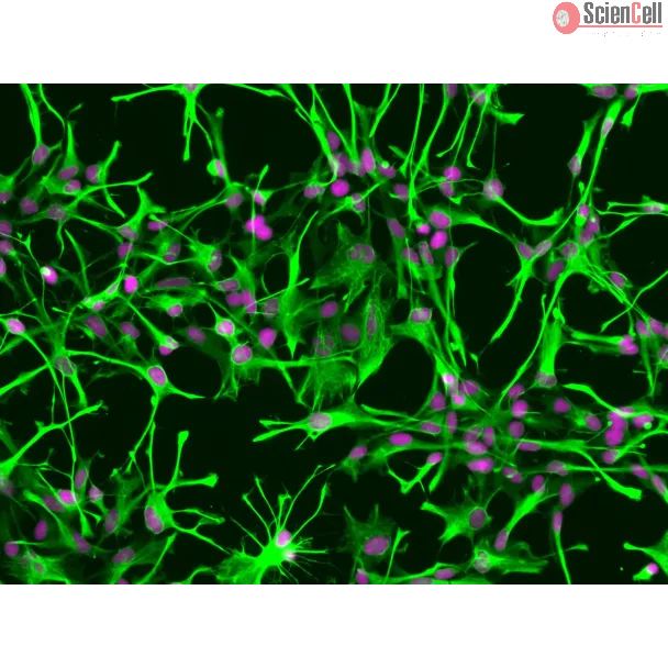
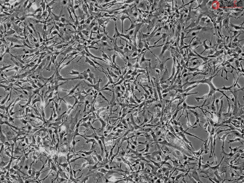
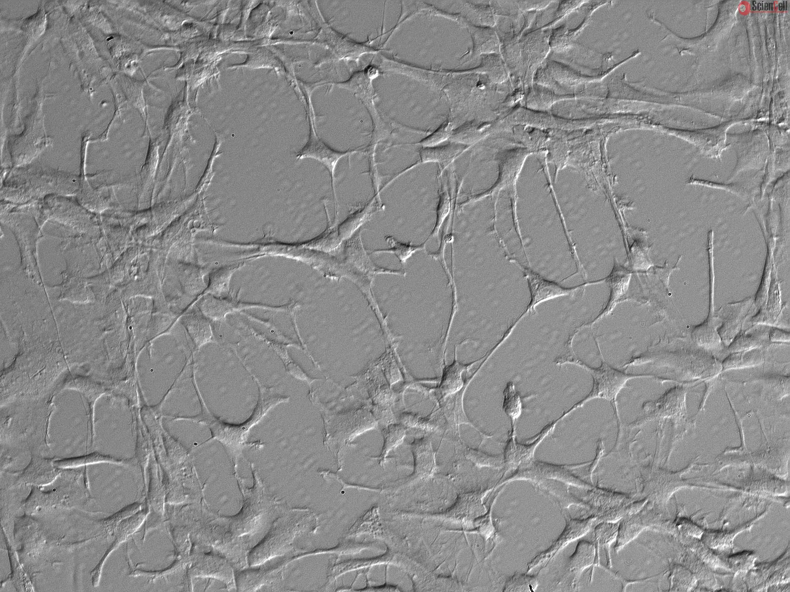
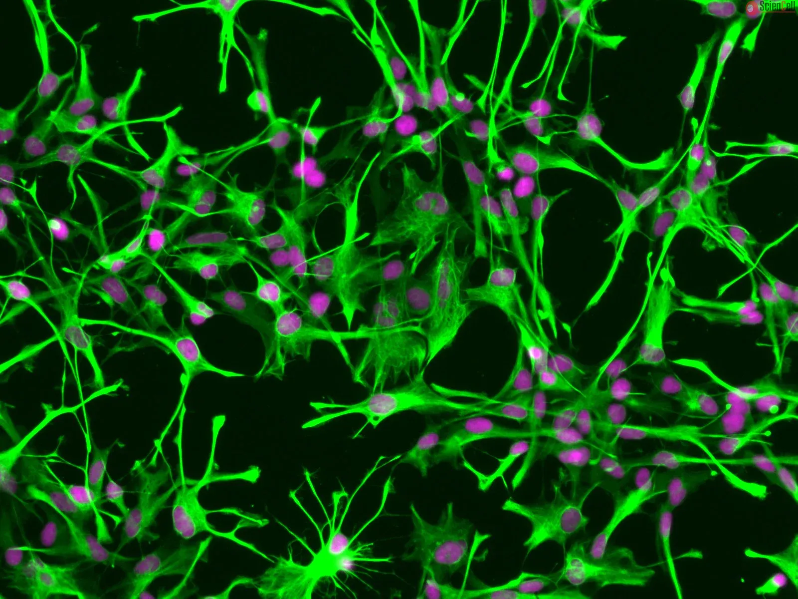
,-1-mg-ml--2.jpg)