Renal proximal tubular epithelial cells (RPTEpiC) play a crucial role in renal function. They reabsorb nearly all the glucose and amino acids in the glomerular filtrate, while allowing substances of no nutritional value to be excreted into the urine. They are also a major site of injury in a variety of congenital, metabolic, and inflammatory diseases. RPTEpiC can produce inflammatory mediators, such as cytokines or chemokines, and actively participate in acute inflammatory processes by affecting and directing leukocyte chemotaxis via the production of IL-8. RPTEpiC express IL-2R alpha and MHC class II antigens during inflammation, after renal transplantation, and during crescentic glomerulonephritis, suggesting that these cells have the capacity to participate in the pathogenesis of immune renal injury. To study the relationship between proximal tubular cells and a variety of renal diseases, the RPTEpiC culture is a useful in vitro model.
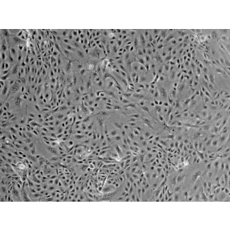
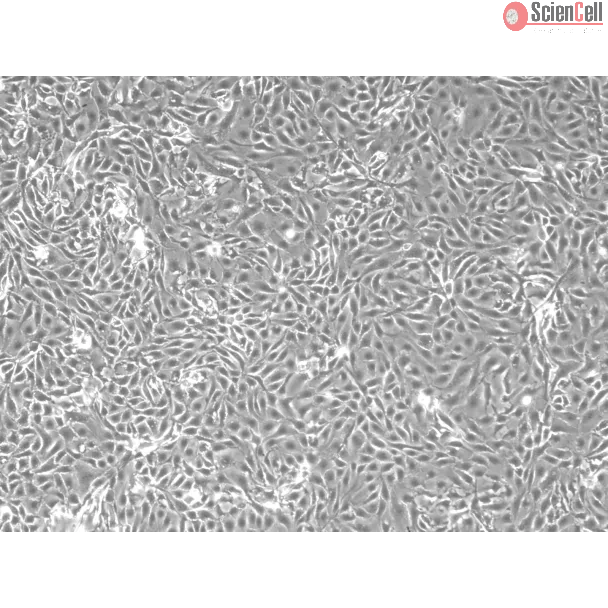
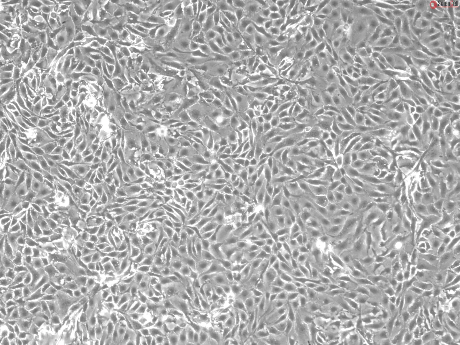
MRPTEpiC from ScienCell Research Laboratories are isolated from CD-1 mouse kidney. MRPTEpiC are cryopreserved at passage one and delivered frozen. Each vial contains >5 x 10⁵ cells in 1 ml volume. MRPTEpiC are characterized by immunofluorescence with antibodies specific to cytokeratin-18. MRPTEpiC are negative for mycoplasma, bacteria, yeast, and fungi. MRPTEpiC are guaranteed to further culture under the conditions provided by ScienCell Research Laboratories; however, MRPTEpiC are not recommended for long-term cultures due to limited expansion capacity and senescence after subculturing.
Recommended Medium
It is recommended to use Epithelial Cell Medium-animal (EpiCM-a, Cat. #4131) for culturing MRPTEpiC in vitro.
Cisplatin-induced nephrotoxicity leaded to apoptosis of tubular epithelial cells (ECs) and tubulointerstitial fibrosis through ROS stress and inflammatory cytokines. Tubu... More
Cisplatin-induced nephrotoxicity leaded to apoptosis of tubular epithelial cells (ECs) and tubulointerstitial fibrosis through ROS stress and inflammatory cytokines. Tubulointerstitial fibrosis caused by cisplatin might be via activation of resident fibroblasts and epithelial-mesenchymal transition (EMT) of tubular ECs. Inflammatory niche was crucial for progression of fibroblast activation or EMT. It had been reported that M1/M2 macrophage polarization regulated pro-inflammation or pro-resolving phase in damage repairing. However, the role of macrophage polarization on cisplatin-induced EMT of tubular ECs had not been well elucidated. In this study, we used co-cultured cell model and condition medium to examine the interaction between tubular ECs, fibroblasts and M1/M2 macrophages. Our data showed that cisplatin alone induced incomplete EMT of tubular ECs, whereas fibroblasts co-cultured with cisplatin-treated ECs could lead to fibroblast activation by detection of α-SMA and collagen-1. Moreover, decrease of iNOS and increase of argenase-1 and CD206 expression indicated that macrophages co-cultured with cisplatin-treated ECs would turn to M2 phenotype. Finally, we found that condition medium of M2 macrophages could promote complete EMT of cisplatin-treated ECs. Taken together, cisplatin created an inflammatory niche via tubular ECs to activate fibroblasts and stimulated M2 macrophage polarization. M2 macrophages could turn back to promote EMT of cisplatin-treated ECs. These results revealed the cooperative roles of tubular ECs, fibroblast and M2 macrophages to facilitate the progression of renal fibroblasis. Less
Renal fibrosis is a common pathological feature of all kinds of chronic kidney diseases (CKDs) with uncertain mechanisms. Accumulating evidence demonstrated an important ... More
Renal fibrosis is a common pathological feature of all kinds of chronic kidney diseases (CKDs) with uncertain mechanisms. Accumulating evidence demonstrated an important role of oxidative stress in the pathogenesis of CKD. Here we hypothesized that MnTBAP (manganese (III) tetrakis (4-benzoic acid)porphyrin chloride), a cell-permeable mimic of superoxide dismutase (SOD), may protect against the fibrotic response in CKD by antagonizing oxidative stress. To verify this hypothesis, we performed experiments in tubular epithelial cells and mice with 5/6 nephrectomy (Nx). In mouse tubular epithelial cells, TGF-β1 induced a significant transition to fibrotic phenotype in line with a remarkable mitochondrial dysfunction, which was markedly improved by MnTBAP (1.14 μM) pretreatment. In remnant kidneys of 5/6 Nx mice, tubulointerstitial fibrosis occurred in parallel with mitochondrial abnormality in renal tubular cells. Administration of MnTBAP significantly attenuated the deposition of extracellular matrix as evidenced by the blocked expressions of fibronectin, collagen I, and collagen III. Masson staining also displayed an ameliorated accumulation of collagenous matrix in MnTBAP-treated mice. Moreover, MnTBAP also significantly improved the severity of proteinuria without altering CKD-related hypertension. Collectively, MnTBAP therapy served as a promising strategy in preventing renal fibrosis in CKDs possibly via antagonizing mitochondrial-derived oxidative stress and subsequent protection of mitochondrial function. Less
ScienCell Research Laboratories (SRL) takes pride in being a resource for researchers all over the world. The publications listed here are not meant as an endorsement or confirmation of the reliability of the products.

,-1-mg-ml--2.jpg)