The trophoblast begins at the outer covering of the early blastocyst and provides the route of nourishment between the maternal endometrium and the developing embryo. The trophoblast adhesion to the uterine wall is the requisite first step of implantation and, subsequently, placentation. Human villous tryophoblasts (HVT) covering the villi of the placenta provide the surface for the exchange of oxygen and nutrients with the maternal circulation. They synthesize and release chorionic gonadotropin, placental lactogen and angiogenin and express CXCR4, CCR5 and prolactin gene family. They acquire CCR1 as they differentiate to an invasive phenotype at the villous-anchoring sites. The features of HVT, together with the recent establishment of trophoblast stem cells, make them an ideal genetic platform to study cell differentiation and organogenesis.
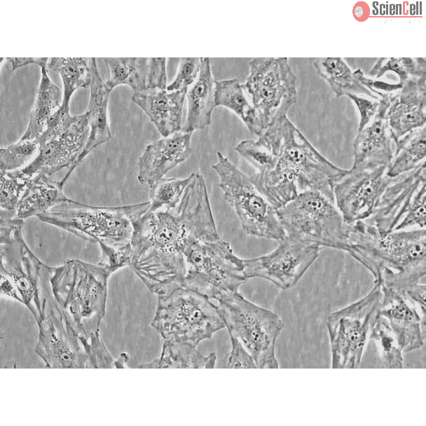
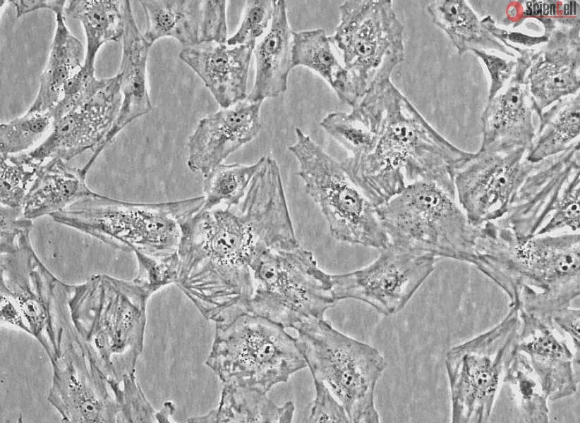
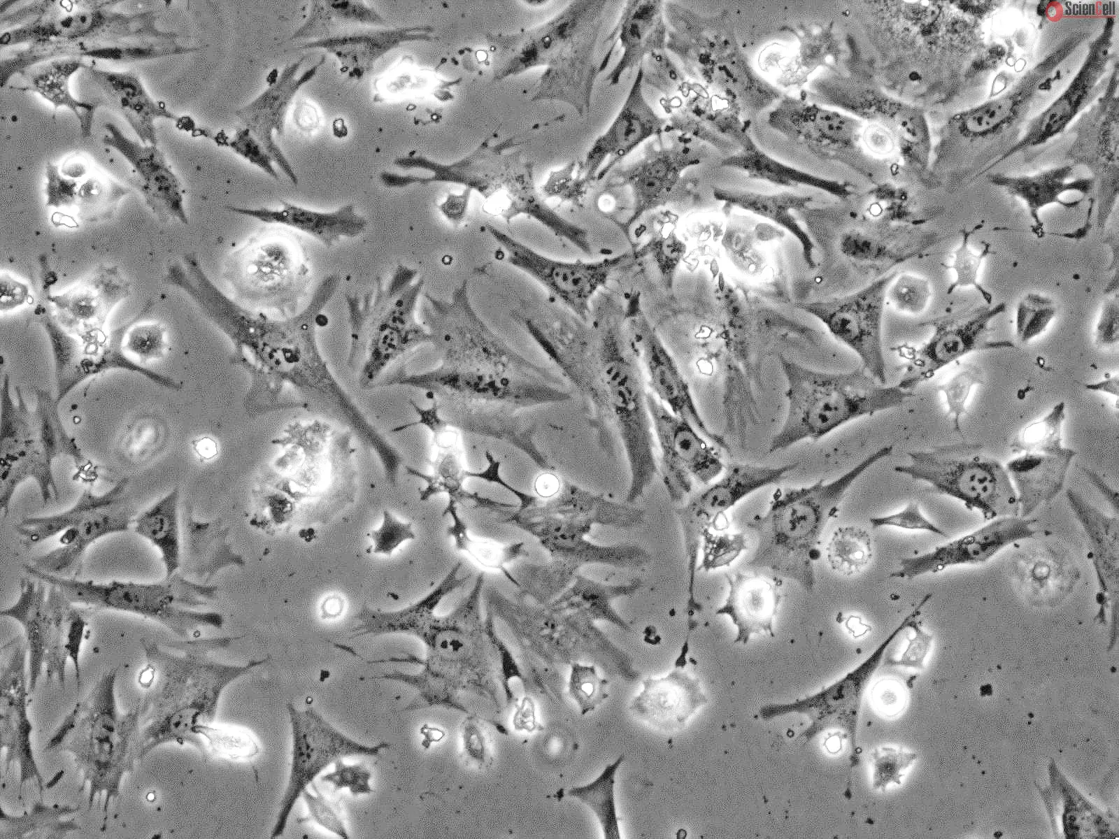
HVT from ScienCell Research Laboratories are isolated from human placental villi. HVT are cryopreserved at passage one and delivered frozen. Each vial contains >1 x 10⁶ cells in 1 ml volume. HVT are characterized by HCG ELISA. HVT are negative for HIV-1, HBV, HCV, mycoplasma, bacteria, yeast and fungi. HVT are guaranteed to further culture under the conditions provided by ScienCell Research Laboratories; however, HVT are not recommended for expanding or long-term cultures since the cells do not proliferate in regular culture.
Recommended Medium
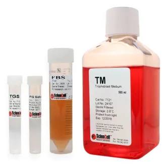
It is recommended to use Trophoblast Medium (TM, Cat. #7121) for the culturing of HVT in vitro.
Product Sheets
 7120
7120
Search for a Certificate of Analysis by Lot Number below:
Virus–host interactions play vital roles in viral replication and virus-induced pathogenesis. Viruses rely entirely upon host cells to reproduce progeny viruses; howeve... More
Virus–host interactions play vital roles in viral replication and virus-induced pathogenesis. Viruses rely entirely upon host cells to reproduce progeny viruses; however, host factors positively or negatively regulate virus replication by interacting with viral proteins. The elucidation of virus–host protein interaction not only provides a better understanding of the molecular mechanisms by which host cells combat viral infections, but also facilitates the development of new anti-viral therapeutics. Identification of relevant host factors requires techniques that enable comprehensive characterization of virus–host protein interactions. In this study, we developed a proteomic approach to systematically identify human protein kinases that interact potently with viral proteins. For this purpose, we synthesized 412 full-length human protein kinases using the wheat germ cell-free protein synthesis system, and screened them for their association with a virus protein using the amplified luminescent proximity homogenous assay (AlphaScreen). Using this system, we attempted to discover a robust anti-viral host restriction mechanism targeting virus protein X (Vpx) of HIV-2. The screen identified H11/HSPB8 as a Vpx-binding protein that negatively regulates the stability and function of Vpx. Indeed, overexpression of H11/HSPB8 promoted the degradation of Vpx via the ubiquitin–proteasome pathway and inhibited its interaction with SAMHD1, a host restriction factor responsible for blocking replication of HIV. Conversely, targeted knockdown of H11/HSPB8 in human trophoblast cells, which ordinarily express high levels of this protein, restored the expression and function of Vpx, making the cells highly susceptible to viral replication. These results demonstrate that our proteomic approach represents a powerful tool for revealing virus–host interaction not yet identified by conventional methods. Furthermore, we showed that H11/HSPB8 could be a potential host regulatory factor that may prevent placental infection of HIV-2 during pregnancy. Less
Microparticles (MPs) are sub-micron membrane vesicles (100–1000 nm) shed from normal and pathologic cells due to stimulation or apoptosis. MPs can be found in the perip... More
Microparticles (MPs) are sub-micron membrane vesicles (100–1000 nm) shed from normal and pathologic cells due to stimulation or apoptosis. MPs can be found in the peripheral blood circulation of healthy individuals, whereas elevated concentrations are found in pregnancy and in a variety of diseases. Also, MPs participate in physiological processes, e.g., coagulation, inflammation, and angiogenesis. Since their clinical properties are important, we have developed a new methodology based on nano-imaging that provides significant new data on MPs nanostructure, their composition and function. We are among the first to characterize by direct-imaging cryogenic transmitting electron microscopy (cryo-TEM) the near-to-native nanostructure of MP systems isolated from different cell types and stimulation procedures. We found that there are no major differences between the MP systems we have studied, as most particles were spherical, with diameters from 200 to 400 nm. However, each MP population is very heterogeneous, showing diverse morphologies. We investigated by cryo-TEM the effects of standard techniques used to isolate and store MPs, and found that either high-g centrifugation of MPs for isolation purposes, or slow freezing to –80°C for storage introduce morphological artifacts, which can influence MP nanostructure, and thus affect the efficiency of these particles as future diagnostic tools. Less
ScienCell Research Laboratories (SRL) takes pride in being a resource for researchers all over the world. The publications listed here are not meant as an endorsement or confirmation of the reliability of the products.




,-1-mg-ml--2.jpg)