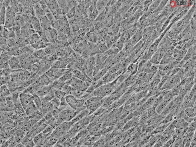
Vascular smooth muscle cells (SMC) are primary contributors to the development of arterial disease. The ability of vascular SMC to switch to a proliferative phenotype is one of the main factors in the development and progression of vascular disease. Vascular smooth muscle cells express ICAM1 and VCAM-1, which may contribute to the inflammatory reaction in the vascular wall and may actively be involved in the progression of vascular disease. Vascular SMC in culture play an important role in vascular disease research and can be used to identify new therapeutic targets to treat arterial disease
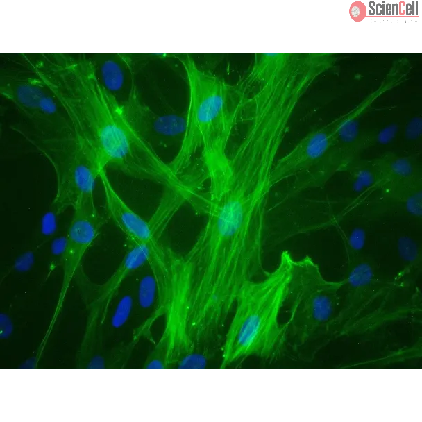
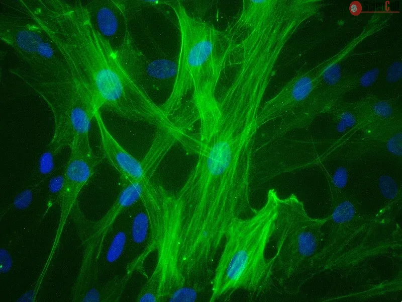
HUVSMC from ScienCell Research Laboratories are isolated from human umbilical veins. HUVSMC are cryopreserved at passage one and delivered frozen. Each vial contains >5 x 10⁵ cells in 1 ml volume. HUVSMC are characterized by immunofluorescence with antibodies specific to α-smooth muscle actin. HUVSMC are negative for HIV-1, HBV, HCV, mycoplasma, bacteria, yeast and fungi. HUVSMC are guaranteed to further expand for 15 population doublings under the conditions provided by ScienCell Research Laboratories.
Recommended Medium
It is recommended to use Smooth Muscle Cell Medium (SMCM, Cat. #1101) for the culturing of HUVSMC in vitro.
Vascular calcification is an important risk factor associated with mortality among patients with chronic kidney disease. Intracellular cholesterol metabolism is involved ... More
Vascular calcification is an important risk factor associated with mortality among patients with chronic kidney disease. Intracellular cholesterol metabolism is involved in the process of vascular cell calcification. In this study, we investigated the role of UbiA prenyltransferase domain containing 1 (UBIAD1) in intracellular cholesterol metabolism and vascular cell calcification, and identified its subcellular location. Primary human umbilical vein smooth muscle cells (HUVSMCs) were incubated with either growth medium (1.4 mmol/L Pi) or calcification medium (CM) (3.0 mmol/L Pi). Under treatment with CM, HUVSMCs were further incubated with exogenous cholesterol, or menaquinone-4, a product of UBIAD1. The plasmid and small interfering RNA were transfected in HUVSMCs to alter the expression of UBIAD1. Matrix calcium quantitation, alkaline phosphatase activity, intracellular cholesterol level and menaquinone-4 level were measured. The expression of several genes involved in cholesterol metabolism were analyzed. Using an anti-UBIAD1 antibody, an endoplasmic reticulum marker and a Golgi marker, the subcellular location of UBIAD1 in HUVSMCs was analyzed. CM increased matrix calcium, alkaline phosphatase activity and intracellular cholesterol level, and reduced UBIAD1 expression and menaquinone-4 level. Addition of cholesterol contributed to increased matrix calcification and alkaline phosphatase activity in a dose-dependent manner. Elevated expression of UBIAD1 or menaquinone-4 in HUVSMCs treated with CM significantly reduced intracellular cholesterol level, matrix calcification and alkaline phosphatase activity, but increased menaquinone-4 level. Elevated expression of UBIAD1 or menaquinone-4 reduced the gene expression of sterol regulatory element-binding protein-2, and increased gene expression of ATP binding cassette transporters A1, which are in charge of cholesterol synthesis and efflux. UBIAD1 co-localized with the endoplasmic reticulum marker and the Golgi marker in HUVSMCs. In conclusion, high intracellular cholesterol content contributes to phosphate-induced vascular cell differentiation and calcification. UBIAD1 or menaquinone-4 could decrease vascular cell differentiation and calcification, probably via its potent role of inversely modulating cellular cholesterol. Less
Autophagy, a type II programmed cell death, is essential for cell survival under stress, e.g. lung injury, and bone marrow-derived mesenchymal stem cells (BM-MSCs) have g... More
Autophagy, a type II programmed cell death, is essential for cell survival under stress, e.g. lung injury, and bone marrow-derived mesenchymal stem cells (BM-MSCs) have great potential for cell therapy. However, the mechanisms underlying the BM-MSC activation of autophagy to provide a therapeutic effect in ischaemia/reperfusion-induced lung injury (IRI) remain unclear. Thus, we investigate the activation of autophagy in IRI following transplantation with BM-MSCs. Seventy mice were pre-treated with BM-MSCs before they underwent lung IRI surgery in vivo. Human pulmonary micro-vascular endothelial cells (HPMVECs) were pre-conditioned with BM-MSCs by oxygen-glucose deprivation/reoxygenation (OGD) in vitro. Expression markers for autophagy and the phosphoinositide 3-kinase/protein kinase B (PI3K/Akt) signalling pathway were analysed. In IRI-treated mice, administration of BM-MSCs significantly attenuated lung injury and inflammation, and increased the level of autophagy. In OGD-treated HPMVECs, co-culture with BM-MSCs attenuated endothelial permeability by decreasing the level of cell death and enhanced autophagic activation. Moreover, administration of BM-MSCs decreased the level of PI3K class I and p-Akt while the expression of PI3K class III was increased. Finally, BM-MSCs-induced autophagic activity was prevented using the inhibitor LY294002. Administration of BM-MSCs attenuated lung injury by improving the autophagy level via the PI3K/Akt signalling pathway. These findings provide further understanding of the mechanisms related to BM-MSCs and will help to develop new cell-based therapeutic strategies in lung injury. Less
Increasing evidence supports the hypothesis that inflammatory reactions serves an important function in the formation, progression and plaque rupture of atherosclerosis. ... More
Increasing evidence supports the hypothesis that inflammatory reactions serves an important function in the formation, progression and plaque rupture of atherosclerosis. Interleukin (IL)‑1 primarily induces inflammation and is closely associated with the inflammatory environment and the formation of atherosclerosis. The present study aimed to establish an in vitro model for the evaluation of drug efficacy in the intervention of atherosclerosis from the inflammatory perspective, and to observe the anti‑inflammatory effects of tanshinone IIA and andrographolide on atherosclerosis. The IL‑1β‑induced inflammation‑activated endothelial cell (EC)‑smooth muscle cell (SMC)‑mononuclear cell (MC) co‑culture model was established, based on the changes in a series of atherosclerosis‑associated inflammatory markers secreted by ECs and SMCs. The expression of connexin in ECs, adhesion of MCs and changes in inflammatory signalling molecules were selected as evaluation indices for the inflammatory microenvironment of atherosclerosis. The use of this model revealed that tanshinone IIA exhibited significant efficacy against atherosclerosis and its inflammatory reactions. Inflammatory reactions were regarded as the primary mechanism underlying atherosclerosis. The established model simulated a series of relevant changes in the arterial wall under the inflammatory cytokines with oxidized low‑density lipoprotein during the atherosclerotic process. The present study presented a reliable method for the identification of drugs with potential anti‑inflammatory activity in atherosclerosis, for investigating the mechanisms of action, considering the improvement of the inflammatory state and the increase in plaque stability observed. Less
ScienCell Research Laboratories (SRL) takes pride in being a resource for researchers all over the world. The publications listed here are not meant as an endorsement or confirmation of the reliability of the products.

,-1-mg-ml--2.jpg)