There is a growing demand for in vitro assays for toxicity screening in three-dimensional (3D) environments. In this study, 3D cell culture using magnetic levitation was ... More
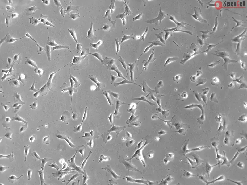
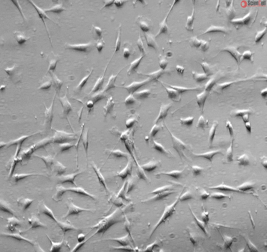
There is a growing demand for in vitro assays for toxicity screening in three-dimensional (3D) environments. In this study, 3D cell culture using magnetic levitation was used to create an assay in which cells were patterned into 3D rings that close over time. The rate of closure was determined from time-lapse images taken with a mobile device and related to drug concentration. Rings of human embryonic kidney cells (HEK293) and tracheal smooth muscle cells (SMCs) were tested with ibuprofen and sodium dodecyl sulfate (SDS). Ring closure correlated with the viability and migration of cells in two dimensions (2D). Images taken using a mobile device were similar in analysis to images taken with a microscope. Ring closure may serve as a promising label-free and quantitative assay for high-throughput in vivo toxicity in 3D cultures. Less
There is a growing demand for in vitro assays for toxicity screening in three-dimensional (3D) environments. In this study, 3D cell culture using magnetic levitation was ... More
There is a growing demand for in vitro assays for toxicity screening in three-dimensional (3D) environments. In this study, 3D cell culture using magnetic levitation was used to create an assay in which cells were patterned into 3D rings that close over time. The rate of closure was determined from time-lapse images taken with a mobile device and related to drug concentration. Rings of human embryonic kidney cells (HEK293) and tracheal smooth muscle cells (SMCs) were tested with ibuprofen and sodium dodecyl sulfate (SDS). Ring closure correlated with the viability and migration of cells in two dimensions (2D). Images taken using a mobile device were similar in analysis to images taken with a microscope. Ring closure may serve as a promising label-free and quantitative assay for high-throughput in vivo toxicity in 3D cultures. Less
Heme oxygenase-1 (HO-1) is known as an oxidative stress protein that is up-regulated by various stimuli. HO-1 has been shown to protect cells against oxidative damage. Ci... More
Heme oxygenase-1 (HO-1) is known as an oxidative stress protein that is up-regulated by various stimuli. HO-1 has been shown to protect cells against oxidative damage. Cigarette smoke is a potential inflammatory mediator that causes chronic obstructive pulmonary disease and asthma. In this study, we report that cigarette smoke particle-phase extract (CSPE) is an inducer of HO-1 expression mediated through various signaling pathways in human tracheal smooth muscle cells (HTSMCs). CSPE-induced HO-1 protein, mRNA expression, and promoter activity were attenuated by pretreatment with a ROS scavenger (N-acetyl-l-cysteine) and inhibitors of c-Src (PP1), NADPH oxidase [diphenylene iodonium chloride (DPI) and apocynin (APO)], MEK1/2 (U0126), p38 MAPK (SB202190), and JNK1/2 (SP600125) or transfection with siRNAs for Src, p47(phox), NOX2, p42, p38, JNK2, or NF-E2-related factor 2 (Nrf2). CSPE-stimulated translocation of p47(phox) and Nrf2, ROS production, and NADPH oxidase activity was attenuated by transfection with siRNAs for Src, p47(phox), and NOX2 or pretreatment with PP1, DPI, or APO. Furthermore, CSPE-induced NOX2, c-Src, and p47(phox) complex formation was revealed by immunoprecipitation using an anti-NOX2, anti-p47(phox), or anti-c-Src Ab followed by Western blot against anti-NOX2, anti-p47(phox), or anti-c-Src Abs. These results demonstrate that CSPE-induced ROS generation is mediated through a c-Src/NADPH oxidase/MAPK pathway and in turn initiates the activation of Nrf2 and ultimately induces HO-1 expression in HTSMCs. Less
Cytosolic phospholipase A(2) (cPLA(2)) plays a pivotal role in mediating agonist-induced arachidonic acid (AA) release for prostaglandin (PG) synthesis during inflammatio... More
Cytosolic phospholipase A(2) (cPLA(2)) plays a pivotal role in mediating agonist-induced arachidonic acid (AA) release for prostaglandin (PG) synthesis during inflammation triggered by IL-1beta. However, the mechanisms underlying IL-1beta-induced cPLA(2) expression and PGE(2) synthesis in human tracheal smooth muscle cells (HTSMCs) remain unknown. IL-1beta-induced cPLA(2) protein and mRNA expression, PGE(2) production, or phosphorylation of p42/p44 MAPK, p38 MAPK, and JNK1/2, which was attenuated by pretreatment with the inhibitors of MEK1/2 (U0126), p38 MAPK (SB202190), and JNK1/2 (SP600125) or transfection with siRNAs of MEK1, p42, p38, and JNK2. IL-1beta-induced cPLA(2) expression was also inhibited by pretreatment with a NF-kappaB inhibitor, helenalin or transfection with siRNA of NIK, IKKalpha, or IKKbeta. IL-beta-induced NF-kappaB translocation was blocked by pretreatment with helenalin, but not U0126, SB202190, and SP600125. In addition, transfection with p300 siRNA blocked cPLA(2) expression induced by IL-1beta. Moreover, p300 was associated with the cPLA(2) promoter, which was dynamically linked to histone H4 acetylation stimulated by IL-1beta. These results suggest that in HTSMCs, activation of MAPKs, NF-kappaB, and p300 are essential for IL-1beta-induced cPLA(2) expression and PGE(2) secretion. Copyright 2010 Wiley-Liss, Inc. Less
Up-regulation of cytosolic phospholipase A2 (cPLA2) by cigarette smoke extract (CSE) may play a critical role in airway inflammatory diseases. However, the mechanisms und... More
Up-regulation of cytosolic phospholipase A2 (cPLA2) by cigarette smoke extract (CSE) may play a critical role in airway inflammatory diseases. However, the mechanisms underlying CSE-induced cPLA2 expression in human tracheal smooth muscle cells (HTSMCs) remain unknown. CSE induced cPLA2 protein and mRNA expression, and ROS generation was attenuated by pretreatment with a reactive oxygen species (ROS) scavenger (N-acetylcysteine), or inhibitors of NADPH oxidase (diphenyleneiodonium chloride, apocynin) and transfection with p47phox siRNA, suggesting that CSE-induced cPLA2 expression was mediated through NADPH oxidase activation and ROS production in HTSMCs. Furthermore, CSE-induced cPLA2 expression was attenuated by pretreatment with the inhibitors of MEK1/2 (U0126), p38 MAPK (SB202190), and JNK (SP600125), which were further confirmed by transfection with siRNAs of JNK1, p42, and p38 to down-regulate the expression of respective proteins and reduce cPLA2 expression. Induction of cPLA2 by CSE was attenuated by selective inhibitors of NF-kappaB (helenalin) and AP-1 (curcumin). Moreover, promoter assays revealed that increases of cPLA2, NF-kappaB, and AP-1 luciferase activities stimulated by CSE were attenuated by these inhibitors. These results suggest that in HTSMCs, CSE induced NADPH oxidase activation leading to phosphorylation of p42/p44 MAPK, p38 MAPK, and JNK. These reactions induced nuclear transcription NF-kappaB and AP-1 activities which were essential for CSE-induced cPLA2 gene expression. Less
Lipopolysaccharide (LPS) has been shown to up-regulate the expression of vascular cell adhesion molecule (VCAM)-1 which contributes to the occurrence of airway inflammato... More
Lipopolysaccharide (LPS) has been shown to up-regulate the expression of vascular cell adhesion molecule (VCAM)-1 which contributes to the occurrence of airway inflammatory diseases. Genetic analysis reveals the existence of activator protein-1 (AP-1) binding site on VCAM-1 promoter region. However, the role of AP-1 in LPS-induced VCAM-1 expression in human tracheal smooth muscle cells (HTSMCs) is not known. Here, we show that LPS increased VCAM-1 expression and adhesiveness of HTSMCs through AP-1, since pretreatment with an AP-1 inhibitor tanshinone attenuated LPS-induced VCAM-1 expression and leukocytes adhesion. The implication of AP-1 in LPS-induced VCAM-1 expression was confirmed by animal studies showing that pretreatment of mice with tanshinone attenuated LPS-induced VCAM-1 mRNA expression in airway tissues and accumulation of leukocytes in bronchoalveolar lavage. By using the pharmacological inhibitors and transfection with siRNA of PKC, p42, p38, or JNK2, LPS-induced expression of c-Fos was mediated through protein kinase C (PKC), p42/p44 MAPK and p38 MAPK. While, c-Jun expression was mediated through PKC and mitogen-activated protein kinases (MAPKs, p42/p44 MAPK, p38 MAPK and JNK) in HTSMCs. Pretreatment with the inhibitors of PKCs or MAPKs attenuated LPS-stimulated nuclear translocation and VCAM-1 promoter binding abilities of AP-1, which attenuated promoter activity and gene expression of VCAM-1 and the adhesiveness between HTSMCs and leukocytes. These results indicated that differential regulation of AP-1 through PKCs-dependent MAPKs activation plays central roles in LPS-induced VCAM-1 expression. The altered modulation of this axis with inhibitors or siRNAs may contribute to the improvement of airway inflammatory diseases. Less
Histone acetylation regulated by histone acetyltransferases (HATs) and histone deacetylases (HDACs) plays a critical role in the expression of inflammatory genes, such as... More
Histone acetylation regulated by histone acetyltransferases (HATs) and histone deacetylases (HDACs) plays a critical role in the expression of inflammatory genes, such as vascular cell adhesion molecule-1 (VCAM-1). Oxidative processes have been shown to induce VCAM-1 expression. Here, we investigated the mechanisms underlying IL-1beta-induced VCAM-1 expression in human tracheal smooth muscle cells (HTSMCs). Our results showed that IL-1beta enhanced HTSMCs-monocyte adhesion through up-regulation of VCAM-1, which was inhibited by pretreatment with selective inhibitors of PKCalpha (Gö6976), c-Src (PP1), NADPH oxidase [diphenylene iodonium (DPI) and apocynin (APO)], intracellular calcium chelator (BAPTA/AM), PI-PLC (U73122), CaM (calmidazolium chloride), CaM kinase II (KN62), p300 (garcinol), NF-kappaB (Bay11-7082), HDAC (trichostatin A), and ROS scavenger [N-acetyl-L-cysteine (NAC)] or transfection with siRNAs of MyD88, PKCalpha, Src, p47(phox), p300, and HDAC4. Moreover, IL-1beta stimulated NF-kappaB and CaMKII phosphorylation through MyD88-dependent PI-PLC/PKCalpha/c-Src/ROS and PI-PLC/Ca2+/CaM pathways, respectively. Activation of NF-kappaB and CaMKII may eventually lead to the acetylation of histone residues and phosphorylation of histone deacetylases. These findings suggested that IL-1beta induced VCAM-1 expression via these multiple signaling pathways in HTSMCs. Blockade of these pathways may reduce monocyte adhesion via VCAM-1 suppression and attenuation of the inflammatory responses in airway diseases. Less
Exposure to cigarette smoke extract (CSE) leads to airway or lung inflammation, which may be mediated through cyclooxygenase-2 (COX-2) expression and its product prostagl... More
Exposure to cigarette smoke extract (CSE) leads to airway or lung inflammation, which may be mediated through cyclooxygenase-2 (COX-2) expression and its product prostaglandin E2 (PGE2) synthesis. The aim of this study was to investigate the molecular mechanisms underlying CSE-induced COX-2 expression in human tracheal smooth muscle cells (HTSMCs). Here, we describe that COX-2 induction is dependent on PKCalpha/c-Src/EGFR, PDGFR/PI3K/Akt/NF-kappaB signaling in HTSMCs. CSE stimulated the phosphorylation of c-Src, EGFR, PDGFR, and Akt, which were inhibited by pretreatment with the inhibitor of PKCalpha (Gö6976 or Gö6983), c-Src (PP1), EGFR (AG1478), PDGFR (AG1296), or PI3K (LY294002). Moreover, CSE induced a significant increase in COX-2 expression, which was reduced by pretreatment with these inhibitors or transfection with siRNA of PKCalpha, Src, or Akt. Furthermore, CSE-stimulated NF-kappaB p65 phosphorylation and translocation were also attenuated by pretreatment with Gö6976, PP1, AG1478, AG1296, or LY294002. CSE-induced COX-2 expression was also mediated through the recruitment of p300 associated with NF-kappaB in HTSMCs, revealed by coimmunoprecipitation and Western blot analysis. In addition, pretreatment with the inhibitors of NF-kappaB (helenalin) and p300 (garcinol) or transfection with p65 siRNA and p300 siRNA markedly inhibited CSE-regulated COX-2 expression. However, CSE-induced PGE2 generation was reduced by pretreatment with the inhibitor of COX-2 (NS-398). These results demonstrated that in HTSMCs, CSE-induced COX-2-dependent PGE2 generation was mediated through PKCalpha/c-Src/EGFR, PDGFR/PI3K/Akt leading to the recruitment of p300 with NF-kappaB complex. Less
Up-regulation of vascular cell adhesion molecule-1 (VCAM-1) involves adhesions between both circulating and resident leukocytes and the human tracheal smooth muscle cells... More
Up-regulation of vascular cell adhesion molecule-1 (VCAM-1) involves adhesions between both circulating and resident leukocytes and the human tracheal smooth muscle cells (HTSMCs) during airway inflammatory reaction. We have demonstrated previously that tumor necrosis factor (TNF)-alpha-induced VCAM-1 expression is regulated by mitogen-activated protein kinases, nuclear factor-kappaB, and p300 activation in HTSMCs. In addition to this pathway, phosphorylation of Akt and CaM kinase II has been implicated in histone acetyltransferase and histone deacetylase 4 (HDAC4) activation. Here, we investigated whether these different mechanisms participated in TNF-alpha-induced VCAM-1 expression and enhanced neutrophil adhesion. TNF-alpha significantly increased HTSMC-neutrophil adhesions, and this effect was associated with increased expression of VCAM-1 on the HTSMCs and was blocked by the selective inhibitors of Src [4-amino-5-(4-methylphenyl)-7-(t-butyl)pyrazolo[3,4-d]-pyrimidine (PP1)], epidermal growth factor receptor [EGFR; 4-(3'-chloroanilino)-6,7-dimethoxy-quinazoline, (AG1478)], phosphatidylinositol 3-kinase (PI3K) [2-(4-morpholinyl)-8-phenyl-1(4H)-benzopyran-4-one hydrochloride(LY294002) and wortmannin],calcium[1,2-bis(2-aminophenoxy) ethane-N,N,N',N'-tetraacetic acid-acetoxymethyl ester; BAPTA-AM], phosphatidylinositol-phospholipase C (PLC) [1-[6-[[17beta-methoxyestra-1,3,5(10)-trien-17-yl]amino]hexyl]-1H-pyrrole-2,5-dione (U73122)], protein kinase C (PKC) [12-(2-cyanoethyl)-6,7,12, 13-tetrahydro-13-methyl-5-oxo-5H-indolo(2,3-a)pyrrolo(3,4-c)-carbazole (Gö6976), rottlerin, and 3-1-[3-(amidinothio)propyl-1H-indol-3-yl]-3-(1-methyl-1H-indol-3-yl) maleimide (bisindolylmaleimide IX) (Ro 31-8220)], CaM (calmidazolium chloride), CaM kinase II [(8R(*),9S(*),11S(*))-(-)-9-hydroxy-9-methoxycarbonyl-8-methyl-14-n-propoxy-2,3,9, 10-tetrahydro-8,11-epoxy, 1H,8H, 11H-2,7b,11a-triazadibenzo[a,g]cycloocta[cde]trinden-1-one (KT5926) and 1-[N,O-bis(5-isoquinolinesulfonyl)-N-methyl-l-tyrosyl]-4-phenylpiperazine (KN62)], p300 (curcumin), and HDAC (trichostatin A) or transfection with short interfering RNAs for Src, Akt, PKCalpha, PKCmu, and HDAC4. At gene regulation level, reverse-transcriptase polymerase chain reaction and promoter assays revealed that expression of VCAM-1 was also attenuated by these signaling molecule inhibitors. Moreover, TNF-alpha induced Akt and CaM kinase II phosphorylation via cascades through Src/EGFR/PI3K and PLC/calcium/CaM, respectively. Finally, activation of Akt and CaM kinase II may eventually lead to the acetylation of histone residues and phosphorylation of histone deacetylase. These findings revealed that TNF-alpha induced VCAM-1 expression via multiple signaling pathways. Blockade of these pathways may be selectively targeted to reduce neutrophil adhesion via VCAM-1 suppression and attenuation of the inflammatory responses in airway diseases. Less
Matrix metalloproteinases (MMPs) are responsible for degradation of extracellular matrix and play important roles in cell migration, proliferation, and tissue remodeling ... More
Matrix metalloproteinases (MMPs) are responsible for degradation of extracellular matrix and play important roles in cell migration, proliferation, and tissue remodeling related to airway inflammation. Interleukin-1beta (IL-1beta) has been shown to induce MMP-9 production in many cell types and contribute to airway inflammatory responses. However, the mechanisms underlying MMP-9 expression induced by IL-1beta in human tracheal smooth muscle cells (HTSMCs) remain unclear. Here, we investigated the roles of p42/p44 MAPK, p38 MAPK, JNK, and NF-kappaB pathways for IL-1beta-induced MMP-9 production in HTSMCs. IL-1beta induced production of MMP-9 protein and mRNA in a time- and concentration-dependent manner determined by zymographic, Western blotting, and RT-PCR analyses, which was attenuated by inhibitors of MEK1/2 (U0126), p38 MAPK (SB202190), JNK (SP600125), and NF-kappaB (helenalin), and transfection with dominant negative mutants of MEK1/2, p38 and JNK, respectively. IL-1beta-stimulated phosphorylation of p42/p44 MAPK, p38 MAPK, and JNK was attenuated by pretreatment with U0126, SB202190, SP600125, or transfection with these dominant negative mutants of MEK, ERK, p38 and JNK, respectively. Furthermore, IL-1beta-stimulated translocation of NF-kappaB into the nucleus and degradation of IkappaB-alpha was blocked by helenalin. Finally, the reporter gene assay revealed that MAPKs and NF-kappaB are required for IL-1beta-induced MMP-9 luciferase activity in HTSMCs. MMP-9 promoter activity was enhanced by IL-1beta in HTSMCs transfected with MMP-9-Luc, which was inhibited by helenalin, U0126, SB202190, and SP600125. Taken together, the transcription factor NF-kappaB, p42/p44 MAPK, p38 MAPK, and JNK that are involved in MMP-9 expression in HTSMCs exposed to IL-1beta have now been identified. Less
Background and objectives: In asthma, airway smooth muscle cell (ASMC) hyperplasia plays an important role in airway remodelling. Increased expression of matrix metallopr... More
Background and objectives: In asthma, airway smooth muscle cell (ASMC) hyperplasia plays an important role in airway remodelling. Increased expression of matrix metalloproteinases-9 (MMP-9), a disintegrin and metalloprotease 33 (ADAM33) in ASMCs are also relevant to asthmatic airway remodelling. 1,25-dihydroxyvitamin D(3) (1,25-(OH)(2)D(3)) has potent antiproliferative properties in vitro in various cell types; however, its role in ASMCs is not well understood. This study investigated the effect of 1,25-(OH)(2)D(3) on passively sensitized human bronchial (airway) smooth muscle cell (HASMC) proliferation and MMP-9 and ADAM33 expressions. Methods: The effect of 1,25-(OH)(2)D(3) on cell proliferation was examined by 3-(4,5-dimethylthiazole-2-yl)-2,5-diphenyltetrazolium-bromide colorimetry assay; cell cycle analysis by flow cytometry; and immunocytochemical staining for proliferating cell nuclear antigen (PCNA). The expression of MMP-9 and ADAM33 in HASMCs was investigated by real-time quantitative PCR and Western Blot analysis. Results: 1,25-(OH)(2)D(3) effectively suppressed passively sensitized HASMC proliferation, proliferating cell nuclear antigen expression and G(1)/S transition in HASMCs passively sensitized with asthmatic serum. Further analysis showed that 1,25-(OH)(2)D(3) significantly down-regulated the expressions of protein for MMP-9 and ADAM33, as well as their mRNA levels in passively sensitized HASMCs. Conclusions: 1,25-(OH)(2)D(3) has direct inhibitory effects on passively sensitized HASMCs in vitro, including inhibition of cell proliferation and expression of MMP-9 and ADAM33, suggesting a possible beneficial role for 1,25-(OH)(2)D(3) in preventing and treating asthmatic airway remodelling. Less
,-1-mg-ml--2.jpg)