Secondhand smoke (SHS) is a confirmed lung carcinogen that introduces thousands of toxic chemicals into the lungs. SHS contains chemicals that have been implicated in cau... More
Secondhand smoke (SHS) is a confirmed lung carcinogen that introduces thousands of toxic chemicals into the lungs. SHS contains chemicals that have been implicated in causing oxidative DNA damage in the airway epithelium. Although DNA repair is considered a key defensive mechanism against various environmental attacks, such as cigarette smoking, the associations of individual repair enzymes with susceptibility to lung cancer are largely unknown. This study investigated the role of NEIL2, a DNA glycosylase excising oxidative base lesions, in human lung cells treated with sidestream smoke (SSS), the main component of SHS. To do so, we generated NEIL2 knockdown cells using siRNA-technology and exposed them to SSS-laden medium. Representative SSS chemical compounds in the medium were analyzed by mass spectrometry. An increased production of reactive oxygen species (ROS) in SSS-exposed cells was detected through the fluorescent detection and the induction of HIF-1α. The long amplicon–quantitative PCR (LA-QPCR) assay detected significant dose-dependent increases of oxidative DNA damage in the HPRT gene of cultured human pulmonary fibroblasts (hPF) and BEAS-2B epithelial cells exposed to SSS for 24 h. These data suggest that SSS exposure increased oxidative stress, which could contribute to SSS-mediated toxicity. siRNA knockdown of NEIL2 in hPF and HEK 293 cells exposed to SSS for 24 h resulted in significantly more oxidative DNA damage in HPRT and POLB than in cells with control siRNA. Taken together, our data strongly suggest that decreased repair of oxidative DNA base lesions due to an impaired NEIL2 expression in non-smokers exposed to SSS would lead to accumulation of mutations in genomic DNA of lung cells over time, thus contributing to the onset of SSS-induced lung cancer. Less
Background: Electronic cigarettes (EC) deliver aerosol by heating fluid containing nicotine. Cartomizer EC combine the fluid chamber and heating element in a single unit.... More
Background: Electronic cigarettes (EC) deliver aerosol by heating fluid containing nicotine. Cartomizer EC combine the fluid chamber and heating element in a single unit. Because EC do not burn tobacco, they may be safer than conventional cigarettes. Their use is rapidly increasing worldwide with little prior testing of their aerosol. Objectives: We tested the hypothesis that EC aerosol contains metals derived from various components in EC. Methods: Cartomizer contents and aerosols were analyzed using light and electron microscopy, cytotoxicity testing, x-ray microanalysis, particle counting, and inductively coupled plasma optical emission spectrometry. Results: The filament, a nickel-chromium wire, was coupled to a thicker copper wire coated with silver. The silver coating was sometimes missing. Four tin solder joints attached the wires to each other and coupled the copper/silver wire to the air tube and mouthpiece. All cartomizers had evidence of use before packaging (burn spots on the fibers and electrophoretic movement of fluid in the fibers). Fibers in two cartomizers had green deposits that contained copper. Centrifugation of the fibers produced large pellets containing tin. Tin particles and tin whiskers were identified in cartridge fluid and outer fibers. Cartomizer fluid with tin particles was cytotoxic in assays using human pulmonary fibroblasts. The aerosol contained particles >1 µm comprised of tin, silver, iron, nickel, aluminum, and silicate and nanoparticles (<100 nm) of tin, chromium and nickel. The concentrations of nine of eleven elements in EC aerosol were higher than or equal to the corresponding concentrations in conventional cigarette smoke. Many of the elements identified in EC aerosol are known to cause respiratory distress and disease. Conclusions: The presence of metal and silicate particles in cartomizer aerosol demonstrates the need for improved quality control in EC design and manufacture and studies on how EC aerosol impacts the health of users and bystanders. Less
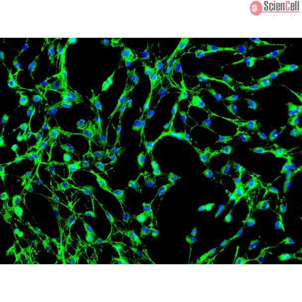
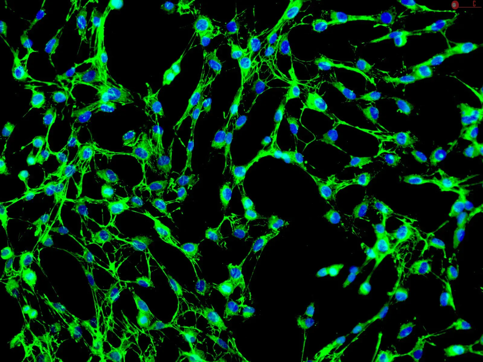
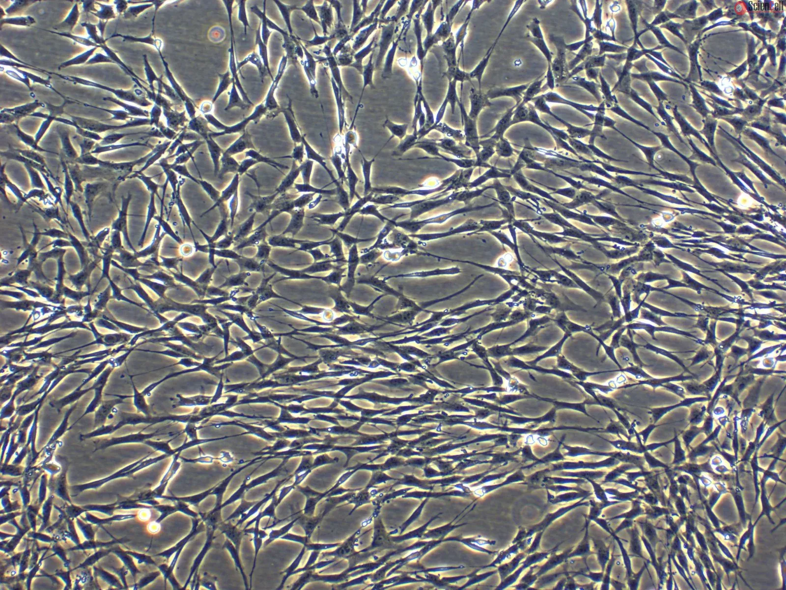
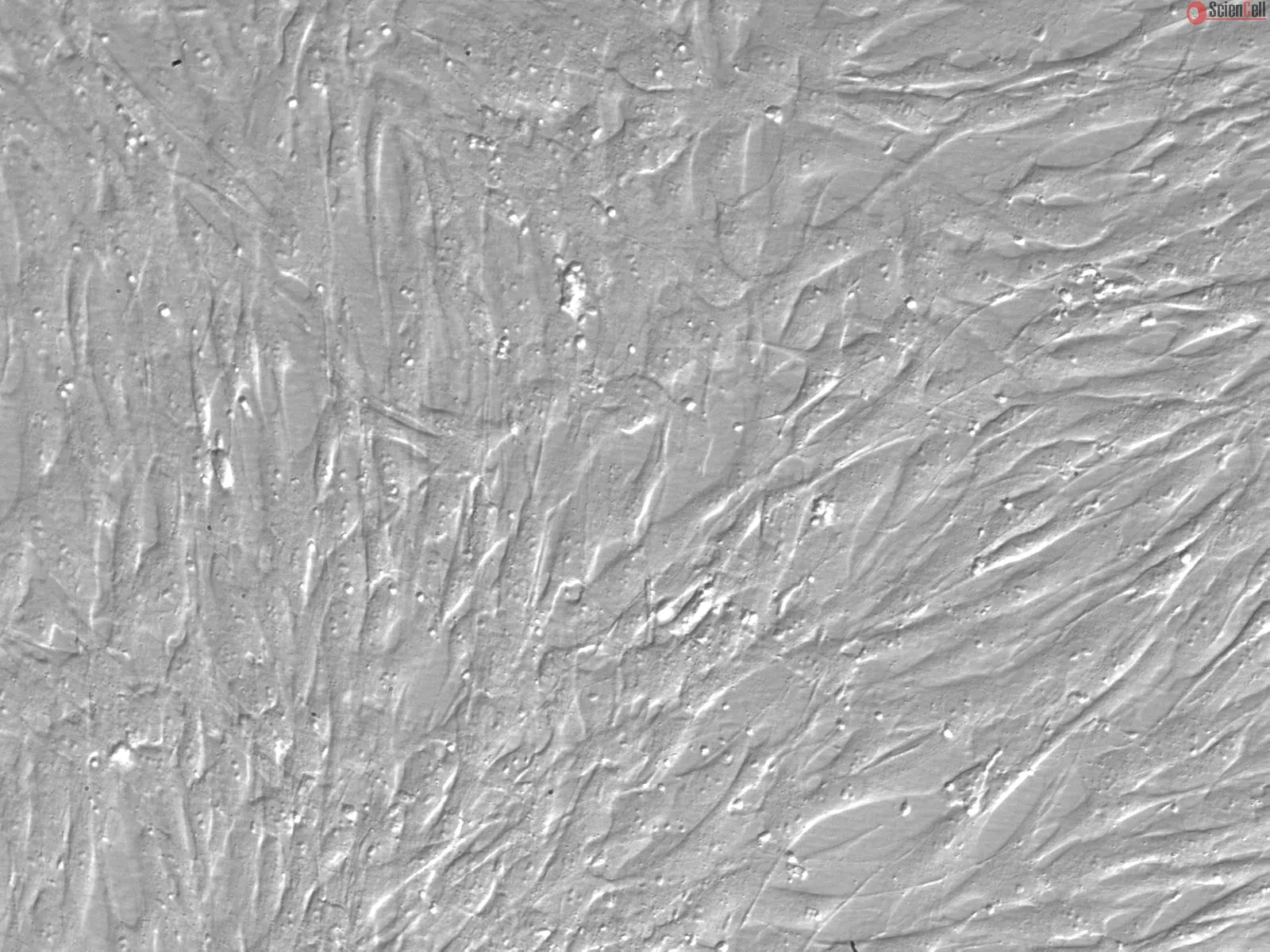
Factors contributing to the development of a fibrotic vascular scar and pulmonary vascular remodeling leading to chronic thromboembolic pulmonary hypertension (CTEPH) are... More
Factors contributing to the development of a fibrotic vascular scar and pulmonary vascular remodeling leading to chronic thromboembolic pulmonary hypertension (CTEPH) are still unknown. This study investigates the potential contribution of multipotent progenitor cells and myofibroblasts to the development and progression of CTEPH. Histological examination of endarterectomized tissues from patients with CTEPH identified significant neointimal formation. Morphological heterogeneity was observed in cells isolated from these tissues, including a network-like growth pattern and the formation of colony-forming unit-fibroblast-like colonies (CFU-F). Cells typically coexpressed intermediate filaments vimentin and smooth muscle α-actin. Cells were characterized by immunofluorescence and quantitated by fluorescent-activated cell sorting (FACS) for the presence of cell surface markers typical of mesenchymal progenitor cells; cells were >99% CD44+ CD73+, CD90+, CD166+; >80% CD29+; 45–99% CD105+; CD34− and CD45−. Cells were capable of adipogenic and osteogenic differentiation, determined by Oil Red O and Alizarin Red staining, respectively. Additionally, a population of Stro-1+ cells, a marker of bone marrow-derived stromal cells (4.2%), was sorted by FACS and also capable of adipogenic and osteogenic differentiation. In conclusion, this study is the first to identify a myofibroblast cell phenotype to be predominant within endarterectomized tissues, contributing extensively to the vascular lesion/clot. This cell may arise from transdifferentiation of adventitial fibroblasts or differentiation of mesenchymal progenitor cells. The unique microenvironment created by the stabilized clot is likely a factor in stimulating such cellular changes. These findings will be critical in establishing future studies in the development of novel and much needed therapeutic approaches for pulmonary hypertension. Keywords: progenitor cells, chronic thromboembolic pulmonary hypertension, occlusion, remodeling Less
The discovery of microRNAs (miRNAs) as a new class of regulators of gene expression has triggered an explosion of research activities, but has left many unanswered questi... More
The discovery of microRNAs (miRNAs) as a new class of regulators of gene expression has triggered an explosion of research activities, but has left many unanswered questions about how this regulation functions and how it is integrated with other regulatory mechanisms. A number of miRNAs have been found to be present in plasma and other body fluids of humans and mice in surprisingly high concentrations. This observation was unexpected in two respects: first, the fact that these molecules are present at all outside the cell at significant concentrations and second, that these molecules appear to be stable outside of the cell. In light of this it has been suggested that the biological function of miRNAs may also extend outside of the cell and mediate cell-cell communication. We report here that after serum deprivation several human cell lines tested promptly export a substantial amount of miRNAs into the culture medium and the export process is largely energy dependent. The exported miRNAs are found both within and outside of the 16.5 and 120 K centrifugation pellets which contain most of the known cell-derived vesicles, the microvesicles and exosomes. We have identified some candidate proteins involved in this system, and one of these proteins may also play a role in protecting extracellular miRNAs from degradation. Our results point to a hitherto unrecognized and uncharacterized miRNA trafficking system in mammalian cells that is consistent with the cell-cell communication hypothesis. Less


,-1-mg-ml--2.jpg)