A key aim of therapy for multiple sclerosis (MS) is to promote the regeneration of oligodendrocytes and remyelination in the central nervous system (CNS). The present stu... More
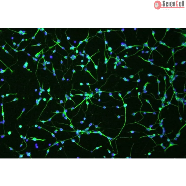
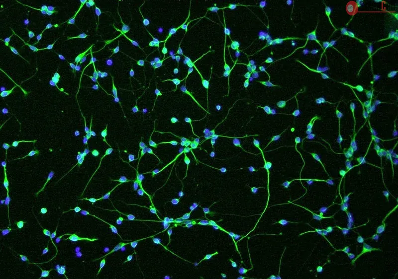
A key aim of therapy for multiple sclerosis (MS) is to promote the regeneration of oligodendrocytes and remyelination in the central nervous system (CNS). The present study provides evidence that the vitamin K-dependent protein growth arrest specific 6 (Gas6) promotes such repair in in vitro cultures of mouse optic nerve and cerebellum. We first determined expression of Gas6 and TAM (Tyro3, Axl, Mer) receptors in the mouse CNS, with all three TAM receptors increasing in expression through postnatal development, reaching maximal levels in the adult. Treatment of cultured mouse optic nerves with Gas6 resulted in significant increases in oligodendrocyte numbers as well as expression of myelin basic protein (MBP). Gas6 stimulation also resulted in activation of STAT3 in optic nerves as well as downregulation of multiple genes involved in MS development, including matrix metalloproteinase-9 (MMP9), which may decrease the integrity of the blood-brain barrier and is found upregulated in MS lesions. The cytoprotective effects of Gas6 were examined in in vitro mouse cerebellar slice cultures, where lysolecithin was used to induce demyelination. Cotreatment of cerebellar slices with Gas6 significantly attenuated demyelination as determined by MBP immunostaining, and Gas6 activated Tyro3 receptor through its phosphorylation. In conclusion, these results demonstrate that Gas6/TAM signaling stimulates the generation of oligodendrocytes and increased myelin production via Tyro3 receptor in the adult CNS, including repair after demyelinating injury. Furthermore, the effects of Gas6 on STAT3 signaling and matrix MMP9 downregulation indicate potential glial cell repair and immunoregulatory roles for Gas6, indicating that Gas6-TAM signaling could be a potential therapeutic target in MS and other neuropathologies. Keywords: TAM receptors; Tyro3; multiple sclerosis; myelin; oligodendrocyte; receptor tyrosine kinase. © The Author(s) 2016. Less
Glioblastoma multiforme (GBM) is inherently invasive, and it is from the invasive cell population that the tumor recurs. The GBM invasion transcriptome reveals over-expre... More
Glioblastoma multiforme (GBM) is inherently invasive, and it is from the invasive cell population that the tumor recurs. The GBM invasion transcriptome reveals over-expression of various autocrine factors that could act as motility drivers, such as autotaxin (ATX). Some of these factors could also have paracrine roles, modulating the behavior of cells in the peri-tumoral brain parenchyma. ATX generates lysophosphatidic acid (LPA), which signals through LPA receptors expressed by GBM as well as in astrocytes, oligodendrocytes (ODC) and microglia; their activation manifest cell specific effects. ATX stimulates invasion of GBM cells in vitro and ex vivo invasion assays. ATX activity enhances GBM adhesion in cells expressing the LPA1 receptor, as well as stimulating rac activation. GBM secreted ATX can also have paracrine effects: ATX activity results in reduced ODC adhesion. ODC monolayer invasion showed that U87 and U251 GBM cells expressing ATX invaded through an ODC monolayer significantly more than cells depleted of ATX or cells expressing inactive ATX, suggesting that GBM cells secreting ATX find ODCs less of a barrier than cells that do not express ATX. Secreted factors that drive GBM invasion can have autocrine and paracrine roles; one stimulates GBM motility and the other results in ODC dis-adhesion. Less
DNA hypermethylation-mediated gene silencing is a frequent and early contributor to aberrant cell growth and invasion in cancer. Malignant gliomas are the most common pri... More
DNA hypermethylation-mediated gene silencing is a frequent and early contributor to aberrant cell growth and invasion in cancer. Malignant gliomas are the most common primary brain tumors in adults and the second most common tumor in children. Morbidity and mortality are high in glioma patients because tumors are resistant to treatment and are highly invasive into surrounding brain tissue rendering complete surgical resection impossible. Invasiveness is regulated by the interplay between secreted proteases (eg, cathepsins) and their endogenous inhibitors (cystatins). In our previous studies we identified cystatin E/M (CST6) as a frequent target of epigenetic silencing in glioma. Cystatin E/M is a potent inhibitor of cathepsin B, which is frequently overexpressed in glioma. Here, we study the expression of cystatin E/M in normal brain and show that it is highly and moderately expressed in oligodendrocytes and astrocytes, respectively, but not in neurons. Consistent with this, the CST6 promoter is hypomethylated in all normal samples using methylation-specific PCR, bisulfite genomic sequencing, and pyrosequencing. In contrast, 78% of 28 primary brain tumors demonstrated reduced/absent cystatin E/M expression using a tissue microarray and this reduced expression correlated with CST6 promoter hypermethylation. Interestingly, CST6 was expressed in neural stem cells (NSC) and markedly induced upon differentiation, whereas a glioma tumor initiating cell (TIC) line was completely blocked for CST6 expression by promoter methylation. Analysis of primary pediatric brain tumor-derived lines also showed CST6 downregulation and methylation in nearly 100% of 12 cases. Finally, ectopic expression of cystatin E/M in glioma lines reduced cell motility and invasion. These results demonstrate that epigenetic silencing of CST6 is frequent in adult and pediatric brain tumors and occurs in TICs, which are thought to give rise to the tumor. CST6 methylation may therefore represent a novel prognostic marker and therapeutic target specifically altered in TICs. Less
With little improvement in the poor prognosis for humans with high-grade glioma brain tumors, alternative therapeutic strategies are needed. As such, selective replicatio... More
With little improvement in the poor prognosis for humans with high-grade glioma brain tumors, alternative therapeutic strategies are needed. As such, selective replication-competent oncolytic viruses may be useful as a potential treatment modality. Here we test the hypothesis that defects in the interferon (IFN) pathway could be exploited to enhance the selective oncolytic profile of vesicular stomatitis virus (VSV) in glioblastoma cells. Two green fluorescent protein-expressing VSV strains, recombinant VSV and the glioma-adapted recombinant VSV-rp30a, were used to study infection of a variety of human glioblastoma cell lines compared to a panel of control cells, including normal human astrocytes, oligodendrocyte precursor cells, and primary explant cultures from human brain tissue. Infection rate, cell viability, viral replication, and IFN-alpha/beta-related gene expression were compared in the absence and presence of IFN-alpha or polyriboinosinic polyribocytidylic acid [poly(I:C)], a synthetic inducer of the IFN-alpha/beta pathway. Both VSV strains caused rapid and total infection and death of all tumor cell lines tested. To a lesser degree, normal cells were also subject to VSV infection. In contrast, IFN-alpha or poly(I:C) completely attenuated the infection of all primary control brain cells, whereas most glioblastoma cell lines treated with IFN-alpha or poly(I:C) showed little or no sign of protection and were killed by VSV. Together, our results demonstrate that activation of the interferon pathway protects normal human brain cells from VSV infection while maintaining the vulnerability of human glioblastoma cells to viral destruction. Less
Accumulating evidence suggests that human herpesvirus type 6 (HHV-6) plays a pathogenic role in diseases of the central nervous system including multiple sclerosis (MS). ... More
Accumulating evidence suggests that human herpesvirus type 6 (HHV-6) plays a pathogenic role in diseases of the central nervous system including multiple sclerosis (MS). Recent studies have indicated that HHV-6 DNA is detected with high frequency in MS lesions compared to normal-appearing white matter, implicating a role for HHV-6 in MS pathogenesis. It appears that T cells, which infiltrate into the brain in MS patients, and resident oligodendrocytes harbor HHV-6 virus in MS lesions. Because T cells infected with HHV-6 have elevated proinflammatory gene expression, we hypothesized that HHV-6 could be indirectly cytotoxic to glial cells, including oligodendrocytes. Supernatants from SupT1 cells infected with HHV-6 variant A (GS or U1102) or variant B (Z29) significantly reduced MO3.1 cell proliferation by 75% +/- 10%, 78% +/- 8% or 51% +/- 9%, respectively. HHV-6 viral supernatants (GS or U1102 or Z29) significantly increased MO3.1 or primary human oligodendrocyte precursor cells (OPCs) cell death, whereas primary human fetal astrocytes were not affected. Removal of HHV-6 virions or proteins by trypsin treatment from culture supernatants did not reverse the loss in oligodendrocyte proliferation or viability. Supernatants from HHV-6 GS or U1102 cultures were significantly more cytotoxic to MO3.1 cells or OPCs compared to supernatants from T cells infected with Z29. Dying oligodendrocytes did not have an apoptotic-like phenotype and toxicity was not inhibited by general inhibitor of apoptosis, ZVAD. Further, oligodendrocytes had minimal caspase-3 activation even in the presence of staurosporine, suggesting that cell death followed caspase-independent pathways. These results indicate that HHV-6 is indirectly cytotoxic to oligodendrocytes and that cell death is driven primarily by caspase-independent pathways. Less


,-1-mg-ml--2.jpg)