Fibroblasts are mesenchymal cells derived from the embryonic mesoderm. They have been extensively used for a wide range of cellular and molecular studies, as they are one of easiest types of cells to grow in culture. Their durability also makes them amenable to a variety of manipulations ranging from studies employing gene transfection to microinjection. In general, fibroblasts secrete a non-rigid extracellular matrix which is rich in type I and/or type III collagen. There is evidence showing that fibroblasts in various organs are intrinsically different. In the lymphoid compartment, fibroblasts construct a unique conduit system with intriguing size exclusion, which plays a fundamental role in regulating immune response and homeostasis of tissue fluids.
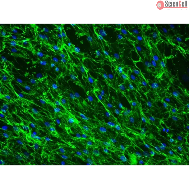
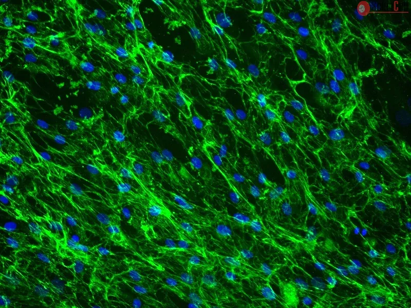
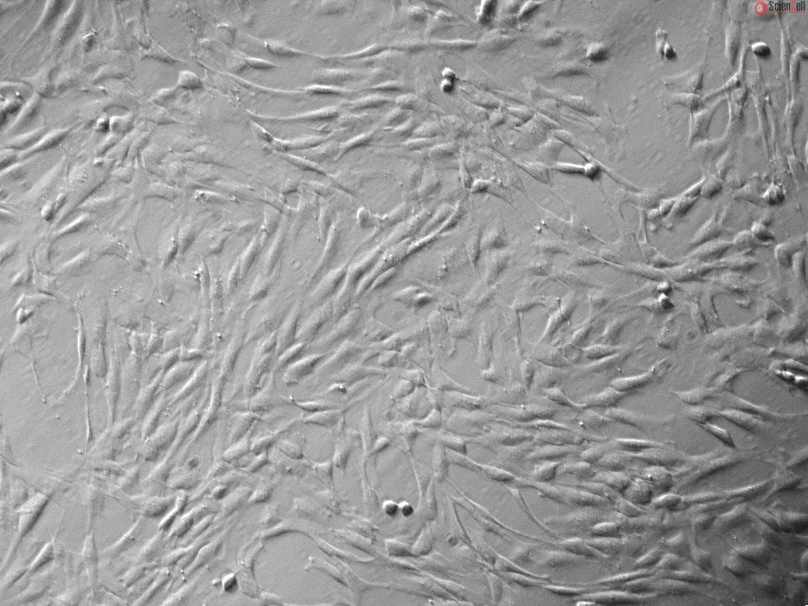
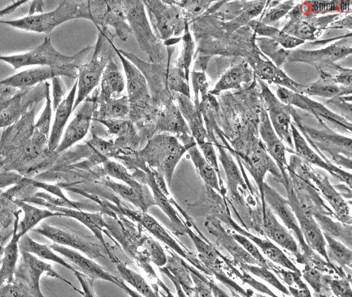
HLF from ScienCell Research Laboratories are isolated from human lymph node. HLF are cryopreserved at passage one and delivered frozen. Each vial contains >5 x 10⁵ cells in 1 ml volume. HLF are characterized by their spindle-shaped morphology and immunofluorescence with antibody specific to fibronectin. HLF are negative for HIV-1, HBV, HCV, mycoplasma, bacteria, yeast, and fungi. HLF are guaranteed to further expand for 15 population doublings under the conditions provided by ScienCell Research Laboratories.
Recommended Medium
It is recommended to use Fibroblast Medium (FM, Cat. #2301) for culturing HLF in vitro.
Product Sheets
 2530
2530
Search for a Certificate of Analysis by Lot Number below:
This work focused on generating a three-dimensional (3D) in vitro dynamic model to study chronic lymphocytic leukemia (CLL) cell dissemination, homing, and mechanisms of ... More
This work focused on generating a three-dimensional (3D) in vitro dynamic model to study chronic lymphocytic leukemia (CLL) cell dissemination, homing, and mechanisms of therapy resistance. We used a gelatin-based, hard porous biomaterial as a support matrix to develop 3D tissue-like models of the human lymph node and bone marrow, which were matured inside bioreactors under dynamic perfusion of medium. Comparing static and dynamic cultures of these 3D constructs revealed that perfusion promoted a tissue-like internal organization of cells, characterized by the expression of specific functional markers and deposition of an intricate extracellular matrix protein network. Recirculation of CLL cells within the dynamic system led to changes in leukemic cell behavior and in the expression of key markers involved in tumor progression. These findings suggest that the model is well suited for investigating the pathophysiological mechanisms of CLL and potentially other hematological malignancies. Less
Several types of cancer spread through the lymphatic system via the sentinel lymph nodes (LNs). Such LN-draining primary tumors, modified by tumor factors, lead to the fo... More
Several types of cancer spread through the lymphatic system via the sentinel lymph nodes (LNs). Such LN-draining primary tumors, modified by tumor factors, lead to the formation of a metastatic niche associated with an increased number of Foxp3+ regulatory T cells (Tregs). These cells are expected to contribute to the elaboration of an immune-suppressive environment. Activated Tregs express glycoprotein A repetitions predominant (GARP), which binds and presents latent transforming growth factor beta 1 (TGF-β1) at their surface. GARP is also expressed by other non-immune cell types poorly described in LNs. Here, we mapped GARP expression in non-immune cells in human and mouse metastatic LNs. The mining of available (human and murine) scRNA-Seq datasets revealed GARP expression by blood (BEC)/lymphatic (LEC) endothelial, fibroblastic, and perivascular cells. Consistently, through immunostaining and in situ RNA hybridization approaches, GARP was detected in and around blood and lymphatic vessels, in (αSMA+) fibroblasts, and in perivascular cells associated with an abundant matrix. Strikingly, GARP was detected in LECs forming the subcapsular sinus and high endothelial venules (HEVs), two vascular structures localized at the interface between LNs and the afferent lymphatic and blood vessels. Altogether, we here provide the first distribution maps for GARP in human and murine LNs.
Keywords:
LRRC32; GARP mRNA; glycoprotein A repetitions predominant (GARP); transforming growth factor beta 1 (TGF-β1); lymph node; tumor microenvironment; metastases; cancer Less
Fibroblastic reticular cells (FRCs) are stromal cells in secondary lymphoid organs, the major sites for HIV-1 infection of CD4+ T cells. Although FRCs regulate T cell sur... More
Fibroblastic reticular cells (FRCs) are stromal cells in secondary lymphoid organs, the major sites for HIV-1 infection of CD4+ T cells. Although FRCs regulate T cell survival, proliferation, and migration, whether they play any role in HIV-1 spread has not been studied. Here, we show that FRCs enhance HIV-1 spread via trans-infection in which FRCs capture HIV-1 and facilitate infection of T cells that come into contact with FRCs. FRCs mediate trans-infection in both two- and three-dimensional culture systems and in a manner dependent on the virus producer cells. This producer cell dependence, which was also observed for virus spread in secondary lymphoid tissues ex vivo, is accounted for by CD44 incorporated into virus particles and hyaluronan bound to such CD44 molecules. This virus-associated hyaluronan interacts with CD44 expressed on FRCs, thereby promoting virus capture by FRCs. Overall, our results reveal a novel role for FRCs in promoting HIV-1 spread. Less
ScienCell Research Laboratories (SRL) takes pride in being a resource for researchers all over the world. The publications listed here are not meant as an endorsement or confirmation of the reliability of the products.





,-1-mg-ml--2.jpg)