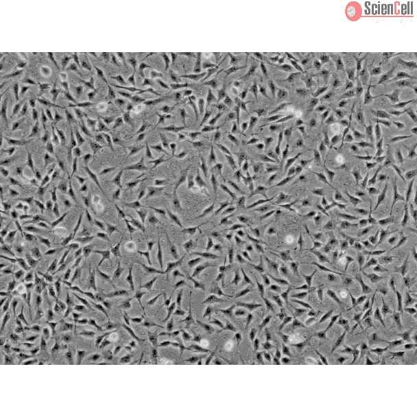
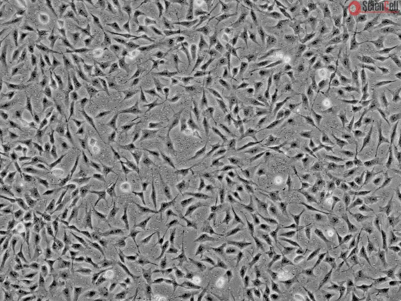
The keratocytes, or corneal fibroblasts, are highly specialized cells that are sandwiched between orthogonally arranged layers of collagen lamellae in the corneal stroma. They play a key role in maintaining the structure and transparency of the cornea, as they are the source of stromal collagen and proglycans. They also play important roles in corneal wound healing and tissue repair, and are known to undergo phenotypic transformations in wounds due to the influence of growth factors and cytokines. Under normal conditions, the keratocytes in the adult cornea are relatively quiescent cells. In the event of corneal injury or trauma, however, the keratocytes differentiate into active, synthesizing cells and rapidly replace damaged stromal matrix. Cultured human keratocytes express functional IL-4Rs and IL-17R on the cell surface, suggesting that these cells may contribute to the role of IL-4 and IL-17 as mediators of allergic reactions in the cornea. Changes in gene expression were observed in keratocytes after interleukin-1 treatment, which provides important insight into gene expression and suggests novel therapeutic targets for the control of corneal inflammation.
HK from ScienCell Research Laboratories are isolated from human cornea. HK are cryopreserved at passage one and delivered frozen. Each vial contains >5 x 10⁵ cells in 1 ml volume. HK are characterized by fibroblast morphology and immunofluorescence with antibody specific to fibronectin. HK are negative for HIV-1, HBV, HCV, mycoplasma, bacteria, yeast and fungi. HK are guaranteed to further expand for 15 population doublings under the conditions provided by ScienCell Research Laboratories.
Recommended Medium
It is recommended to use Fibroblast Medium (FM, Cat. #2301) for culturing HK in vitro.
Background: Mutations in a novel gene, UBIAD1, were recently found to cause the autosomal dominant eye disease Schnyder corneal dystrophy (SCD). SCD is characterized by a... More
Background: Mutations in a novel gene, UBIAD1, were recently found to cause the autosomal dominant eye disease Schnyder corneal dystrophy (SCD). SCD is characterized by an abnormal deposition of cholesterol and phospholipids in the cornea resulting in progressive corneal opacification and visual loss. We characterized lesions in the UBIAD1 gene in new SCD families and examined protein homology, localization, and structure.Methodology/principal findings: We characterized five novel mutations in the UBIAD1 gene in ten SCD families, including a first SCD family of Native American ethnicity. Examination of protein homology revealed that SCD altered amino acids which were highly conserved across species. Cell lines were established from patients including keratocytes obtained after corneal transplant surgery and lymphoblastoid cell lines from Epstein-Barr virus immortalized peripheral blood mononuclear cells. These were used to determine the subcellular localization of mutant and wild type protein, and to examine cholesterol metabolite ratios. Immunohistochemistry using antibodies specific for UBIAD1 protein in keratocytes revealed that both wild type and N102S protein were localized sub-cellularly to mitochondria. Analysis of cholesterol metabolites in patient cell line extracts showed no significant alteration in the presence of mutant protein indicating a potentially novel function of the UBIAD1 protein in cholesterol biochemistry. Molecular modeling was used to develop a model of human UBIAD1 protein in a membrane and revealed potentially critical roles for amino acids mutated in SCD. Potential primary and secondary substrate binding sites were identified and docking simulations indicated likely substrates including prenyl and phenolic molecules.Conclusions/significance: Accumulating evidence from the SCD familial mutation spectrum, protein homology across species, and molecular modeling suggest that protein function is likely down-regulated by SCD mutations. Mitochondrial UBIAD1 protein appears to have a highly conserved function that, at least in humans, is involved in cholesterol metabolism in a novel manner. Less
Peroxisome proliferator-activated receptor α (PPARα) agonism in ocular inflammation has not been thoroughly investigated. The objective of this investigation was to det... More
Peroxisome proliferator-activated receptor α (PPARα) agonism in ocular inflammation has not been thoroughly investigated. The objective of this investigation was to determine the effect of WY-14 643, a selective PPARα agonist, on inflammatory cytokine release in human ocular cells. Stimulation of primary human corneal epithelial cells, keratocytes, and retinal endothelial cells with 1 to 10 ng/mL interleukin 1β (IL-1β) resulted in a significant increase in numerous inflammatory cytokines, including IL-6, IL-8, and tumor necrosis factor α (TNF-α); and dexamethasone was able to significantly inhibit these effects. However, WY-14 643 did not effectively block IL-1β-induced cytokine release in ocular cells; rather, significant increases in IL-1β-induced inflammatory cytokines were observed in these cells but not in aortic smooth muscle cells. WY-14 643 also significantly upregulated vascular endothelial growth factor (VEGF) expression in corneal epithelial cells and keratocytes. These studies demonstrate for the first time that PPARα agonism may be proinflammatory and proangiogenic in a variety of ocular cells and suggest that therapeutic applications of such agents in ophthalmology may be limited. Less
ScienCell Research Laboratories (SRL) takes pride in being a resource for researchers all over the world. The publications listed here are not meant as an endorsement or confirmation of the reliability of the products.


,-1-mg-ml--2.jpg)