Background: Esophageal squamous cell carcinoma (ESCC) is one of the most common causes of cancer mortality in the gastrointestinal tract. Promoter hypermethylation of tum... More
Background: Esophageal squamous cell carcinoma (ESCC) is one of the most common causes of cancer mortality in the gastrointestinal tract. Promoter hypermethylation of tumor suppressor genes contributes to gene inactivation during development of ESCC. Aim: To identify novel methylation-silenced genes in ESCC. Methods: Genome-wide microarrays were applied to search for genes that were markedly upregulated after treatment with 5-aza-2'-deoxycytidine (5-Aza-dC) and that were markedly decreased in tumor tissue compared with paired adjacent nontumor tissue. Reverse-transcription polymerase chain reaction (PCR), immunohistochemistry, methylation-specific PCR, and bisulfite genomic sequencing were employed to investigate expression and methylation of candidate genes in five human ESCC cell lines, two human immortalized normal esophageal epithelial cell lines, primary ESCC tumor tissues, and paired adjacent nontumor tissues. Results: GPX3 was selected as a novel candidate hypermethylated gene in ESCC through microarray analysis. In most ESCC cell lines, GPX3 messenger RNA (mRNA) expression was downregulated and the CpG island of GPX3 promoter was methylated. Demethylation treatment with 5-Aza-dC restored GPX3 mRNA expression. Methylation of GPX3 promoter was more frequent in ESCC tumor tissues (71.4%) than in adjacent nontumor tissues (10.7%) (P < 0.001), and methylation of GPX3 promoter correlated significantly with GPX3 mRNA downregulation. Finally, GPX3 protein expression was also significantly lower in ESCC tumor tissues than in adjacent nontumor tissues. Conclusion: GPX3 is downregulated through promoter hypermethylation in ESCC, which may be a potential biomarker of ESCC. Less
Polo-like kinase 1 (PLK1) is overexpressed in various human cancers. However, the biological functions and the post-transcriptional regulations of PLK1 in esophageal canc... More
Polo-like kinase 1 (PLK1) is overexpressed in various human cancers. However, the biological functions and the post-transcriptional regulations of PLK1 in esophageal cancer (EC) are still unknown. The purposes of this study are to determine whether PLK1 can be a molecular target of EC therapy, and to identify a microRNA targeting PLK1. We performed loss-of- and gain-of-function experiments regarding cell proliferation, cell cycle, apoptosis, in vivo tumor formation, and luciferase reporter assays, using siRNAs against PLK1 and microRNA. PLK1 protein was expressed in all 11 EC cell lines, but not in normal esophageal epithelial cells (HEEpiC). Knock-down of PLK1 in EC cells induced G2/M arrest (p<0.001) in cell cycle assay, and reduced cell proliferation (p=0.019) and tumor formation ability in vivo (p<0.0001). MiR-593*, identified as a microRNA targeting PLK1 by a database search, was less expressed especially in six EC cell lines than HEEpiC cells. Moreover, miR-593* expression level was inversely correlated with PLK1 mRNA level in 48 clinical tissue specimens of EC (p=0.006). Introduction of synthetic miR-593* suppressed PLK1 expression by 69–73%, reduced cell proliferation (p=0.008), and increased cell proportion of G2/M phase (p=0.01) in HSA/c (an EC cells), whereas a miR-593* inhibitor up-regulated PLK1 expression by 11–55%. Additionally, luciferase assay demonstrated that miR-593* interacted two binding sites in the PLK1 3′-UTR and reduced 56.8–71.5% of luciferase activity by degrading luciferase mRNA in HSA/c cells. In conclusion, PLK1 is post-transcriptionally regulated by miR-593*, and could be a promising molecular target for EC treatment. Keywords: esophageal cancer, PLK1, microRNA, miR-593* Less
The importance of microRNAs (miRNAs) in human malignancies has been well recognized. Here, we report that the expression of microRNA-210 (miR-210) is down-regulated in hu... More
The importance of microRNAs (miRNAs) in human malignancies has been well recognized. Here, we report that the expression of microRNA-210 (miR-210) is down-regulated in human esophageal squamous cell carcinoma and derived cell lines. Marked decreases in the level of miR-210 were observed especially in poorly differentiated carcinomas. We found that miR-210 inhibits cancer cell survival and proliferation by inducing cell death and cell cycle arrest in G1/G0 and G2/M. Finally, we identified fibroblast growth factor receptor-like 1 (FGFRL1) as a target of miR-210 in esophageal squamous cell carcinoma and demonstrated that FGFRL1 accelerates cancer cell proliferation by preventing cell cycle arrest in G1/G0. Taken together, our findings show an important role for miR-210 as a tumor-suppressive microRNA with effects on cancer cell proliferation. Keywords: Cell Cycle, Cell Death, MicroRNA, Tumor, Tumor Suppressor, FGFRL1, Esophageal Squamous Cell Carcinoma, miR-210, Tumor Differentiation Less
INTRODUCTION: The incidence of Barrett esophageal adenocarcinoma (BEAC) has been increasing at an alarming rate in western countries. In this study, we have evaluated the... More
INTRODUCTION: The incidence of Barrett esophageal adenocarcinoma (BEAC) has been increasing at an alarming rate in western countries. In this study, we have evaluated the therapeutic potential of sulforaphane (SFN), an antioxidant derived from broccoli, in BEAC. METHODS: BEAC cells were treated with SFN, alone or in combination with chemotherapeutic, paclitaxel, or telomerase-inhibiting agents (MST-312, GRN163L), and live cell number determined at various time points. The effect on drug resistance/chemosensitivity was evaluated by rhodamine efflux assay. Apoptosis was detected by annexin V labeling and Western blot analysis of poly(ADP-ribose) polymerase cleavage. Effects on genes implicated in cell cycle and apoptosis were determined by Western blot analyses. To evaluate the efficacy in vivo, BEAC cells were injected subcutaneously in severe combined immunodeficient mice, and after the appearance of palpable tumors, mice were treated with SFN. RESULTS: SFN induced both time- and dose-dependent decline in cell survival, cell cycle arrest, and apoptosis. The treatment with SFN also suppressed the expression of multidrug resistance protein, reduced drug efflux, and increased anticancer activity of other antiproliferative agents including paclitaxel. A significant reduction in tumor volume was also observed by SFN in a subcutaneous tumor model of BEAC. Anticancer activity could be attributed to the induction of caspase 8 and p21 and down-regulation of hsp90, a molecular chaperon required for activity of several proliferation-associated proteins. CONCLUSIONS: These data indicate that a natural product with antioxidant properties from broccoli has great potential to be used in chemoprevention and treatment of BEAC. Less
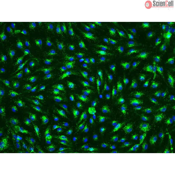
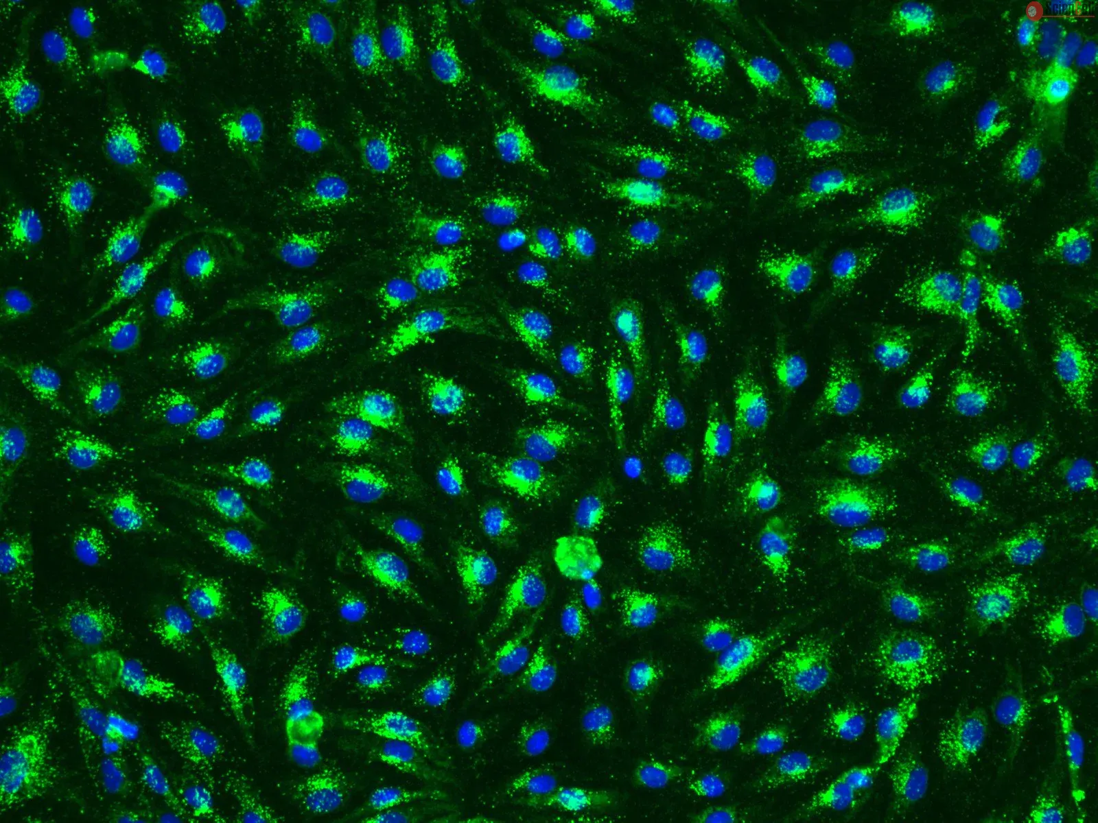
Human esophageal epithelial cells play a key role in esophageal inflammation in response to acidic pH during gastroesophageal reflux disease (GERD), increasing secretion ... More
Human esophageal epithelial cells play a key role in esophageal inflammation in response to acidic pH during gastroesophageal reflux disease (GERD), increasing secretion of IL-6 and IL-8. The mechanisms underlying IL-6 and IL-8 expression and secretion in esophageal epithelial cells after acid stimulation are not well characterized. We investigated the role of PKC, MAPK, and NF-kappaB signaling pathways and transcriptional regulation of IL-6 and IL-8 expression in HET-1A cells exposed to acid. Exposure of HET-1A cells to pH 4.5 induced NF-kappaB activity and enhanced IL-6 and IL-8 secretion and mRNA and protein expression. Acid stimulation of HET-1A cells also resulted in activation of MAPKs and PKC (alpha and epsilon). Curcumin, as well as inhibitors of NF-kappaB (SN-50), PKC (chelerythrine), and p44/42 MAPK (PD-098059) abolished the acid-induced expression of IL-6 and IL-8. The JNK inhibitor SP-600125 blocked expression/secretion of IL-6 but only partially attenuated IL-8 expression. The p38 MAPK inhibitor SB-203580 did not inhibit IL-6 expression but exerted a stronger inhibitory effect on IL-8 expression. Together, these data demonstrate that 1) acid is a potent inducer of IL-6 and IL-8 production in HET-1A cells; 2) MAPK and PKC signaling play a key regulatory role in acid-mediated IL-6 and IL-8 expression via NF-kappaB activation; and 3) the anti-inflammatory plant compound curcumin inhibits esophageal activation in response to acid. Thus IL-6 and IL-8 expression by acid may contribute to the pathobiology of mucosal injury in GERD, and inhibition of the NF-kappaB/proinflammatory cytokine pathways may emerge as important therapeutic targets for treatment of esophageal inflammation. Less
Esophageal adenocarcinoma arises in the backdrop of Barrett metaplasia-dysplasia sequence, with the vast majority of patients presenting with late stage malignancy. Mesot... More
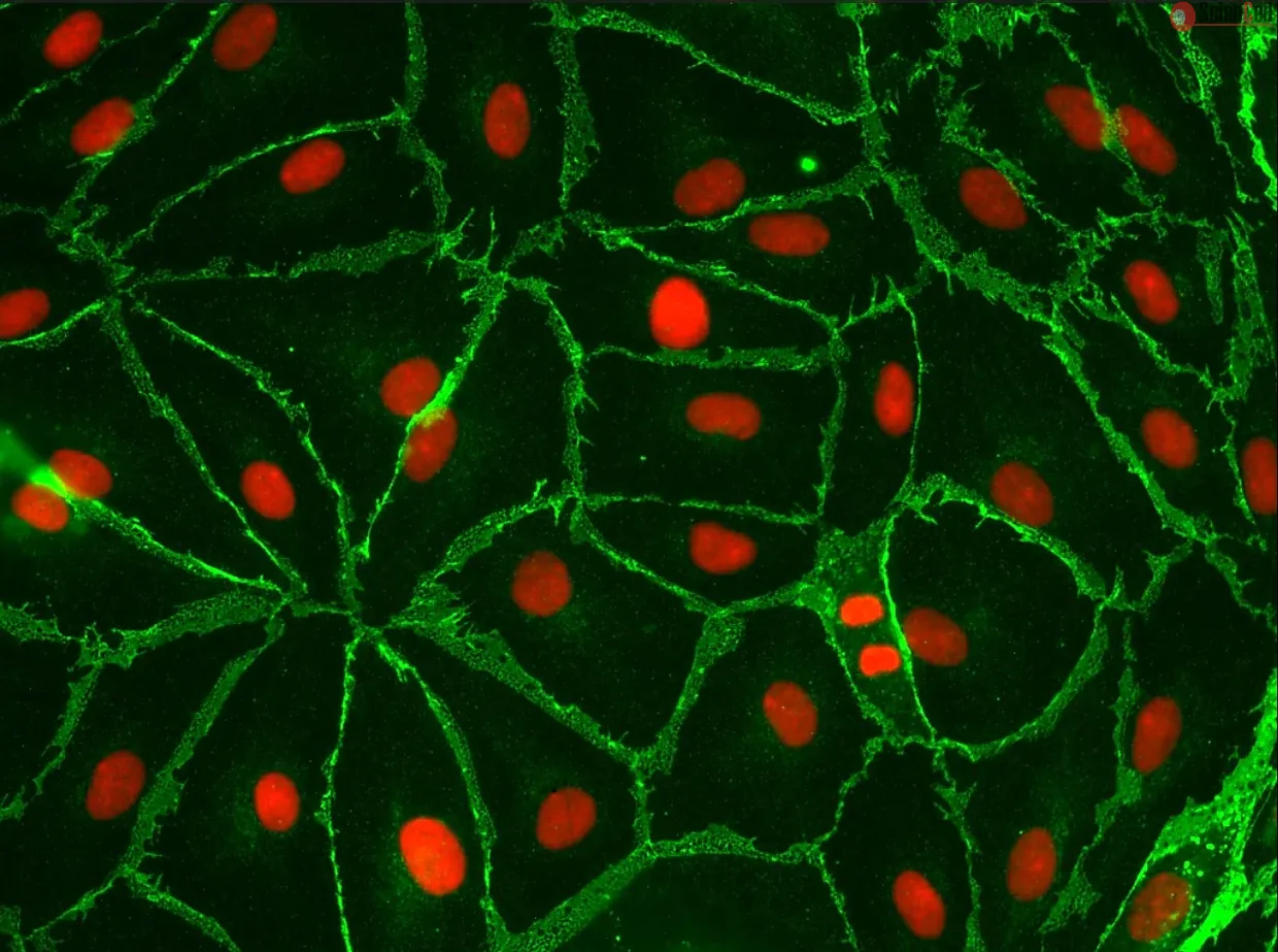
Esophageal adenocarcinoma arises in the backdrop of Barrett metaplasia-dysplasia sequence, with the vast majority of patients presenting with late stage malignancy. Mesothelin, a GPI-anchored protein, is aberrantly overexpressed on the surface of many solid cancers. Mesothelin expression was assessed in esophageal tissue microarrays (TMAs) encompassing the entire histologic spectrum of Barrett-associated dysplasia and adenocarcinoma. Mesothelin expression was observed in 24/84 (29%) of invasive adenocarcinomas, and in 5/34 (15%) lymph node metastases. In contrast, normal squamous and cardia mucosa, as well as non-invasive Barrett lesions, failed to label with mesothelin. Mesothelin was expressed in the esophageal adenocarcinoma cell line JHU-EsoAd1, but not in primary human esophageal epithelial cells. Anti-mesothelin antibody conjugated CdSe/CDS/ZnS quantum rods (QRs) were synthesized as described (Young et al, NanoLetters, 2007), and confocal bio-imaging confirmed robust binding to JHU-EsoAd1 cells. Anti-mesothelin antibody conjugated nanoparticles can be useful for the diagnosis and therapy of mesothelin-overexpressing esophageal adenocarcinomas. Less
Human pituitary tumor-transforming 1 (PTTG1)/securin is a putative oncoprotein that is overexpressed in various tumor types. However, the involvement of PTTG1 in gastroin... More
Human pituitary tumor-transforming 1 (PTTG1)/securin is a putative oncoprotein that is overexpressed in various tumor types. However, the involvement of PTTG1 in gastrointestinal cancer development and progression remains unclear. In this study, we investigated the clinical significance and biological effects of PTTG1 in esophageal squamous cell carcinoma (ESCC). Immunohistochemical studies performed on 113 primary ESCC specimens revealed a high prevalence of PTTG1 overexpression (60.2%), which was significantly associated with lymph node metastasis (regional, P = 0.042; distant, P = 0.005), advanced tumor stage (P = 0.028), and poorer overall survival (P = 0.017, log-rank test; P = 0.044, Cox proportional hazard model). Eleven ESCC cell lines expressed PTTG1 protein at levels 2.4 to 6.6 times higher than those in normal esophageal epithelial cells (HEEpiC). PTTG1 protein expression was confined to the nucleus in HEEpiC cells but present in both the cytoplasm and nucleus in ESCC cells. Two small interfering RNAs (siRNA) inhibited PTTG1 mRNA and protein expression in three ESCC cell lines by 77% to 97%. In addition, PTTG1 down-regulation by these siRNAs significantly reduced cell motility in all three ESCC cell lines (P < 0.01) in vitro, as well as popliteal lymph node metastases of ESCC cells in nude mice (P = 0.020). Global gene expression profiling suggested that several members of the Ras and Rho gene families, including RRAS, RHOG, ARHGAP1, and ARHGADIA, represented potential downstream genes in the PTTG1 pathway. Taken together, these findings suggest that PTTG1 overexpression promotes cell motility and lymph node metastasis in ESCC patients, leading to poorer survival. Thus, PTTG1 constitutes a potential biomarker and therapeutic target in ESCCs with lymph node metastases. Less
Purpose: The aims of this study were to investigate telomere function in normal and Barrett's esophageal adenocarcinoma (BEAC) cells purified by laser capture microdissec... More
Purpose: The aims of this study were to investigate telomere function in normal and Barrett's esophageal adenocarcinoma (BEAC) cells purified by laser capture microdissection (LCM) and to evaluate the impact of telomerase inhibition in cancer cells in vitro and in vivo. Experimental Design: Epithelial cells were purified from surgically resected esophagi. Telomerase activity was measured by modified “Telomeric Repeat Amplification Protocol” and telomere length determined by Real-Time PCR assay. To evaluate the impact of telomerase inhibition, adenocarcinoma cell lines were continuously treated with a specific telomerase inhibitor (GRN163L) and live cell number determined weekly. Apoptosis was evaluated by annexin labeling and senescence by beta-galactosidase staining. For in vivo studies, SCID-mice were subcutaneously inoculated with adenocarcinoma cells and following appearance of palpable tumors, injected intraperitoneally with saline or GRN163L. Results: Telomerase activity was significantly elevated whereas telomeres were shorter in BEAC cells relative to normal esophageal epithelial cells. The treatment of adenocarcinoma cells with telomerase inhibitor, GRN163L, led to loss of telomerase activity, reduction in telomere length, and growth arrest through induction of both the senescence and apoptosis. GRN163L induced cell death could also be expedited by addition of chemotherapeutic agents, doxorubicin and ritonavir. Finally, the treatment with GRN163L led to a significant reduction in tumor volume in a subcutaneous tumor model. Conclusions: We show that telomerase activity is significantly elevated whereas telomeres are shorter in BEAC and suppression of telomerase inhibits proliferation of adenocarcinoma cells both in vitro and in vivo. Keywords: Telomerase, Telomere, Laser Capture, Barrett's, Adenocarcinoma Less