Melanocytes are neural crest-derived cells that produce melanin via melanogenesis. Melanocytes localize to several tissues including the epidermis, eye, inner ear and leptomeninges. Dysregulation of melanocyte migration, proliferation, or survival during embryonic development thus causes congenital disorders in those tissues as seen in Tietz syndrome, Waardenburg syndrome, and piebaldism. In the bottom layer of skin epidermis, melanocytes synthesize and transfer dark-colored melanin to surrounding keratinocytes to give skin pigmentation. Melanin also blocks UV-B light to protect the hypodermis from solar exposure-induced photodamage. Progress in culture techniques, along with an improved understanding of melanocyte biology, has led to a successful culture system to model melanomas, inner ear homeostasis, vitiligo, and
mitochondrial dysfunction in Duchenne Muscular Dystrophy.
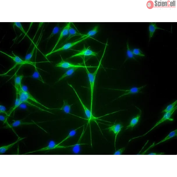
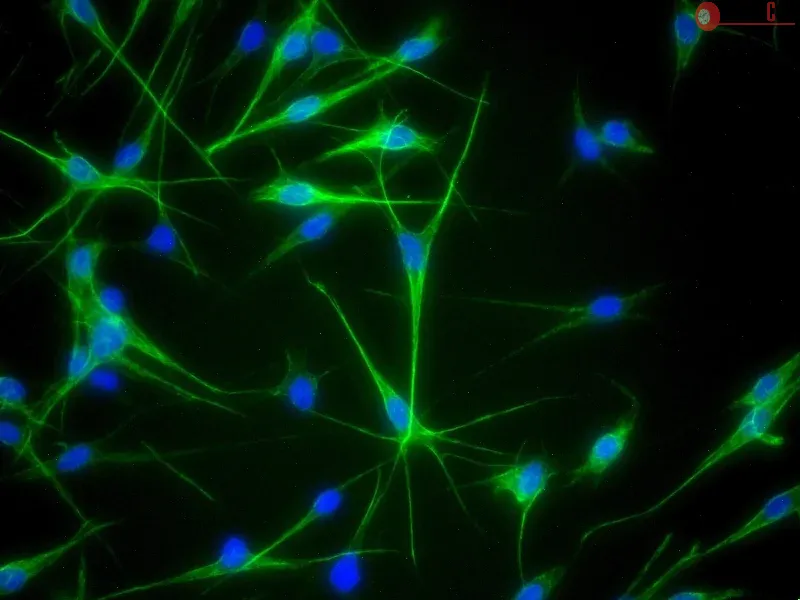
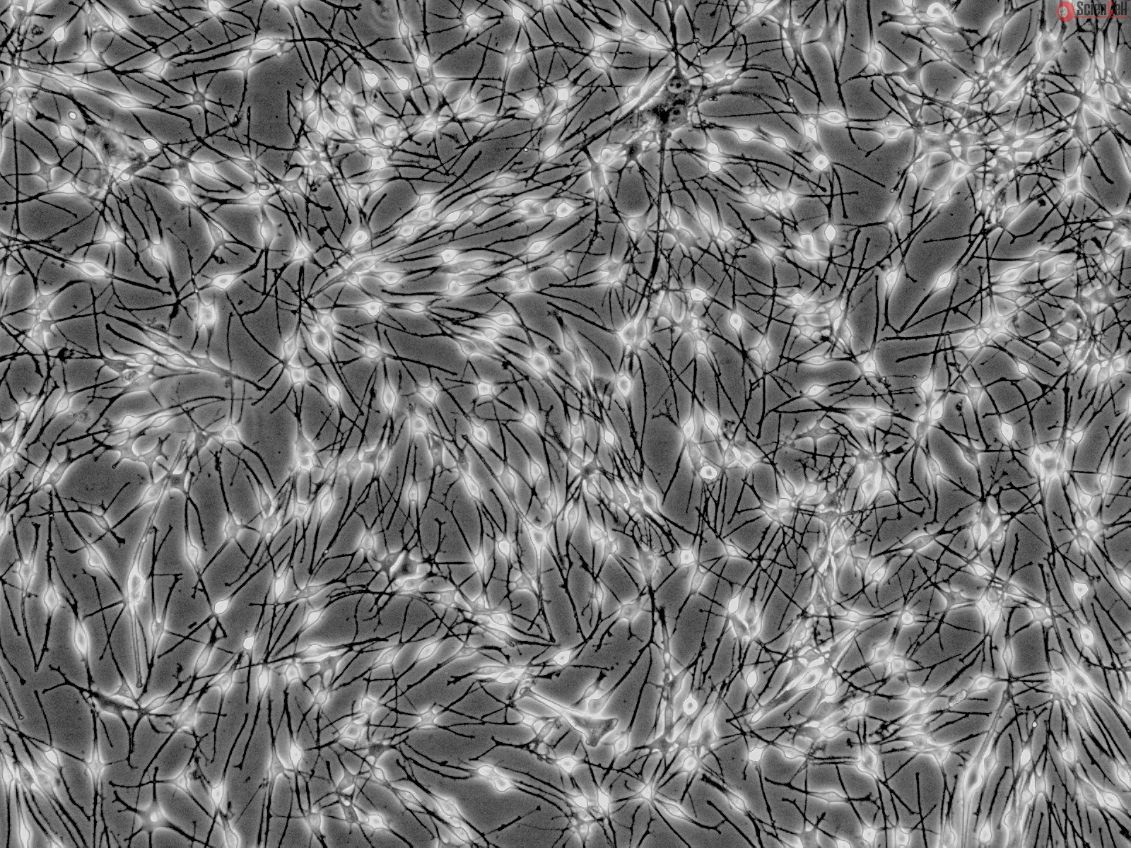
HEM-m from ScienCell Research Laboratories is isolated from neonatal human skin. HEM-m are cryopreserved at passage one and delivered frozen. Each vial contains >5 x 10⁵ cells in 1 ml volume. HEM-m are characterized by immunofluorescence with antibodies specific to S100β and/or NGF-receptor (p75). HEM-m are negative for HIV-1, HBV, HCV, mycoplasma, bacteria, yeast, and fungi. HEM-m are guaranteed to further expand for 15 population doublings under
the conditions provided by ScienCell Research Laboratories.
Recommended Medium
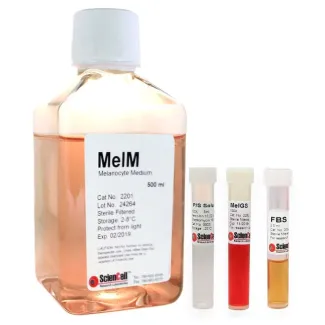
It is recommended to use Melanocyte Medium (MelM, Cat. #2201) for culturing HEM-m in vitro.
Product Sheets
 2210
2210
Search for a Certificate of Analysis by Lot Number below:
Grifolin, a secondary metabolite isolated from the fresh fruiting bodies of the mushroom Albatrellus confluens, has been shown to inhibit the growth of some cancer cell l... More
Grifolin, a secondary metabolite isolated from the fresh fruiting bodies of the mushroom Albatrellus confluens, has been shown to inhibit the growth of some cancer cell lines in vitro by induction of apoptosis in previous studies of our group. However, the mechanisms of action are not completely understood. An apoptosis-related gene expression profiling analysis provided a clue that death-associated protein kinase 1 (dapk1) gene was upregulated at least twofold in response to grifolin treatment in nasopharyngeal carcinoma cell CNE1. Here, we further investigated the role of DAPK1 in apoptotic effect induced by grifolin. We observed that protein as well as mRNA level of DAPK1 was induced by grifolin in a dose-dependent manner in nasopharyngeal carcinoma cell CNE1. We found that grifolin increased both Ser392 and Ser20 phosphorylation levels of transcription factor p53 protein, which could promote its transcriptional activity. Moreover, induced by grifolin, the recruitment of p53 to dapk1 gene promoter was confirmed to enhance markedly using EMSA and ChIP assays analysis. The involvement of DAPK1 in grifolin-induced apoptosis was supported by the studies that introducing siRNA targeting DAPK1 to CNE1 cells remarkably interfered grifolin-caused apoptotic effect as well as the activation of caspase-3. Grifolin induced upregulation of DAPK1 via p53 was also observed in tumour cells derived from human breast cancer and human colon cancer. The findings suggest that upregulation of DAPK1 via p53-DAPK1 pathway is an important mechanism of grifolin contributing to its ability to induce apoptotic effect. Since growing evidence found a significant loss of DAPK1 expression in a large variety of tumour types, grifolin may represent a promising candidate in the intervention of cancer via targeting DAPK1. Copyright © 2010 Elsevier Ltd. All rights reserved. Less
The majority of melanoma cells do not express argininosuccinate synthetase (ASS), and hence cannot synthesize arginine from citrulline. Their growth and proliferation dep... More
The majority of melanoma cells do not express argininosuccinate synthetase (ASS), and hence cannot synthesize arginine from citrulline. Their growth and proliferation depend on exogenous supply of arginine. Arginine degradation using arginine deiminase (ADI) leads to growth inhibition and eventually cell death while normal cells which express ASS can survive. This notion has been translated into clinical trial. Pegylated ADI (ADI-PEG20) has shown antitumor activity in melanoma. However, the sensitivity to ADI is different among ASS(−) melanoma cells. We have investigated and reviewed the signaling pathways which are affected by arginine deprivation and their consequences which lead to cell death. We have found that arginine deprivation inhibits mTOR signaling but leads to activation of MEK and ERK with no changes in BRAF. These changes most likely lead to autophagy, a possible mechanism to survive by recycling intracellular arginine. However apoptosis does occur which can be both caspase-dependent or independent. In order to increase the therapeutic efficacy of this form of treatment, one should consider adding other agent(s) which can drive the cells toward apoptosis or inhibit the autophagic process. Keywords: Arginine deiminase, argininosuccinate synthetase, melanoma Less
Background: Malignant melanoma is resistant to almost all conventional forms of chemotherapy. Recent evidence suggests that anti-apoptotic proteins of the Bcl-2 family ar... More
Background: Malignant melanoma is resistant to almost all conventional forms of chemotherapy. Recent evidence suggests that anti-apoptotic proteins of the Bcl-2 family are overexpressed in melanoma and may contribute to melanoma's striking resistance to apoptosis. ABT-737, a small-molecule inhibitor of Bcl-2, Bcl-xl and Bcl-w, has demonstrated efficacy in several forms of leukemia, lymphoma as well as solid tumors. However, overexpression of Mcl-1, a frequent observance in melanoma, is known to confer ABT-737 resistance. Methodology/Principal Findings Here we report that knockdown of Mcl-1 greatly reduces cell viability in combination with ABT-737 in six different melanoma cell lines. We demonstrate that the cytotoxic effect of this combination treatment is due to apoptotic cell death involving not only caspase-9 activation but also activation of caspase-8, caspase-10 and Bid, which are normally associated with the extrinsic pathway of apoptosis. Caspase-8 (and caspase-10) activation is abrogated by inhibition of caspase-9 but not by inhibitors of the death receptor pathways. Furthermore, while caspase-8/-10 activity is required for the full induction of cell death with treatment, the death receptor pathways are not. Finally, we demonstrate that basal levels of caspase-8 and Bid correlate with treatment sensitivity. Conclusions/Significance Our findings suggest that the combination of ABT-737 and Mcl-1 knockdown represents a promising, new treatment strategy for malignant melanoma. We also report a death receptor-independent role for extrinsic pathway proteins in treatment response and suggest that caspase-8 and Bid may represent potential markers of treatment sensitivity. Less
ScienCell Research Laboratories (SRL) takes pride in being a resource for researchers all over the world. The publications listed here are not meant as an endorsement or confirmation of the reliability of the products.

,-1-mg-ml--2.jpg)