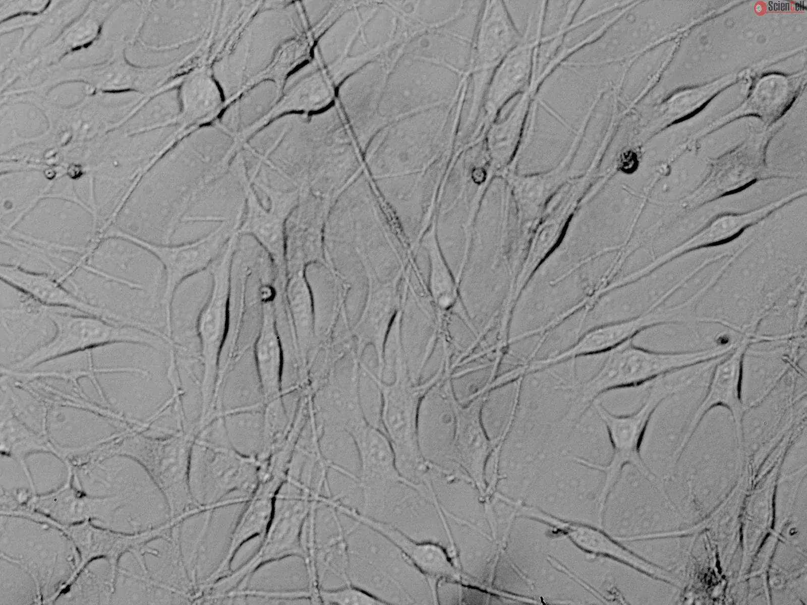
Melanocytes are neural crest-derived cells that produce melanin via melanogenesis. Melanocytes localize to several tissues including the epidermis, eye, inner ear and leptomeninges. Dysregulation of melanocyte migration, proliferation, or survival during embryonic development thus causes congenital disorders in those tissues as seen in Tietz syndrome, Waardenburg syndrome, and piebaldism. In the bottom layer of skin epidermis, melanocytes synthesize and transfer dark-colored melanin to surrounding keratinocytes to give skin pigmentation. Melanin also blocks UV-B light to protect the hypodermis from solar exposure-induced photodamage. Progress in culture techniques, along with an improved understanding of melanocyte biology, has led to a successful culture system to model melanomas, inner ear homeostasis, vitiligo, and
mitochondrial dysfunction in Duchenne Muscular Dystrophy.
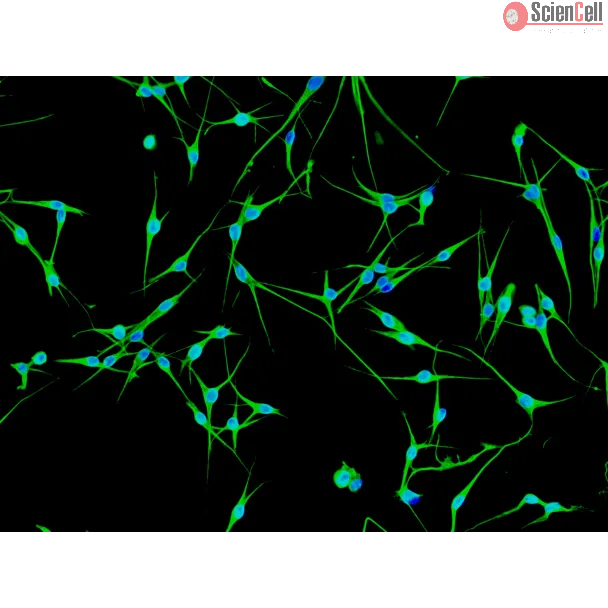
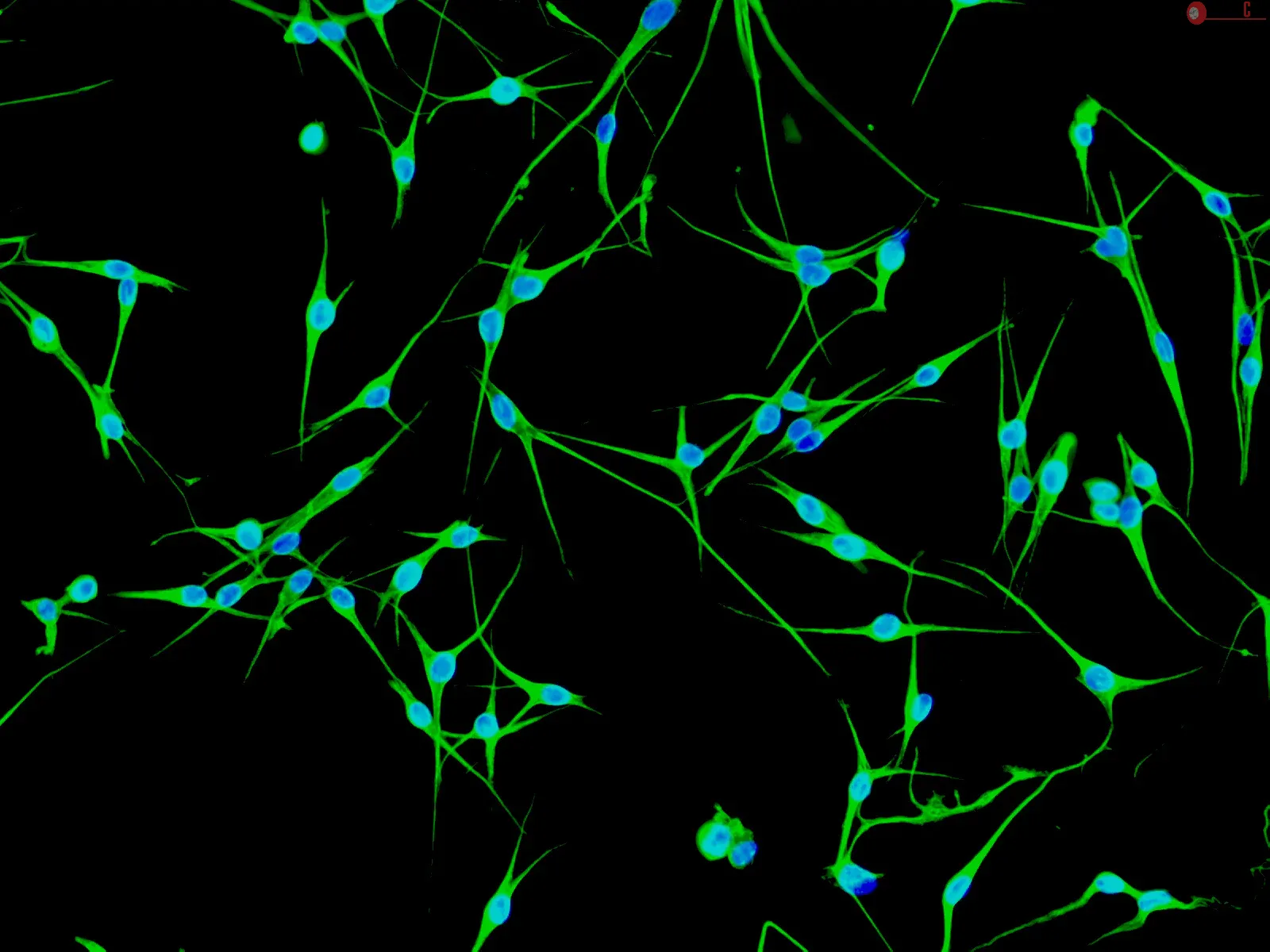
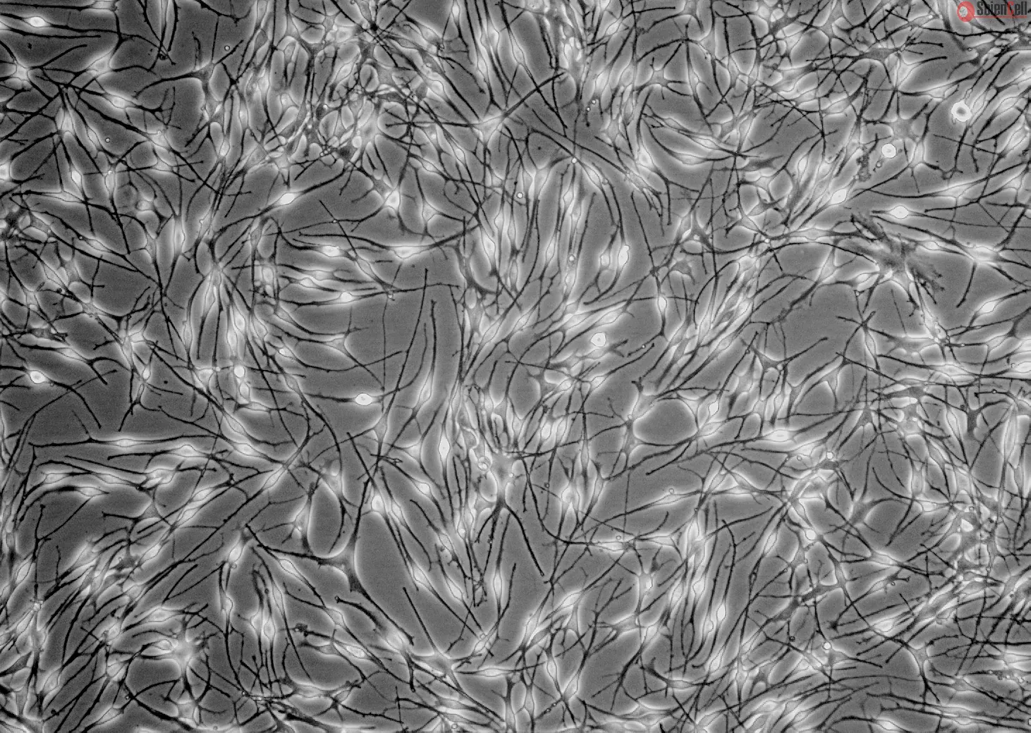
HEM-d from ScienCell Research Laboratories is isolated from neonatal human skin. HEM-d are cryopreserved at passage one and delivered frozen. Each vial contains >5 x 10⁵ cells in 1 ml volume. HEM-d are characterized by immunofluorescence with antibodies specific to S100β and/or NGF-receptor (p75). HEM-d are negative for HIV-1, HBV, HCV, mycoplasma, bacteria, yeast, and fungi. HEM-d are guaranteed to further expand for 10 population doublings under the
conditions provided by ScienCell Research Laboratories.
Recommended Medium
It is recommended to use Melanocyte Medium (MelM, Cat. #2201) for culturing HEM-d in vitro.
Most metastatic melanoma patients fail to respond to available therapy, underscoring the need for novel approaches to identify new effective treatments. In this study, we... More
Most metastatic melanoma patients fail to respond to available therapy, underscoring the need for novel approaches to identify new effective treatments. In this study, we screened 2,000 compounds from the Spectrum Library at a concentration of 1 micromol/L using two chemoresistant melanoma cell lines (M-14 and SK-Mel-19) and a spontaneously immortalized, nontumorigenic melanocyte cell line (melan-a). We identified 10 compounds that inhibited the growth of the melanoma cells yet were largely nontoxic to melanocytes. Strikingly, 4 of the 10 compounds (mebendazole, albendazole, fenbendazole, and oxybendazole) are benzimidazoles, a class of structurally related, tubulin-disrupting drugs. Mebendazole was prioritized to further characterize its mechanism of melanoma growth inhibition based on its favorable pharmacokinetic profile. Our data reveal that mebendazole inhibits melanoma growth with an average IC(50) of 0.32 micromol/L and preferentially induces apoptosis in melanoma cells compared with melanocytes. The intrinsic apoptotic response is mediated through phosphorylation of Bcl-2, which occurs rapidly after treatment with mebendazole in melanoma cells but not in melanocytes. Phosphorylation of Bcl-2 in melanoma cells prevents its interaction with proapoptotic Bax, thereby promoting apoptosis. We further show that mebendazole-resistant melanocytes can be sensitized through reduction of Bcl-2 protein levels, showing the essential role of Bcl-2 in the cellular response to mebendazole-mediated tubulin disruption. Our results suggest that this screening approach is useful for identifying agents that show promise in the treatment of even chemoresistant melanoma and identifies mebendazole as a potent, melanoma-specific cytotoxic agent. Less
Vascular endothelial growth factor (VEGF) is a cytokine and endothelial cell (EC) mitogen that has been studied for its role in angiogenesis of malignant tumors. Elevated... More
Vascular endothelial growth factor (VEGF) is a cytokine and endothelial cell (EC) mitogen that has been studied for its role in angiogenesis of malignant tumors. Elevated quantities of VEGF in the serum and plasma of patients have been correlated with the presence of cancer and metastasis. Since VEGF induces hyperpermeability of EC monolayers, this protein can be detected in vitro with a whole cell-based biosensor. This biosensor consists of a confluent monolayer of human umbilical vein endothelial cells (HUVECs) attached to a cellulose triacetate (CTA) membrane of an ion-selective electrode (ISE). Previous studies regarding this biosensor have shown that when the biosensor was exposed to a model toxin, such as histamine, the response of the biosensor served as an indirect measurement of the presence of histamine. Similarly, the biosensor responds to the presence of VEGF, but is much more sensitive because VEGF is known to be 50,000-fold more potent than histamine when inducing EC hyperpermeability. The ISE response increased with increasing VEGF concentration. Since lower concentrations required more exposure time, the detection limit was established as a function of exposure time (2-10 h). The practical applicability of the biosensor was also established with cultured human melanoma cells WM793 (nonmetastatic) and 1205LU (metastatic). The resultant change in the potential values revealed significant production of VEGF from the 1205LU cells. A VEGF ELISA was performed to confirm the VEGF concentration in each sample. The biosensor closely predicted the concentrations determined through the ELISA. These results support the use of a cell-based ISE as a quick screening method for the presence of VEGF. Less
ScienCell Research Laboratories (SRL) takes pride in being a resource for researchers all over the world. The publications listed here are not meant as an endorsement or confirmation of the reliability of the products.


,-1-mg-ml--2.jpg)