Melanocytes are neural crest-derived cells that produce melanin via melanogenesis. Melanocytes localize to several tissues including the epidermis, eye, inner ear and leptomeninges. Dysregulation of melanocyte migration, proliferation, or survival during embryonic development thus causes congenital disorders in those tissues as seen in Tietz syndrome, Waardenburg syndrome, and piebaldism. In the bottom layer of skin epidermis, melanocytes synthesize and transfer dark-colored melanin to surrounding keratinocytes to give skin pigmentation. Melanin also blocks UV-B light to protect the hypodermis from solar exposure-induced photodamage. Progress in culture techniques, along with an improved understanding of melanocyte biology, has led to a successful culture system to model melanomas, inner ear homeostasis, vitiligo, and
mitochondrial dysfunction in Duchenne Muscular Dystrophy.
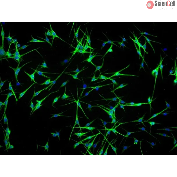
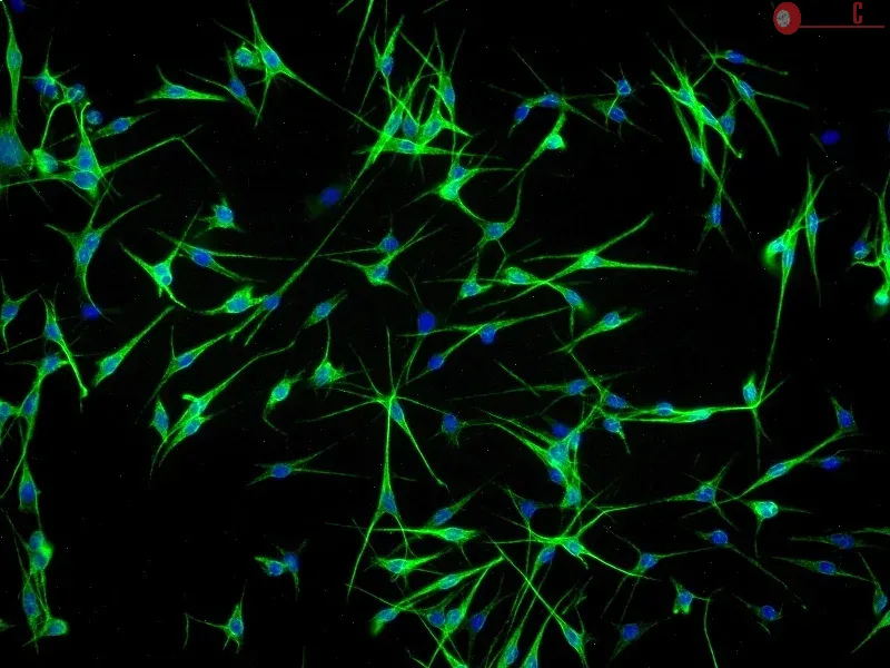
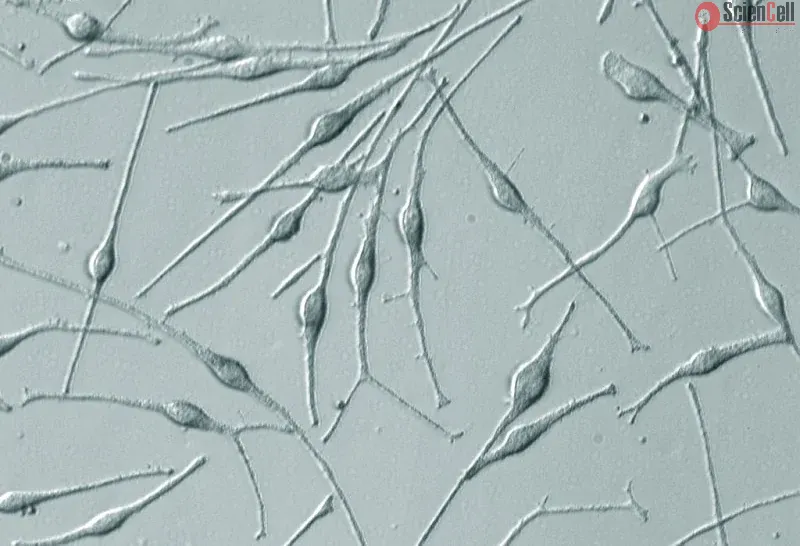
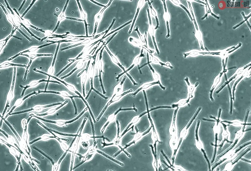
HEM-a from ScienCell Research Laboratories is isolated from adult human skin. HEM-a are cryopreserved at passage one and delivered frozen. Each vial contains >5 x 10⁵ cells in 1 ml volume. HEM-a are characterized by immunofluorescence with antibodies specific to S100β and/or NGF-receptor (p75). HEM-a are negative for HIV-1, HBV, HCV, mycoplasma, bacteria, yeast, and fungi. HEM-a are guaranteed to further expand for 10 population doublings under the conditions provided by ScienCell Research Laboratories.
Recommended Medium
It is recommended to use Melanocyte Medium (MelM, Cat. #2201) for culturing HEM-a in vitro.
Background: One of the most important and often neglected physiological stimuli contributing to the differentiation of vascular endothelial cells (ECs) into a blood-brain... More
Background: One of the most important and often neglected physiological stimuli contributing to the differentiation of vascular endothelial cells (ECs) into a blood-brain barrier (BBB) phenotype is shear stress (SS). With the use of a well established humanized dynamic in vitro BBB model and cDNA microarrays, we have profiled the effect of SS in the induction/suppression of ECs genes and related functions. Results: Specifically, we found a significant upregulation of tight and adherens junctions proteins and genes. Trans-endothelial electrical resistance (TEER) and permeability measurements to know substances have shown that SS promoted the formation of a tight and highly selective BBB. SS also increased the RNA level of multidrug resistance transporters, ion channels, and several p450 enzymes. The RNA level of a number of specialized carrier-mediated transport systems (e.g., glucose, monocarboxylic acid, etc.) was also upregulated.RNA levels of modulatory enzymes of the glycolytic pathway (e.g., lactate dehydrogenase) were downregulated by SS while those involved in the Krebs cycle (e.g., lactate and other dehydrogenases) were upregulated. Measurements of glucose consumption versus lactate production showed that SS negatively modulated the glycolytic bioenergetic pathways of glucose metabolism in favor of the more efficient aerobic respiration. BBB ECs are responsive to inflammatory stimuli. Our data showed that SS increased the RNA levels of integrins and vascular adhesion molecules. SS also inhibited endothelial cell cycle via regulation of BTG family proteins encoding genes. This was paralleled by significant increase in the cytoskeletal protein content while that of membrane, cytosol, and nuclear sub-cellular fractions decreased. Furthermore, analysis of 2D gel electrophoresis (which allows identifying a large number of proteins per sample) of EC proteins extracted from membrane sub-cellular endothelial fractions showed that SS increased the expression levels of tight junction proteins. In addition, regulatory enzymes of the Krebb's cycle (aerobic glucose metabolism) were also upregulated. Furthermore, the expression pattern of key protein regulators of the cell cycle and parallel gene array data supported a cell proliferation inhibitory role for SS. Conclusions: Genomic and proteomic analyses are currently used to examine BBB function in healthy and diseased brain and characterize this dynamic interface. In this study we showed that SS plays a key role in promoting the differentiation of vascular endothelial cells into a truly BBB phenotype. SS affected multiple aspect of the endothelial physiology spanning from tight junctions formation to cell division as well as the expression of multidrug resistance transporters. BBB dysfunction has been observed in many neurological diseases, but the causes are generally unknown. Our study provides essential insights to understand the role played by SS in the BBB formation and maintenance. Less
Background: Centratherum anthelminticum (L.) Kuntze (scientific synonyms: Vernonia anthelmintica; black cumin) is one of the ingredients of an Ayurvedic preparation, call... More
Background: Centratherum anthelminticum (L.) Kuntze (scientific synonyms: Vernonia anthelmintica; black cumin) is one of the ingredients of an Ayurvedic preparation, called “Kayakalp”, commonly applied to treat skin disorders in India and Southeast Asia. Despite its well known anti-inflammatory property on skin diseases, the anti-cancer effect of C. anthelminticum seeds on skin cancer is less documented. The present study aims to investigate the anti-cancer effect of Centratherum anthelminticum (L.) seeds chloroform fraction (CACF) on human melanoma cells and to elucidate the molecular mechanism involved. Methods: A chloroform fraction was extracted from C. anthelminticum (CACF). Bioactive compounds of the CACF were analyzed by liquid chromatography-tandem mass spectrometry (LC-MS/MS). Human melanoma cell line A375 was treated with CACF in vitro. Effects of CACF on growth inhibition, morphology, stress and survival of the cell were examined with MTT, high content screening (HSC) array scan and flow cytometry analyses. Involvement of intrinsic or extrinsic pathways in the CACF-induced A375 cell death mechanism was examined using a caspase luminescence assay. The results were further verified with different caspase inhibitors. In addition, Western blot analysis was performed to elucidate the changes in apoptosis-associated molecules. Finally, the effect of CACF on the NF-κB nuclear translocation ability was assayed. Results: The MTT assay showed that CACF dose-dependently inhibited cell growth of A375, while exerted less cytotoxic effect on normal primary epithelial melanocytes. We demonstrated that CACF induced cell growth inhibition through apoptosis, as evidenced by cell shrinkage, increased annexin V staining and formation of membrane blebs. CACF treatment also resulted in higher reactive oxygen species (ROS) production and lower Bcl-2 expression, leading to decrease mitochondrial membrane potential (MMP). Disruption of the MMP facilitated the release of mitochondrial cytochrome c, which activates caspase-9 and downstream caspase-3/7, resulting in DNA fragmentation and up-regulation of p53 in melanoma cells. Moreover, CACF prevented TNF-α-induced NF-κB nuclear translocation, which further committed A375 cells toward apoptosis. Conclusions: Together, our findings suggest CACF as a potential therapeutic agent against human melanoma malignancy. Keywords: Centratherum anthelminticum, Melanoma, Caspase cascade, Apoptosis, Bcl-2, p53, NF-κB Less
Background: Genomic instability due to UV radiation is one of the leading causes for melanoma. Histone acetyltransferase p300 plays an indispensible role in DNA repair an... More
Background: Genomic instability due to UV radiation is one of the leading causes for melanoma. Histone acetyltransferase p300 plays an indispensible role in DNA repair and maintenance of genomic integrity. The present study was performed to analyze the correlation between p300 expression, melanoma progression and patient survival. Methods: Tissue microarray and immunohistochemical analysis was employed to study the expression of p300 in melanoma patients. A total of 358 melanoma patients (250 primary melanoma and 108 metastatic melanoma) were used for the study. Kaplan-Meier, univariate and multivariate Cox regression analysis, and receiver-operating characteristic curves, were used to elucidate the prognostic significance of p300 expression. Results: Our results demonstrate that p300 is expressed in both nucleus and cytoplasm but the nuclear expression of p300 is predominant. The progression of disease from dysplastic nevi to primary melanoma and to metastatic melanoma was associated with decreased nuclear and increased cytoplasmic p300 expression. Especially, the loss of nuclear and gain in cytoplasmic p300 was correlated with the progression of melanoma from AJCC stage II to stage III, which requires the migration and metastasis of cancer cells from primary sites to lymph nodes. Similarly, decrease in nuclear, and increase in cytoplasmic p300 expression correlated with worse survival of melanoma patients. Nuclear p300 but not cytoplasmic p300 could predict the patient survival independent of AJCC stage, age and gender. Conclusion: Loss of nuclear p300 expression is an indicator of worse patient survival and is an independent prognostic marker for melanoma. Less
ScienCell Research Laboratories (SRL) takes pride in being a resource for researchers all over the world. The publications listed here are not meant as an endorsement or confirmation of the reliability of the products.
,-1-mg-ml--2.jpg)


.webp)

