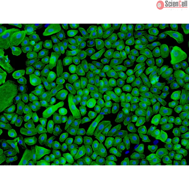
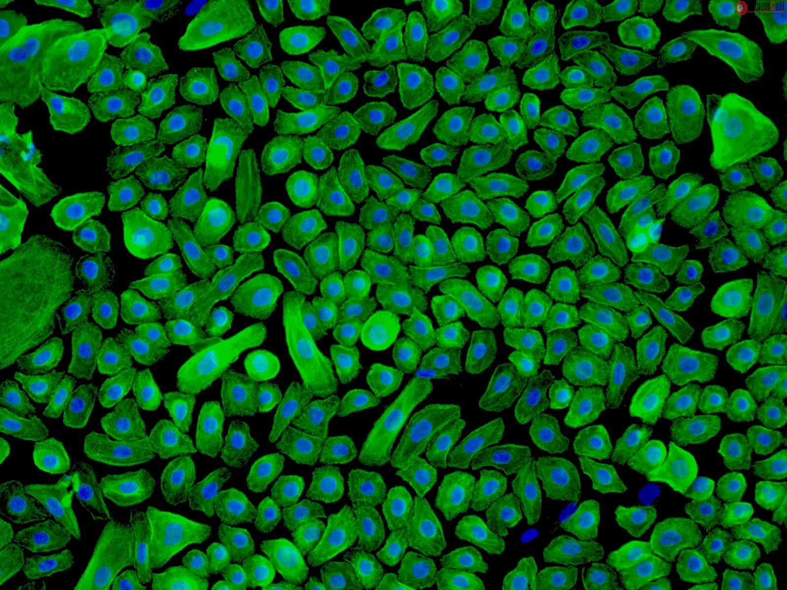
The epidermal layer of the skin provides an essential function as a protective barrier against insults from the external environment. The predominant cell type in the epidermis is keratinocytes and they are located in the stratified squamous epithelia. Keratinocytes are named after keratin, which is the most abundant protein in this cell. Progenitors of keratinocytes reside and divide in the basal layer of the epidermis. They then differentiate, migrate towards the surface of epidermis, and eventually withdraw from the cell cycle permanently. Keratinocyte proliferation, differentiation, and programmed cell death are complex and carefully choreographed processes. Apart from their protective functions, keratinocytes express adhesion molecules and cytokines, further suggesting an implication in skin innate immunity, tissue homeostasis, wound healing, cancer development, and skin-based gene-therapy.
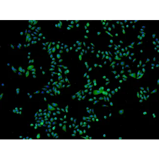
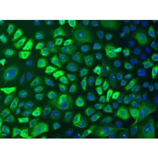
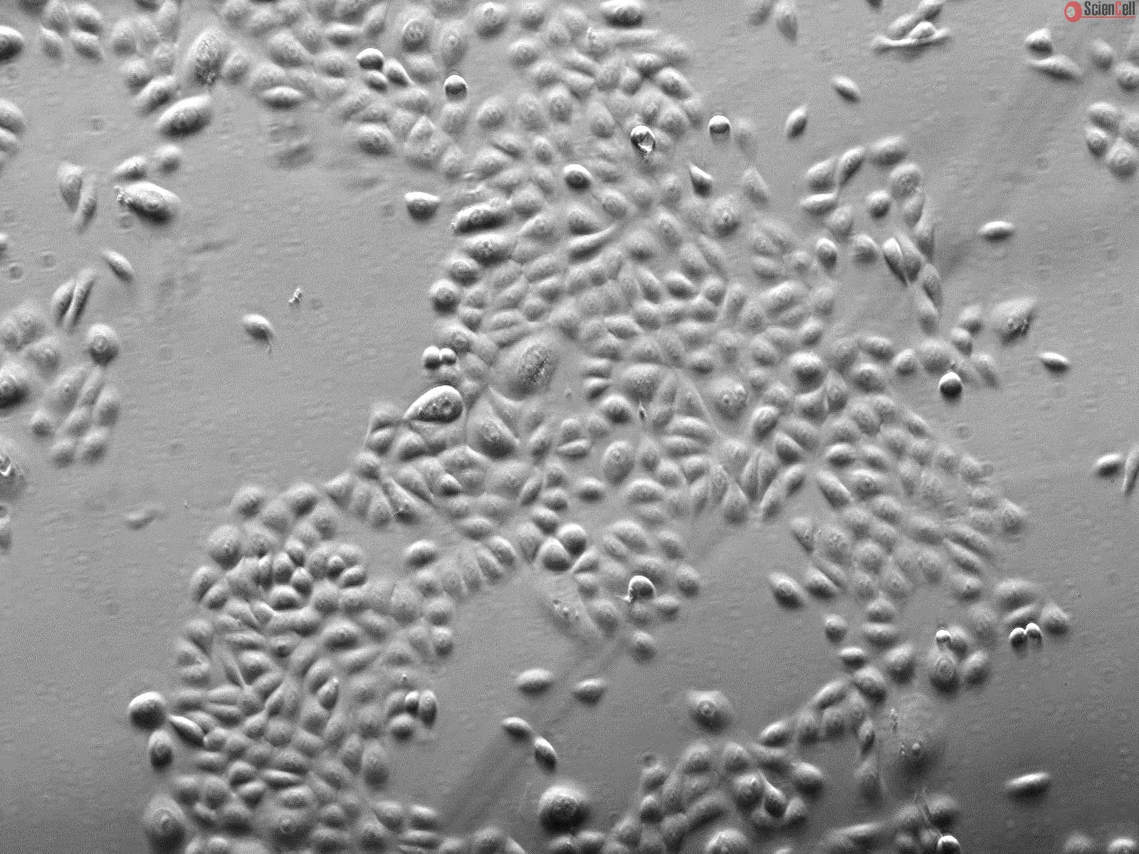
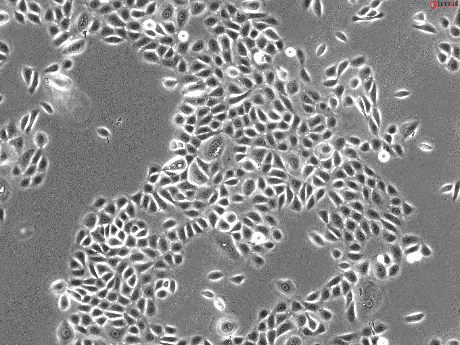
HEK from ScienCell Research Laboratories are isolated from neonatal human skin. HEK are cryopreserved at passage one and delivered frozen. Each vial contains >5 x 10⁵ cells in 1 ml volume. HEK are characterized by immunofluorescence with antibodies specific to cytokeratin-18 and/or cytokeratin-19. HEK are negative for HIV-1, HBV, HCV, mycoplasma, bacteria, yeast, and fungi. HEK are guaranteed to further expand for 15 population doublings under the conditions provided by ScienCell Research Laboratories.
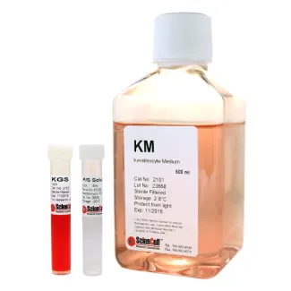
Recommended Medium
It is recommended to use Keratinocyte Medium (KM, Cat. #2101) for culturing HEK in vitro.
IL-31 is a novel T(h) type 2 cytokine that can induce pruritus and dermatitis in mice resembling human atopic dermatitis (AD). Eosinophil infiltration in skin lesions is ... More
IL-31 is a novel T(h) type 2 cytokine that can induce pruritus and dermatitis in mice resembling human atopic dermatitis (AD). Eosinophil infiltration in skin lesions is a predominant pathological feature of AD. In the present study, we investigated the effects of IL-31 on the activation of human eosinophils and epidermal keratinocytes. Eosinophils and keratinocytes were cultured either together or separately in the presence or absence of IL-31 stimulation. IL-31 could significantly induce the release of pro-inflammatory cytokines IL-1beta, IL-6 and AD-related chemokines CXCL1, CXCL8, CCL2 and CCL18 from eosinophils, via functional cell surface IL-31 receptor. Such induction was further enhanced upon the co-culture of eosinophils and keratinocytes, in which eosinophils were the main source for releasing pro-inflammatory cytokines and chemokines. The presence of transwell inserts in co-culture system demonstrated that the direct interaction between eosinophils and keratinocytes was required for IL-31-induced cytokine and chemokine release. Cell surface expression of adhesion molecule CD18 on eosinophils and intercellular adhesion molecule-1 on keratinocytes was up-regulated in the co-culture, and levels were further enhanced upon IL-31 stimulation. The interaction between eosinophils and keratinocytes under IL-31 stimulation was differentially mediated through intracellular mitogen-activated protein kinases, nuclear factor-kappaB and phosphatidylinositol 3-kinase-Akt pathways. The above findings suggest a crucial immunopathological role of IL-31 in AD through activation of eosinophils-keratinocytes system. Less
The authors determined the effect of cyclic guanosine 3′,5′-monophosphate (cGMP) on hydrogen peroxide (H2O2)-induced barrier dysfunction in bovine lung microvascular ... More
The authors determined the effect of cyclic guanosine 3′,5′-monophosphate (cGMP) on hydrogen peroxide (H2O2)-induced barrier dysfunction in bovine lung microvascular endothelial cell (BLMVEC) monolayers and compared the results to bovine pulmonary artery endothelial cells (BPAECs). In BLMVECs, H2O2 (250 μM) caused a 31.9% ± 4.8% decrease in transendothelial electrical resistance (TER) associated with increased actin stress fiber formation, intercellular gaps, and intracellular calcium concentration ([Ca2+]i). The cGMP analogue 8-(p-chlorophenylthio)-cGMP (8p-CPT-cGMP; 30 or 50 μM) prevented the H2O2-induced decrease in TER (p < .001) as well as the cytoskeletal rearrangement and intercellular gap formation. 8-pCPT-cGMP (50 μM) attenuated the peak (418.8 ± 42.1 versus 665.2 ± 38.0 nmol/L; p < .001) and eliminated the sustained increase in [Ca2+]i (193.5 ± 21.3 versus 418.8 ± 42.1 nmol/L; p < .001) caused by H2O2. 8-pCPT-cGMP also increased TER (14.2% ± 2.2%; p < .05) and decreased [Ca2+]i (201.2 ± 12.5 vs. 214.4 ± 12.1 nmol/L; p < .03) before H2O2. In BPAECs, 8p-CPT-cGMP significantly attenuated H2O2-induced increases in permeability and [C2+]i but less effectively than in BLMVECs. These results suggest that in BLMVECs, cGMP countered the adverse effects of H2O2 on barrier function by preventing actin cytoskeletal rearrangement and attenuating the increase in [Ca2+]i. Less
ScienCell Research Laboratories (SRL) takes pride in being a resource for researchers all over the world. The publications listed here are not meant as an endorsement or confirmation of the reliability of the products.


,-1-mg-ml--2.jpg)